 Antibody at 1/1000 dilution.")


| 製品: | Phospho-IRAK1 (Thr387) Antibody |
| カタログ: | AF8009 |
| タンパク質の説明: | Rabbit polyclonal antibody to Phospho-IRAK1 (Thr387) |
| アプリケーション: | WB IHC |
| Cited expt.: | WB |
| 反応性: | Human, Mouse, Rat |
| 予測: | Pig, Zebrafish, Bovine, Horse, Sheep, Rabbit, Dog |
| 分子量: | 77kDa(Observed); 77kD(Calculated). |
| ユニプロット: | P51617 |
| RRID: | AB_2840072 |

製品説明
*The optimal dilutions should be determined by the end user. For optimal experimental results, antibody reuse is not recommended.
*Tips:
WB: For western blot detection of denatured protein samples. IHC: For immunohistochemical detection of paraffin sections (IHC-p) or frozen sections (IHC-f) of tissue samples. IF/ICC: For immunofluorescence detection of cell samples. ELISA(peptide): For ELISA detection of antigenic peptide.
引用形式: Affinity Biosciences Cat# AF8009, RRID:AB_2840072.
折りたたみ/展開
AA48924; Il1rak; Interleukin 1 receptor associated kinase 1; Interleukin-1 receptor-associated kinase 1; IRAK; IRAK-1; Irak1; IRAK1-S; IRAK1_HUMAN; mPLK; OTTHUMP00000026014; OTTHUMP00000026015; OTTHUMP00000026020; OTTHUMP00000180621; Pelle; Pelle homolog; Pelle-like protein kinase; Plpk;
免疫原
A synthesized peptide derived from human IRAK1 around the phosphorylation site of Thr387.
Isoform 1 and isoform 2 are ubiquitously expressed in all tissues examined, with isoform 1 being more strongly expressed than isoform 2.
- P51617 IRAK1_HUMAN:
- Protein BLAST With
- NCBI/
- ExPASy/
- Uniprot
MAGGPGPGEPAAPGAQHFLYEVPPWVMCRFYKVMDALEPADWCQFAALIVRDQTELRLCERSGQRTASVLWPWINRNARVADLVHILTHLQLLRARDIITAWHPPAPLPSPGTTAPRPSSIPAPAEAEAWSPRKLPSSASTFLSPAFPGSQTHSGPELGLVPSPASLWPPPPSPAPSSTKPGPESSVSLLQGARPFPFCWPLCEISRGTHNFSEELKIGEGGFGCVYRAVMRNTVYAVKRLKENADLEWTAVKQSFLTEVEQLSRFRHPNIVDFAGYCAQNGFYCLVYGFLPNGSLEDRLHCQTQACPPLSWPQRLDILLGTARAIQFLHQDSPSLIHGDIKSSNVLLDERLTPKLGDFGLARFSRFAGSSPSQSSMVARTQTVRGTLAYLPEEYIKTGRLAVDTDTFSFGVVVLETLAGQRAVKTHGARTKYLKDLVEEEAEEAGVALRSTQSTLQAGLAADAWAAPIAMQIYKKHLDPRPGPCPPELGLGLGQLACCCLHRRAKRRPPMTQVYERLEKLQAVVAGVPGHSEAASCIPPSPQENSYVSSTGRAHSGAAPWQPLAAPSGASAQAAEQLQRGPNQPVESDESLGGLSAALRSWHLTPSCPLDPAPLREAGCPQGDTAGESSWGSGPGSRPTAVEGLALGSSASSSSEPPQIIINPARQKMVQKLALYEDGALDSLQLLSSSSLPGLGLEQDRQGPEESDEFQS
種類予測
Score>80(red) has high confidence and is suggested to be used for WB detection. *The prediction model is mainly based on the alignment of immunogen sequences, the results are for reference only, not as the basis of quality assurance.
High(score>80) Medium(80>score>50) Low(score<50) No confidence
研究背景
Serine/threonine-protein kinase that plays a critical role in initiating innate immune response against foreign pathogens. Involved in Toll-like receptor (TLR) and IL-1R signaling pathways. Is rapidly recruited by MYD88 to the receptor-signaling complex upon TLR activation. Association with MYD88 leads to IRAK1 phosphorylation by IRAK4 and subsequent autophosphorylation and kinase activation. Phosphorylates E3 ubiquitin ligases Pellino proteins (PELI1, PELI2 and PELI3) to promote pellino-mediated polyubiquitination of IRAK1. Then, the ubiquitin-binding domain of IKBKG/NEMO binds to polyubiquitinated IRAK1 bringing together the IRAK1-MAP3K7/TAK1-TRAF6 complex and the NEMO-IKKA-IKKB complex. In turn, MAP3K7/TAK1 activates IKKs (CHUK/IKKA and IKBKB/IKKB) leading to NF-kappa-B nuclear translocation and activation. Alternatively, phosphorylates TIRAP to promote its ubiquitination and subsequent degradation. Phosphorylates the interferon regulatory factor 7 (IRF7) to induce its activation and translocation to the nucleus, resulting in transcriptional activation of type I IFN genes, which drive the cell in an antiviral state. When sumoylated, translocates to the nucleus and phosphorylates STAT3.
Following recruitment on the activated receptor complex, phosphorylated on Thr-209, probably by IRAK4, resulting in a conformational change of the kinase domain, allowing further phosphorylations to take place. Thr-387 phosphorylation in the activation loop is required to achieve full enzymatic activity.
Polyubiquitinated by TRAF6 after cell stimulation with IL-1-beta by PELI1, PELI2 and PELI3. Polyubiquitination occurs with polyubiquitin chains linked through 'Lys-63'. Ubiquitination promotes interaction with NEMO/IKBKG. Also sumoylated; leading to nuclear translocation.
Cytoplasm. Nucleus. Lipid droplet.
Note: Translocates to the nucleus when sumoylated. RSAD2/viperin recruits it to the lipid droplet (By similarity).
Isoform 1 and isoform 2 are ubiquitously expressed in all tissues examined, with isoform 1 being more strongly expressed than isoform 2.
The ProST region is composed of many proline and serine residues (more than 20 of each) and some threonines. This region is the site of IRAK-1 hyperphosphorylation.
Belongs to the protein kinase superfamily. TKL Ser/Thr protein kinase family. Pelle subfamily.
研究領域
· Environmental Information Processing > Signal transduction > MAPK signaling pathway. (View pathway)
· Environmental Information Processing > Signal transduction > NF-kappa B signaling pathway. (View pathway)
· Human Diseases > Infectious diseases: Bacterial > Pertussis.
· Human Diseases > Infectious diseases: Parasitic > Leishmaniasis.
· Human Diseases > Infectious diseases: Parasitic > Chagas disease (American trypanosomiasis).
· Human Diseases > Infectious diseases: Parasitic > Toxoplasmosis.
· Human Diseases > Infectious diseases: Bacterial > Tuberculosis.
· Human Diseases > Infectious diseases: Viral > Measles.
· Human Diseases > Infectious diseases: Viral > Epstein-Barr virus infection.
· Organismal Systems > Immune system > Toll-like receptor signaling pathway. (View pathway)
· Organismal Systems > Nervous system > Neurotrophin signaling pathway. (View pathway)
参考文献
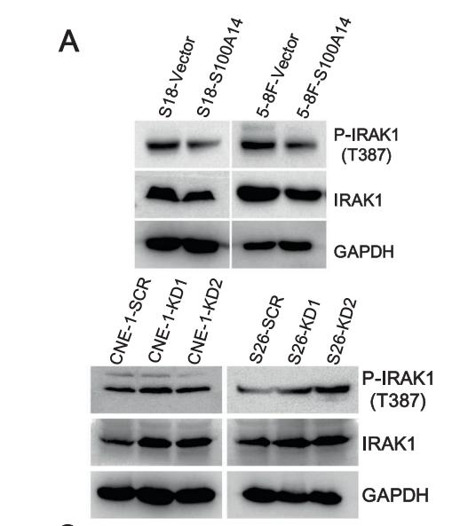
Application: WB Species: Human Sample: nasopharyngeal mucosa and nasopharyngeal carcinoma

Application: WB Species: mouse Sample:

Application: WB Species: Rat Sample:

Application: WB Species: Rat Sample: OPCs

Application: IF/ICC Species: Rat Sample: OPCs
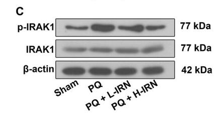
Application: WB Species: Rat Sample: kidney tissues
Restrictive clause
Affinity Biosciences tests all products strictly. Citations are provided as a resource for additional applications that have not been validated by Affinity Biosciences. Please choose the appropriate format for each application and consult Materials and Methods sections for additional details about the use of any product in these publications.
For Research Use Only.
Not for use in diagnostic or therapeutic procedures. Not for resale. Not for distribution without written consent. Affinity Biosciences will not be held responsible for patent infringement or other violations that may occur with the use of our products. Affinity Biosciences, Affinity Biosciences Logo and all other trademarks are the property of Affinity Biosciences LTD.