製品: GAP43 Antibody
カタログ: DF7766
タンパク質の説明: Rabbit polyclonal antibody to GAP43
アプリケーション: WB IHC IF/ICC
反応性: Human, Mouse, Rat
予測: Pig, Bovine, Rabbit, Dog
分子量: 43 kDa; 25kD(Calculated).
ユニプロット: P17677
RRID: AB_2841232

類似製品を見る>>

   サイズ 価格 在庫状況
 100ul $280 在庫あり
 200ul $350 在庫あり

リードタイム: 当日配達

For pricing and ordering contact:
お問い合わせ先

製品説明

ソース:
Rabbit
アプリケーション:
WB 1:1000-3000, IF/ICC 1:100-1:500, IHC 1:50-1:200
*The optimal dilutions should be determined by the end user.
*Tips:

WB: For western blot detection of denatured protein samples. IHC: For immunohistochemical detection of paraffin sections (IHC-p) or frozen sections (IHC-f) of tissue samples. IF/ICC: For immunofluorescence detection of cell samples. ELISA(peptide): For ELISA detection of antigenic peptide.

反応性:
Human,Mouse,Rat
予測:
Pig(88%), Bovine(88%), Rabbit(88%), Dog(88%)
クローナリティ:
Polyclonal
特異性:
GAP43 Antibody detects endogenous levels of total GAP43.
RRID:
AB_2841232
引用形式: Affinity Biosciences Cat# DF7766, RRID:AB_2841232.
コンジュゲート:
Unconjugated.
精製:
The antiserum was purified by peptide affinity chromatography using SulfoLink™ Coupling Resin (Thermo Fisher Scientific).
保存:
Rabbit IgG in phosphate buffered saline , pH 7.4, 150mM NaCl, 0.02% sodium azide and 50% glycerol. Store at -20 °C. Stable for 12 months from date of receipt.
別名:

折りたたみ/展開

Axonal membrane protein GAP 43; Axonal membrane protein GAP-43; B 50; Calmodulin binding protein P 57; F1; GAP 43; GAP43; Growth Associated Protein 43; Growth-associated protein 43; Nerve Growth Related Peptide; Nerve growth related peptide GAP43; NEUM_HUMAN; Neural phosphoprotein B 50; Neural phosphoprotein B-50; Neuromodulin; Neuron growth associated protein 43; PP46; Protein F1; QtrA-11580; QtrA-13071;

免疫原

免疫原:
Uniprot:
遺伝子(ID):
タンパク質配列:
MLCCMRRTKQVEKNDDDQKIEQDGIKPEDKAHKAATKIQASFRGHITRKKLKGEKKDDVQAAEAEANKKDEAPVADGVEKKGEGTTTAEAAPATGSKPDEPGKAGETPSEEKKGEGDAATEQAAPQAPASSEEKAGSAETESATKASTDNSPSSKAEDAPAKEEPKQADVPAAVTAAAATTPAAEDAAAKATAQPPTETGESSQAEENIEAVDETKPKESARQDEGKEEEPEADQEHA

種類予測

種類予測:

Score>80(red) has high confidence and is suggested to be used for WB detection. *The prediction model is mainly based on the alignment of immunogen sequences, the results are for reference only, not as the basis of quality assurance.

Species
Results
Score
Pig
88
Bovine
88
Dog
88
Rabbit
88
Chicken
69
Xenopus
56
Horse
0
Sheep
0
Zebrafish
0
Model Confidence:
High(score>80) Medium(80>score>50) Low(score<50) No confidence

PTMs - P17677 基板として

Site PTM Type Enzyme
T36 Phosphorylation
K37 Ubiquitination
S41 Phosphorylation Q02156 (PRKCE)
T107 Phosphorylation
S109 Phosphorylation
S131 Phosphorylation
S142 Phosphorylation
T144 Phosphorylation
S147 Phosphorylation
T148 Phosphorylation
S151 Phosphorylation
S154 Phosphorylation
T181 Phosphorylation
K216 Methylation

研究背景

機能:

This protein is associated with nerve growth. It is a major component of the motile 'growth cones' that form the tips of elongating axons. Plays a role in axonal and dendritic filopodia induction.

PTMs:

Phosphorylated (By similarity). Phosphorylation of this protein by a protein kinase C is specifically correlated with certain forms of synaptic plasticity (By similarity).

Palmitoylation by ARF6 is essential for plasma membrane association and axonal and dendritic filopodia induction. Deacylated by LYPLA2.

細胞の位置付け:

Cell membrane>Peripheral membrane protein>Cytoplasmic side. Cell projection>Growth cone membrane>Peripheral membrane protein>Cytoplasmic side. Cell junction>Synapse. Cell projection>Filopodium membrane>Peripheral membrane protein. Perikaryon. Cell projection>Dendrite. Cell projection>Axon. Cytoplasm.
Note: Cytoplasmic surface of growth cone and synaptic plasma membranes.

Extracellular region or secreted Cytosol Plasma membrane Cytoskeleton Lysosome Endosome Peroxisome ER Golgi apparatus Nucleus Mitochondrion Manual annotation Automatic computational assertionSubcellular location
サブユニット構造:

Identified in a complex containing FGFR4, NCAM1, CDH2, PLCG1, FRS2, SRC, SHC1, GAP43 and CTTN (By similarity). Interacts (via IQ domain) with calmodulin (By similarity). Binds calmodulin with a greater affinity in the absence of Ca(2+) than in its presence (By similarity).

タンパク質ファミリー:

Belongs to the neuromodulin family.

参考文献

1). SiNiSan ameliorates depression-like behavior in rats by enhancing synaptic plasticity via the CaSR-PKC-ERK signaling pathway. BIOMEDICINE & PHARMACOTHERAPY, 2020 (PubMed: 31958763) [IF=7.5]

Application: WB    Species: rat    Sample: HIP

Fig. 5. |Effects of SNS on synaptic-associated protein in the HIP and PFC of stressed rats. Representative immunoblots for PSD-95, GAP-43, Syn, and Tublin in the HIP(A) and PFC(B) regions. (A) Results of relative protein levels of PSD-95, GAP-43, and Syn in the HIP of rats of each group.

2). Exosomes derived from human induced pluripotent stem cell-derived neural progenitor cells protect neuronal function under ischemic conditions. Neural Regeneration Research, 2021 (PubMed: 33642395) [IF=6.1]

Application: WB    Species: Rat    Sample: neural progenitor cells

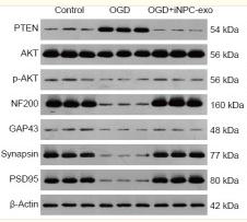
Figure 3 Effect of iPSC-NPC-derived exosomes (OGD+iNPC-exo) on expression of the PTEN/AKT signaling pathway and of synaptic plasticity-related proteins in OGD induced neurons. (A–C) mRNA expression (A) and protein expression (B, C) of the PTEN/AKT signaling pathway and of synaptic plasticity-related proteins (NF200, GAP-43, Synapsin, and PSD95) analyzed by polymerase chain reaction and western blot assay. The mRNA expression is described by the optical density ratio relative to the control group. Protein expression was described by the optical density ratio relative to β-actin. Data are presented as mean ± SD. ***P < 0.001, vs. control group; ###P < 0.001 (one-way analysis of variance with post hoc Bonferroni test). All experiments were repeated three times. GAP43: growth associated protein 43; iPSC-NPCs: induced pluripotent stem cells-derived neural progenitor cells; NF200: neurofilament 200; OGD: oxygen-glucose deprivation; p-Akt: phosphor-Akt; PSD95: postsynaptic density protein 95; PTEN: phosphatase and tensin homolog deleted on chromosome ten.

3). Early-Life Stress Induces Depression-Like Behavior and Synaptic-Plasticity Changes in a Maternal Separation Rat Model: Gender Difference and Metabolomics Study. Frontiers in Pharmacology, 2020 (PubMed: 32174832) [IF=5.6]

Application: WB    Species: Rat    Sample: hippocampus

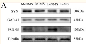
Figure 5 MS reduces the expression of synaptic-plasticity protein. (A) The bands of synaptic-plasticity proteins of SYN, PSD-95, and GAP-43 in the hippocampus by WB. Statistical results indicate the relative protein levels expressed by SYN, GAP-43, and PSD-95. (B) The bands of synaptic-plasticity proteins of SYN, PSD-95, and GAP-43 in cortex by WB. Statistical results indicate the relative protein levels expressed by SYN, GAP-43, and PSD-95. Statistical analyses are performed by two-way ANOVA followed by t-test. Data are presented as mean ± SEM, *p < 0.05, **p < 0.01, n = 3 per group.

4). Erythropoietin-Induced Autophagy Protects Against Spinal Cord Injury and Improves Neurological Function via the Extracellular-Regulated Protein Kinase Signaling Pathway. MOLECULAR NEUROBIOLOGY, 2020 (PubMed: 32647973) [IF=5.1]

Application: IF/ICC    Species: rat    Sample:

Fig. 6 |EPO benefit antiinflammation and axonal regeneration can be neutralized by ERK inhibition, and activator can mimic the effect of EPO. a The axonal special protein GAP43 immunofluorescence staining and histogram at 14 days (magnification × 400). b–e The inflammatory marker proteins immunofluorescence staining and histograms at 14 days, including CD86, GFAP, TNF-α, iNOS(magnification × 400). All data presented as mean ± SEM in each group. *p < 0.05 vs. SCI + saline group, **p < 0.01 vs. SCI + saline group. #p < 0.05 comparison between SCI + EPO group and SCI + EPO + PD98059, ##p < 0.01 comparison between SCI + EPO group and SCI + EPO + PD98059

5). The Effect of Early Maternal Separation Combined With Adolescent Chronic Unpredictable Mild Stress on Behavior and Synaptic Plasticity in Adult Female Rats. Frontiers in Psychiatry, 2021 (PubMed: 33746787) [IF=4.7]

Application: WB    Species: Rat    Sample: Hippocampus

Figure 5 Western blot analysis. The expressions of synaptic plasticity proteins were determined by western blot (A), including PSD-95 (B), GAP-43 (C), and SYN (D). The values represent the mean ± SEM, n = 5. *p < 0.05, **p < 0.01 vs. CON, #p < 0.05, ##p < 0.01 vs. MS. CON, control; MS, maternal separation; CUMS, chronic unpredictable mild stress.

6). Water Treadmill Training Ameliorates Neurite Outgrowth Inhibition Associated with NGR/RhoA/ROCK by Inhibiting Astrocyte Activation following Spinal Cord Injury. Oxidative Medicine and Cellular Longevity, 2021 (PubMed: 35387259)

Application: WB    Species: rat    Sample: spinal cord

Figure 3:| TT promoted axonal outgrowth after SCI. (a)–(c) Double staining of spinal cord sections in each group of rats for GAP43 (green)/NF200 (red)/DAPI (blue). Scale bars are 500 μm (a, b) and 50 μm (c).

Restrictive clause


Affinity Biosciences tests all products strictly. Citations are provided as a resource for additional applications that have not been validated by Affinity Biosciences. Please choose the appropriate format for each application and consult Materials and Methods sections for additional details about the use of any product in these publications.

For Research Use Only.
Not for use in diagnostic or therapeutic procedures. Not for resale. Not for distribution without written consent. Affinity Biosciences will not be held responsible for patent infringement or other violations that may occur with the use of our products. Affinity Biosciences, Affinity Biosciences Logo and all other trademarks are the property of Affinity Biosciences LTD.