, using B-Myb Antibody at 1/1000 dilution.
5ug/NC membrane strip.
Exposure for 10s with Affinity™ ECL Kit(#KF8001).
Bands result from membrane strip incubation.")
, using B-Myb Antibody at 1/1000 dilution.
5ug/NC membrane strip.
Exposure for 10s with Affinity™ ECL Kit(#KF8001).
Bands result from membrane strip incubation.")
Control Products

製品説明
*The optimal dilutions should be determined by the end user. For optimal experimental results, antibody reuse is not recommended.
*Tips:
WB: For western blot detection of denatured protein samples. IHC: For immunohistochemical detection of paraffin sections (IHC-p) or frozen sections (IHC-f) of tissue samples. IF/ICC: For immunofluorescence detection of cell samples. ELISA(peptide): For ELISA detection of antigenic peptide.
引用形式: Affinity Biosciences Cat# DF7817, RRID:AB_2841266.
折りたたみ/展開
B-Myb; BMyB; MGC15600; MYB L2; Myb related protein B; Myb-like protein 2; Myb-related protein B; MybB; MYBB_HUMAN; MYBL 2; Mybl2; v myb avian myeloblastosis viral oncogene homolog like 2; v myb myeloblastosis viral oncogene homolog (avian) like 2; v myb myeloblastosis viral oncogene homolog like 2;
免疫原
A synthesized peptide derived from human B-Myb, corresponding to a region within the internal amino acids.
- P10244 MYBB_HUMAN:
- Protein BLAST With
- NCBI/
- ExPASy/
- Uniprot
MSRRTRCEDLDELHYQDTDSDVPEQRDSKCKVKWTHEEDEQLRALVRQFGQQDWKFLASHFPNRTDQQCQYRWLRVLNPDLVKGPWTKEEDQKVIELVKKYGTKQWTLIAKHLKGRLGKQCRERWHNHLNPEVKKSCWTEEEDRIICEAHKVLGNRWAEIAKMLPGRTDNAVKNHWNSTIKRKVDTGGFLSESKDCKPPVYLLLELEDKDGLQSAQPTEGQGSLLTNWPSVPPTIKEEENSEEELAAATTSKEQEPIGTDLDAVRTPEPLEEFPKREDQEGSPPETSLPYKWVVEAANLLIPAVGSSLSEALDLIESDPDAWCDLSKFDLPEEPSAEDSINNSLVQLQASHQQQVLPPRQPSALVPSVTEYRLDGHTISDLSRSSRGELIPISPSTEVGGSGIGTPPSVLKRQRKRRVALSPVTENSTSLSFLDSCNSLTPKSTPVKTLPFSPSQFLNFWNKQDTLELESPSLTSTPVCSQKVVVTTPLHRDKTPLHQKHAAFVTPDQKYSMDNTPHTPTPFKNALEKYGPLKPLPQTPHLEEDLKEVLRSEAGIELIIEDDIRPEKQKRKPGLRRSPIKKVRKSLALDIVDEDVKLMMSTLPKSLSLPTTAPSNSSSLTLSGIKEDNSLLNQGFLQAKPEKAAVAQKPRSHFTTPAPMSSAWKTVACGGTRDQLFMQEKARQLLGRLKPSHTSRTLILS
種類予測
Score>80(red) has high confidence and is suggested to be used for WB detection. *The prediction model is mainly based on the alignment of immunogen sequences, the results are for reference only, not as the basis of quality assurance.
High(score>80) Medium(80>score>50) Low(score<50) No confidence
研究背景
Transcription factor involved in the regulation of cell survival, proliferation, and differentiation. Transactivates the expression of the CLU gene.
Phosphorylated by cyclin A/CDK2 during S-phase. Phosphorylation at Thr-520 is probably involved in transcriptional activity.
Nucleus.
研究領域
· Cellular Processes > Cell growth and death > Cellular senescence. (View pathway)
· Human Diseases > Infectious diseases: Viral > HTLV-I infection.
参考文献
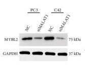
Application: WB Species: Mouse Sample: Tumor tissues
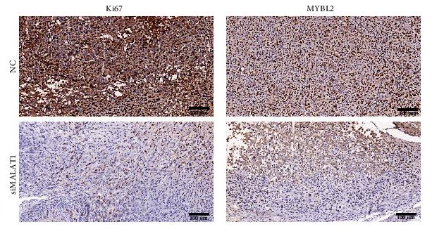
Application: IHC Species: Mouse Sample: Tumor tissues
Restrictive clause
Affinity Biosciences tests all products strictly. Citations are provided as a resource for additional applications that have not been validated by Affinity Biosciences. Please choose the appropriate format for each application and consult Materials and Methods sections for additional details about the use of any product in these publications.
For Research Use Only.
Not for use in diagnostic or therapeutic procedures. Not for resale. Not for distribution without written consent. Affinity Biosciences will not be held responsible for patent infringement or other violations that may occur with the use of our products. Affinity Biosciences, Affinity Biosciences Logo and all other trademarks are the property of Affinity Biosciences LTD.