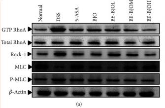
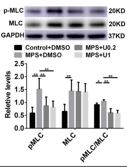
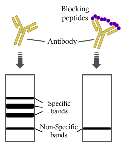
製品: MLC2 Antibody
カタログ: DF7911
タンパク質の説明: Rabbit polyclonal antibody to MLC2
アプリケーション: WB IHC IF/ICC
Cited expt.: WB
反応性: Human, Mouse, Rat
予測: Bovine, Horse, Sheep, Rabbit, Dog, Chicken, Xenopus
分子量: 19 kDa; 19kD(Calculated).
ユニプロット: P10916
RRID: AB_2841328

類似製品を見る>>

   サイズ 価格 在庫状況
 100ul $280 在庫あり
 200ul $350 在庫あり

リードタイム: 当日配達

For pricing and ordering contact:
お問い合わせ先

製品説明

ソース:
Rabbit
アプリケーション:
WB 1:1000-3000, IF/ICC 1:100-1:500, IHC 1:50-1:200
*The optimal dilutions should be determined by the end user. For optimal experimental results, antibody reuse is not recommended.
*Tips:

WB: For western blot detection of denatured protein samples. IHC: For immunohistochemical detection of paraffin sections (IHC-p) or frozen sections (IHC-f) of tissue samples. IF/ICC: For immunofluorescence detection of cell samples. ELISA(peptide): For ELISA detection of antigenic peptide.

反応性:
Human,Mouse,Rat
予測:
Bovine(92%), Horse(100%), Sheep(100%), Rabbit(100%), Dog(100%), Chicken(100%), Xenopus(92%)
クローナリティ:
Polyclonal
特異性:
MLC2 Antibody detects endogenous levels of total MLC2.
RRID:
AB_2841328
引用形式: Affinity Biosciences Cat# DF7911, RRID:AB_2841328.
コンジュゲート:
Unconjugated.
精製:
The antiserum was purified by peptide affinity chromatography using SulfoLink™ Coupling Resin (Thermo Fisher Scientific).
保存:
Rabbit IgG in phosphate buffered saline , pH 7.4, 150mM NaCl, 0.02% sodium azide and 50% glycerol. Store at -20 °C. Stable for 12 months from date of receipt.
別名:

折りたたみ/展開

Cardiac myosin light chain-2; Cardiac ventricular myosin light chain 2; CMH10; MLC 2v; MLC-2; MLC-2v; MLC2; MLRV_HUMAN; MYL 2; MYL2; Myosin light chain 2 regulatory cardiac slow; Myosin light polypeptide 2 regulatory cardiac slow; Myosin regulatory light chain 2; Myosin regulatory light chain 2 ventricular/cardiac muscle isoform; Regulatory light chain of myosin; RLC of myosin; Slow cardiac myosin regulatory light chain 2; ventricular/cardiac muscle isoform;

免疫原

免疫原:

A synthesized peptide derived from human MLC2, corresponding to a region within N-terminal amino acids.

Uniprot:
遺伝子(ID):
タンパク質配列:
MAPKKAKKRAGGANSNVFSMFEQTQIQEFKEAFTIMDQNRDGFIDKNDLRDTFAALGRVNVKNEEIDEMIKEAPGPINFTVFLTMFGEKLKGADPEETILNAFKVFDPEGKGVLKADYVREMLTTQAERFSKEEVDQMFAAFPPDVTGNLDYKNLVHIITHGEEKD

種類予測

種類予測:

Score>80(red) has high confidence and is suggested to be used for WB detection. *The prediction model is mainly based on the alignment of immunogen sequences, the results are for reference only, not as the basis of quality assurance.

Species
Results
Score
Horse
100
Sheep
100
Dog
100
Chicken
100
Rabbit
100
Bovine
92
Xenopus
92
Pig
0
Zebrafish
0
Model Confidence:
High(score>80) Medium(80>score>50) Low(score<50) No confidence

研究背景

機能:

Contractile protein that plays a role in heart development and function (By similarity). Following phosphorylation, plays a role in cross-bridge cycling kinetics and cardiac muscle contraction by increasing myosin lever arm stiffness and promoting myosin head diffusion; as a consequence of the increase in maximum contraction force and calcium sensitivity of contraction force. These events altogether slow down myosin kinetics and prolong duty cycle resulting in accumulated myosins being cooperatively recruited to actin binding sites to sustain thin filament activation as a means to fine-tune myofilament calcium sensitivity to force (By similarity). During cardiogenesis plays an early role in cardiac contractility by promoting cardiac myofibril assembly (By similarity).

PTMs:

N-terminus is methylated by METTL11A/NTM1.

Phosphorylated by MYLK3 and MYLK2; promotes cardiac muscle contraction and function (By similarity). Dephosphorylated by PPP1CB complexed to PPP1R12B (By similarity). The phosphorylated form in adult is expressed as gradients across the heart from endocardium (low phosphorylation) to epicardium (high phosphorylation); regulates cardiac torsion and workload distribution (By similarity).

細胞の位置付け:

Cytoplasm>Myofibril>Sarcomere>A band.

Extracellular region or secreted Cytosol Plasma membrane Cytoskeleton Lysosome Endosome Peroxisome ER Golgi apparatus Nucleus Mitochondrion Manual annotation Automatic computational assertionSubcellular location

研究領域

· Cellular Processes > Cellular community - eukaryotes > Focal adhesion.   (View pathway)

· Cellular Processes > Cellular community - eukaryotes > Tight junction.   (View pathway)

· Cellular Processes > Cell motility > Regulation of actin cytoskeleton.   (View pathway)

· Environmental Information Processing > Signal transduction > Apelin signaling pathway.   (View pathway)

· Human Diseases > Cardiovascular diseases > Hypertrophic cardiomyopathy (HCM).

· Human Diseases > Cardiovascular diseases > Dilated cardiomyopathy (DCM).

· Organismal Systems > Circulatory system > Cardiac muscle contraction.   (View pathway)

· Organismal Systems > Circulatory system > Adrenergic signaling in cardiomyocytes.   (View pathway)

· Organismal Systems > Immune system > Leukocyte transendothelial migration.   (View pathway)

参考文献
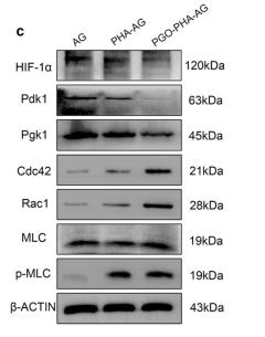
1). Polydopamine-mediated graphene oxide and nanohydroxyapatite-incorporated conductive scaffold with an immunomodulatory ability accelerates periodontal bone regeneration in diabetes. Bioactive Materials, 2022 (PubMed: 35387166) [IF=18.9]

Application: WB    Species: rat    Sample: RAW 264.7 cells

Fig. S16. | Verification of transcriptome analysis of RAW 264.7 cells cultured on various scaffolds. The mRNA expression levels of (a) glycolytic and (b) RhoA/ROCK pathways-related genes in each group. (c) Western blot (WB) analysis of glycolytic and RhoA/ROCK pathways-related proteins HIF-1α, Pdk1, Pgk1, Cdc42, Rac1, and p-MLC/MLC in each group.

2). Biased histamine signaling selectively gates fat preference. Neuron, 2026 (PubMed: 41308645) [IF=14.7]

3). Protective effect of oxyberberine against acute lung injury in mice via inhibiting RhoA/ROCK signaling pathway. BIOMEDICINE & PHARMACOTHERAPY, 2022 (PubMed: 35753262) [IF=6.9]

4). Swiprosin-1 participates in the berberine-regulated AMPK/MLCK pathway to attenuate colitis-induced tight junction damage. Phytomedicine : international journal of phytotherapy and phytopharmacology, 2024 (PubMed: 39369569) [IF=6.7]

5). Integrated identification and mechanism exploration of bioactive ingredients from Salvia miltiorrhiza to induce vascular normalization. Phytomedicine : international journal of phytotherapy and phytopharmacology, 2025 (PubMed: 39892310) [IF=6.7]

6). Primary cilia prevent activation of the cGAS-STING pathway during mouse decidualization. Communications biology, 2025 (PubMed: 40229503) [IF=5.9]

Application: WB    Species: Mouse    Sample:

Fig. 2: Ciliary defects promote actomyosin contraction. A F-Actin immunofluorescence, quantification of fibers length and F-Actin intensity in uterine stromal cells transfected with siScr or siTMEM67 for 24 h and further induced in vitro decidualization for 48 h. Scale bars, 15 μm. B Western blot analysis and quantification of RhoA, p-MLC2, and TMEM67 after stromal cells were transfected with siScr or siTMEM67 for 24 h and further induced in vitro decidualization for 48 h. C Western blot analysis and quantification of RhoA and p-MLC2 in stromal cells under in vitro decidualization for 2 and 3 days. D F-Actin and p-MLC2 immunofluorescence in uterine stromal cells. Scale bar, 25 μm. All images were representative of three biologically independent experiments. All data were presented as means ± SD from three biologically independent experiments. The two-tailed Student’s t-test was used for comparing two groups. One-way ANOVA test was used for comparing more than two groups. *P 

7). Phosphorylated CPI-17 and MLC2 as Biomarkers of Coronary Artery Spasm-Induced Sudden Cardiac Death. International journal of molecular sciences, 2024 (PubMed: 38474189) [IF=5.6]

Application: WB    Species: Rat    Sample:

Figure 4 Effects of CAS on the expression of phosphorylated MLC2 in rat coronary arteries. (A,B) Representative photomicrographs and semi-quantitative data of p-MLC2 protein levels detected via IHC staining. Lower enlarged images are from upper images. n = 6 per group. Scale bar: 100 µm (upper images) or 50 µm (lower images). (C) Representative immunofluores-cent images of p-MLC2 (red) and DAPI (blue) in the coronary arteries of rats. Scale bar: 50 µm. (D) The mean fluorescence intensity of p-MLC2 per section was quantified. n = 6 per group. (E,F) Western blot and quantitative analysis of p-MLC2 protein levels of rat coronary arteries. n = 6 per group. Data presented as mean ± SEM. ** p < 0.01 and *** p < 0.001 vs. control group.

8). High Glucose Aggravates Retinal Endothelial Cell Dysfunction by Activating the RhoA/ROCK1/pMLC/Connexin43 Signaling Pathway. Investigative Ophthalmology & Visual Science, 2022 (PubMed: 35881407) [IF=5.0]

Application: WB    Species: Mice    Sample: mRMVECs

Figure 6.RhoA/ROCK1 signaling regulates Cx43 through the directly interaction between pMLC and Cx43. (A, C) Co-immunoprecipitation of pMLC and Cx43 in mRMVECs with or without high glucose. (B, D, E) The protein expression of pMLC, MLC, and Cx43 in mRMVECs treated with different conditions. ns, no significance. EV, empty vector. *P < 0.05, **P < 0.01, ***P < 0.001, ****P < 0.0001 compared with the control group; ##P < 0.01 compared with the high glucose (30 mM) group. N = 3. The bars are the mean ± SD.

9). Bisphenol A disrupts the neuronal F-actin cytoskeleton by activating the RhoA/ROCK/LIMK pathway in Neuro-2a cells. Toxicology, 2024 (PubMed: 39527977) [IF=4.8]

10). An Inulin-Type Fructan CP-A from Codonopsis pilosula Alleviated 5-Fluorouracil-Induced Intestinal Mucositis via the ERK/MLCK/MLC2 Pathway and Regulation of Gut Microbiota. Pharmaceuticals (Basel, Switzerland), 2024 (PubMed: 38543083) [IF=4.6]

Application: WB    Species: Rat    Sample: IEC-6 cells

Figure 4. Effects of CP-A on ERK/MLCK/MLC2 signaling pathway proteins in 5-FU-induced rats and IEC-6 cells: (a–d) Expression of MLC2, p-MLC2, MLCK, ERK1/2 and p-ERK1/2 in IEC-6 cells. (e) MLCK protein in IEC-6 cells (magnification 100×). (f–i) Expression of MLC2, p-MLC2, MLCK, ERK1/2 and p-ERK1/2 in intestinal. (j) The p-MLC2 protein in IEC-6 cells (magnification 100×). Values are represented as mean ± SEM (n = 3); ## p < 0.01 versus control group, * p < 0.05 and ** p < 0.01 versus 5-FU group.

もっと読みます

Restrictive clause


Affinity Biosciences tests all products strictly. Citations are provided as a resource for additional applications that have not been validated by Affinity Biosciences. Please choose the appropriate format for each application and consult Materials and Methods sections for additional details about the use of any product in these publications.

For Research Use Only.
Not for use in diagnostic or therapeutic procedures. Not for resale. Not for distribution without written consent. Affinity Biosciences will not be held responsible for patent infringement or other violations that may occur with the use of our products. Affinity Biosciences, Affinity Biosciences Logo and all other trademarks are the property of Affinity Biosciences LTD.