, using Phospho-IKK alpha/ beta (Ser180/Ser181) Antibody at 1/1000 dilution.
5ug/NC membrane strip.
Exposure for 2min with Affinity™ ECL Kit(#KF8003).
Bands result from membrane strip incubation.")
 and mouse anti-beta tubulin Ab(#T0023) for 1 hour at 37°C. An AlexaFluor594 conjugated goat anti-rabbit IgG Ab(Red) and an AlexaFluor488 conjugated goat anti-mouse IgG Ab(Green) were used as the secondary antibody.
The nuclear counter stain is DAPI (blue).")

 on the expression of hepatic (A) XBP1, (B) phospho-IKK alpha/beta (Thr 183+Tyr 185), (C) phospho-JNK1/2/3 (Ser180/181) and (D) phospho-IRS1 (Ser 307).")
transforming growth factor (TGF)-b1, (b) phosphorylated (p)-Smad2/3, (c) Smad2/3, (d) Smad4, (e) Smad7, (f) Toll-like receptor 4 (TLR4), (g) myeloid differentiation factor 88 (MyD88), (h) p-inhibitor of nuclear factor-kappa B (NF-jB) kinase b (p-IKKb), (i) IKKb, NF-jB in (j) cytoplasm and in (k)nucleus.")
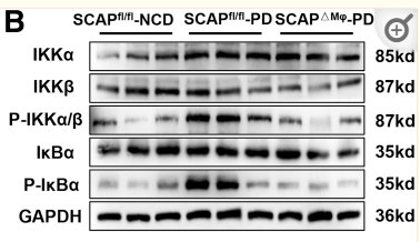
 The expression levels of p-p38, p-JNK, p-ERK1/2, p-IKK-a/b, p-p65, and p65 were analyzed by western blotting.")
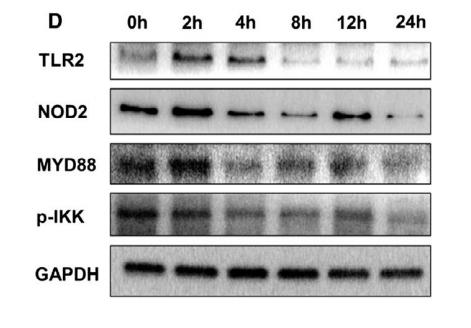
. Cells were administrated by dioscin (15 ng/mL)for 24 h, and then treated with MSU (100 μg/mL) for 12 h.d The protein expressions of TLR4,MyD88, p-IKKβ, IKKβ, p-p65 in cytoplasm, and NF-κB p65 innuclei were measured utilizing western blotting. GAPDH and Histone H3 were acted as internal references, respectively.")
 p-IκB and p-p65 protein levels in HUVECs were determined (n=5).")
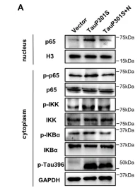
 Relative protein levels of NF-κB p65 (cytoplasm), NF-κB p65 (nucleus), p-IKK Ser180/Ser181, IKK, p-IκB-α Ser32/36, and IκB-α in lung tissues of mice in different groups.")
 Relative mRNA levels of S1pr1 in lung tissues of mice in different groups. (b) Relative protein levels of S1PR1 in lung tissues of mice in different groups. (c) Relative protein levels of NF-κB p65 (cytoplasm), NF-κB p65 (nucleus), p-IKK Ser180/Ser181, IKK, p-IκB-α Ser32/36, and IκB-α in lung tissues of mice in different groups. (d) Representative images of dual immunofluorescence staining with S1PR1 and F4/80 in lung tissues of mice in different groups. (e) A luciferase reporter assay was used to measure the relative luciferase activities of S1PR1-3’-UTR wt and mut in RAW264.7 cells. Results were expressed as means ± SD, *P<0.05.")
 qRT-PCR analysis was performed to detect the relative mRNA expression of cathepsin K, TRAP, NFATc1, and MMP-9 in BMSCs. (b) Western blot analysis was performed to detect the relative protein expression of IκBα, p-IκBα, p65, p-p65, IKKα, p-IKKα, NFATc1, and cathepsin K in BMSCs (n = 3); results are represented as the mean ± SD. ▲P < 0.05, compared to the Con group; ▲▲P < 0.01, compared to the Con group; ∗P < 0.05, compared to the Ti group; ∗∗P < 0.01, compared to the Ti group. BMSCs: bone marrow stromal cells; Ti: titanium particles; Con: control.")
 to ACE2 detected via immunofluorescence. Single channel images of S-RBD, ACE2 and DAPI are shown. Moreover, a single overlapped image (the merge image) was merged and generated using the single channel images for S-RBD, ACE2, and DAPI. b: Effect of β-chitosan on S-RBD binding to ACE2 as determined using flow cytometry. Merge 1 was generated by merging the single channel images of S-RBD and ACE2. c: Western blot analysis of inflammation-related proteins. Cells were treated with S-RBD and/or β-chitosan as shown in Table 1. Relative IOD = IOD/β-actin. Scale bars, 50 μm (white).")
 and upregulated (c) KEGG pathway in overexpression zebrafish bmp8a 3T3-L1 cells. b, d After induction of adipogenic differentiation, the downregulated (b) and upregulated (d) KEGG pathway in overexpression mouse Bmp8a 3T3-L1 cells. e, f Immunoblot analysis and quantification of p-IKKα/β and p-p65 in Mock, LV-ZsGreen1, and LV-bmp8a 3T3-L1 cells (n = 3). g, h Immunoblot analysis and quantification of p-IKKα/β and p-p65 in Mock, LV-ZsGreen1, and LV-Bmp8a 3T3-L1cells. Protein expression levels were quantified by ImageJ software and normalized to total protein (n = 3). i Co-immunoprecipitation and immunoblot analysis of co-transfected with PPARγ and p65 (n = 3). j Schematic drawing of predicted PPRE site in Fabp4 promoter region. k Schematic drawing of WT and PPRE site mutation Luc-report plasmids. l, m Quantification of the activity of Fabp4-promoter (l) and Fabp4-promoter-ΔPPRE (m) luciferase reporters in mouse HEK293T cells transfected with Vector, pCMV-Pparγ, or co-transfected pCMV-Pparγ and pCMV-p65, respectively. Renilla luciferase was used as the internal control (n = 3). Data were from three independent experiments and were analyzed by One-way ANOVA and were presented as mean ± SD (ns not significant, **p")
 Representative bands of NF-κB, p-NF-κB, p-IKK-α, p-IKK-β, IκB, p-IκB, TNF and IL-6wereshown.")
 The serum inflammatory cytokines (i.e., TNF-α, IL-1β, IL-6, and IL-10) in serum were measured by ELISA. (E–J) Representative images of phosphorylation levels of P38, Erk1/2, JNK, NF-κB and IKK α/β were examined by western blot and the fold activation data analysis. Data are expressed as mean ± SD (at least n = 6/group), #p < 0.05 (vs. Sham group), *p < 0.05 (vs. CLP group).")

 Effects of TNFR2 knockdown on migratory and invasive capacities using Transwell assays in PANC-1 and ASPC-1 cells. (C) Western blotting determined that NF-κB signaling-related proteins were overexpressed in the PANC-1 and ASPC-1 cells. (D) Western blotting determined that NF-κB signaling-related proteins were downregulated after TNFR silencing in PANC-1 and ASPC-1 cells.")
 Effects of bilirubin on L-O2 cell morphology (100×). (B) CCK-8 assay of bilirubin-treated L-O2 cells. (C) mRNA expression of p65, IκB-α, and IKK-β in L-O2 cells stimulated by bilirubin. (D) Measurement of p65, p-p65, IKK-β, p-IKK-β, IκB-α, and p-IκB-α protein levels in L-O2 cells by Western blotting. (E) Statistical analysis of p-p65, p-IKK-β, and p-IκB-α protein levels in L-O2 cells (normalized to p65, IKK-β, or IκB-α). At least three replicate samples were used for each experiment. Statistical significance is shown as ** p < 0.01, *** p < 0.001, **** p < 0.0001.")
 Effects of bilirubin on L-O2 cell morphology (100×). (B) CCK-8 assay of bilirubin-treated L-O2 cells. (C) mRNA expression of p65, IκB-α, and IKK-β in L-O2 cells stimulated by bilirubin. (D) Measurement of p65, p-p65, IKK-β, p-IKK-β, IκB-α, and p-IκB-α protein levels in L-O2 cells by western blotting (normalized to p65, IKK-β, or IκB-α).")
 complex. (B) Representative images of immunofluorescence trial for p-NF-κB. The expression of NF-κB, p-NF-κB, and p-IKK α/β detected by western blot and the fold activation data analysis (C-E) (n = 3). In the statistical figure, the red line is the mean and the black horizontal line is the quartile. VC = vitamic C; JAK2 = Janus kinase 2; STAT3 = signal transducer and activator of transcription 3; NF-κB = nuclear factor kappa B; SIMI = sepsis-induced myocardial injury; 3D = three-dimensional.")
 of CD40 or STING. B Co-IP using anti-TRAF2 to pull down STING and immunoblot (IB) detection of CD40 and STING molecules. C Multiplex immunofluorescence demonstrated the interactions of CD40 and STING with TRAF2 (scale bar = 100 μm). D Impact of CD40 and STING stimulation in the NF-κB pathway. E Flow cytometry revealed the degrees of B cell differentiation under different stimuli (APC-CD19, PE-pre-BCR).")
. D Changes in protein ratios of iNOS and Arg1 (n = 6 independent experiments). E-H Western blot detects changes in protein levels of anti-inflammatory cytokines IL-4, IL-10, and TGF-β in mouse brain (n = 6 independent experiments). I-K Western blot detects changes in protein levels of anti-inflammatory cytokines IL-1β and IFN-γ in mouse substantia nigra (n = 6 independent experiments). L-R Western blot detected the protein levels of RANK, RANKL, OPG, p-NIK, NIK, p-IKK, IKK, p-NF-κB2, and NF-κB2 protein level changes (n = 6 independent experiments). iNOS is a marker for neurotoxic microglial, Arg1 (arginase-1) is a marker for neuroprotective microglial. Data are mean ± SEM.")
| 製品: | Phospho-IKK alpha/ beta (Ser180/Ser181) Antibody |
| カタログ: | AF3013 |
| タンパク質の説明: | Rabbit polyclonal antibody to Phospho-IKK alpha/ beta (Ser180/Ser181) |
| アプリケーション: | WB IHC IF/ICC |
| Cited expt.: | WB |
| 反応性: | Human, Mouse, Rat |
| 予測: | Pig, Zebrafish, Bovine, Horse, Sheep, Rabbit, Dog, Chicken, Xenopus |
| 分子量: | 85kDa(Observed); 85kD,87kD(Calculated). |
| ユニプロット: | O15111 | O14920 |
| RRID: | AB_2834452 |

製品説明
*The optimal dilutions should be determined by the end user. For optimal experimental results, antibody reuse is not recommended.
*Tips:
WB: For western blot detection of denatured protein samples. IHC: For immunohistochemical detection of paraffin sections (IHC-p) or frozen sections (IHC-f) of tissue samples. IF/ICC: For immunofluorescence detection of cell samples. ELISA(peptide): For ELISA detection of antigenic peptide.
引用形式: Affinity Biosciences Cat# AF3013, RRID:AB_2834452.
折りたたみ/展開
chuk; CHUK1; Conserved Helix Loop Helix Ubiquitous Kinase; Conserved helix loop ubiquitous kinase; Conserved helix-loop-helix ubiquitous kinase; I Kappa B Kinase 1; I Kappa B Kinase Alpha; I-kappa-B kinase 1; I-kappa-B kinase alpha; IkappaB kinase; IkB kinase alpha subunit; IkBKA; IKK 1; IKK A; IKK a kinase; IKK-A; IKK-alpha; IKK1; IKKA; IKKA_HUMAN; Inhibitor Of Kappa Light Polypeptide Gene Enhancer In B Cells; Inhibitor Of Nuclear Factor Kappa B Kinase Alpha Subunit; Inhibitor of nuclear factor kappa-B kinase subunit alpha; NFKBIKA; Nuclear Factor Kappa B Inhibitor Kinase Alpha; Nuclear factor NF kappa B inhibitor kinase alpha; Nuclear factor NF-kappa-B inhibitor kinase alpha; Nuclear factor NFkappaB inhibitor kinase alpha; Nuclear Factor Of Kappa Light Chain Gene Enhancer In B Cells Inhibitor; TCF-16; TCF16; Transcription factor 16; I kappa B kinase 2; I kappa B kinase beta; I-kappa-B kinase 2; I-kappa-B-kinase beta; IkBKB; IKK beta; IKK-B; IKK-beta; IKK2; IKKB; IKKB_HUMAN; IMD15; Inhibitor of kappa light polypeptide gene enhancer in B cells, kinase beta; Inhibitor of nuclear factor kappa-B kinase subunit beta; NFKBIKB; Nuclear factor NF-kappa-B inhibitor kinase beta;
免疫原
A synthesized peptide derived from human IKK- alpha/ beta around the phosphorylation site of Ser180/181.
Widely expressed.
O14920 IKKB_HUMAN:Highly expressed in heart, placenta, skeletal muscle, kidney, pancreas, spleen, thymus, prostate, testis and peripheral blood.
- O15111 IKKA_HUMAN:
- Protein BLAST With
- NCBI/
- ExPASy/
- Uniprot
MERPPGLRPGAGGPWEMRERLGTGGFGNVCLYQHRELDLKIAIKSCRLELSTKNRERWCHEIQIMKKLNHANVVKACDVPEELNILIHDVPLLAMEYCSGGDLRKLLNKPENCCGLKESQILSLLSDIGSGIRYLHENKIIHRDLKPENIVLQDVGGKIIHKIIDLGYAKDVDQGSLCTSFVGTLQYLAPELFENKPYTATVDYWSFGTMVFECIAGYRPFLHHLQPFTWHEKIKKKDPKCIFACEEMSGEVRFSSHLPQPNSLCSLVVEPMENWLQLMLNWDPQQRGGPVDLTLKQPRCFVLMDHILNLKIVHILNMTSAKIISFLLPPDESLHSLQSRIERETGINTGSQELLSETGISLDPRKPASQCVLDGVRGCDSYMVYLFDKSKTVYEGPFASRSLSDCVNYIVQDSKIQLPIIQLRKVWAEAVHYVSGLKEDYSRLFQGQRAAMLSLLRYNANLTKMKNTLISASQQLKAKLEFFHKSIQLDLERYSEQMTYGISSEKMLKAWKEMEEKAIHYAEVGVIGYLEDQIMSLHAEIMELQKSPYGRRQGDLMESLEQRAIDLYKQLKHRPSDHSYSDSTEMVKIIVHTVQSQDRVLKELFGHLSKLLGCKQKIIDLLPKVEVALSNIKEADNTVMFMQGKRQKEIWHLLKIACTQSSARSLVGSSLEGAVTPQTSAWLPPTSAEHDHSLSCVVTPQDGETSAQMIEENLNCLGHLSTIIHEANEEQGNSMMNLDWSWLTE
- O14920 IKKB_HUMAN:
- Protein BLAST With
- NCBI/
- ExPASy/
- Uniprot
MSWSPSLTTQTCGAWEMKERLGTGGFGNVIRWHNQETGEQIAIKQCRQELSPRNRERWCLEIQIMRRLTHPNVVAARDVPEGMQNLAPNDLPLLAMEYCQGGDLRKYLNQFENCCGLREGAILTLLSDIASALRYLHENRIIHRDLKPENIVLQQGEQRLIHKIIDLGYAKELDQGSLCTSFVGTLQYLAPELLEQQKYTVTVDYWSFGTLAFECITGFRPFLPNWQPVQWHSKVRQKSEVDIVVSEDLNGTVKFSSSLPYPNNLNSVLAERLEKWLQLMLMWHPRQRGTDPTYGPNGCFKALDDILNLKLVHILNMVTGTIHTYPVTEDESLQSLKARIQQDTGIPEEDQELLQEAGLALIPDKPATQCISDGKLNEGHTLDMDLVFLFDNSKITYETQISPRPQPESVSCILQEPKRNLAFFQLRKVWGQVWHSIQTLKEDCNRLQQGQRAAMMNLLRNNSCLSKMKNSMASMSQQLKAKLDFFKTSIQIDLEKYSEQTEFGITSDKLLLAWREMEQAVELCGRENEVKLLVERMMALQTDIVDLQRSPMGRKQGGTLDDLEEQARELYRRLREKPRDQRTEGDSQEMVRLLLQAIQSFEKKVRVIYTQLSKTVVCKQKALELLPKVEEVVSLMNEDEKTVVRLQEKRQKELWNLLKIACSKVRGPVSGSPDSMNASRLSQPGQLMSQPSTASNSLPEPAKKSEELVAEAHNLCTLLENAIQDTVREQDQSFTALDWSWLQTEEEEHSCLEQAS
種類予測
Score>80(red) has high confidence and is suggested to be used for WB detection. *The prediction model is mainly based on the alignment of immunogen sequences, the results are for reference only, not as the basis of quality assurance.
High(score>80) Medium(80>score>50) Low(score<50) No confidence
研究背景
Serine kinase that plays an essential role in the NF-kappa-B signaling pathway which is activated by multiple stimuli such as inflammatory cytokines, bacterial or viral products, DNA damages or other cellular stresses. Acts as part of the canonical IKK complex in the conventional pathway of NF-kappa-B activation and phosphorylates inhibitors of NF-kappa-B on serine residues. These modifications allow polyubiquitination of the inhibitors and subsequent degradation by the proteasome. In turn, free NF-kappa-B is translocated into the nucleus and activates the transcription of hundreds of genes involved in immune response, growth control, or protection against apoptosis. Negatively regulates the pathway by phosphorylating the scaffold protein TAXBP1 and thus promoting the assembly of the A20/TNFAIP3 ubiquitin-editing complex (composed of A20/TNFAIP3, TAX1BP1, and the E3 ligases ITCH and RNF11). Therefore, CHUK plays a key role in the negative feedback of NF-kappa-B canonical signaling to limit inflammatory gene activation. As part of the non-canonical pathway of NF-kappa-B activation, the MAP3K14-activated CHUK/IKKA homodimer phosphorylates NFKB2/p100 associated with RelB, inducing its proteolytic processing to NFKB2/p52 and the formation of NF-kappa-B RelB-p52 complexes. In turn, these complexes regulate genes encoding molecules involved in B-cell survival and lymphoid organogenesis. Participates also in the negative feedback of the non-canonical NF-kappa-B signaling pathway by phosphorylating and destabilizing MAP3K14/NIK. Within the nucleus, phosphorylates CREBBP and consequently increases both its transcriptional and histone acetyltransferase activities. Modulates chromatin accessibility at NF-kappa-B-responsive promoters by phosphorylating histones H3 at 'Ser-10' that are subsequently acetylated at 'Lys-14' by CREBBP. Additionally, phosphorylates the CREBBP-interacting protein NCOA3. Also phosphorylates FOXO3 and may regulate this pro-apoptotic transcription factor. Phosphorylates RIPK1 at 'Ser-25' which represses its kinase activity and consequently prevents TNF-mediated RIPK1-dependent cell death (By similarity).
Phosphorylated by MAP3K14/NIK, AKT and to a lesser extent by MEKK1, and dephosphorylated by PP2A. Autophosphorylated.
(Microbial infection) Acetylation of Thr-179 by Yersinia yopJ prevents phosphorylation and activation, thus blocking the I-kappa-B signaling pathway.
Cytoplasm. Nucleus.
Note: Shuttles between the cytoplasm and the nucleus.
Widely expressed.
The kinase domain is located in the N-terminal region. The leucine zipper is important to allow homo- and hetero-dimerization. At the C-terminal region is located the region responsible for the interaction with NEMO/IKBKG.
Belongs to the protein kinase superfamily. Ser/Thr protein kinase family. I-kappa-B kinase subfamily.
Serine kinase that plays an essential role in the NF-kappa-B signaling pathway which is activated by multiple stimuli such as inflammatory cytokines, bacterial or viral products, DNA damages or other cellular stresses. Acts as part of the canonical IKK complex in the conventional pathway of NF-kappa-B activation. Phosphorylates inhibitors of NF-kappa-B on 2 critical serine residues. These modifications allow polyubiquitination of the inhibitors and subsequent degradation by the proteasome. In turn, free NF-kappa-B is translocated into the nucleus and activates the transcription of hundreds of genes involved in immune response, growth control, or protection against apoptosis. In addition to the NF-kappa-B inhibitors, phosphorylates several other components of the signaling pathway including NEMO/IKBKG, NF-kappa-B subunits RELA and NFKB1, as well as IKK-related kinases TBK1 and IKBKE. IKK-related kinase phosphorylations may prevent the overproduction of inflammatory mediators since they exert a negative regulation on canonical IKKs. Phosphorylates FOXO3, mediating the TNF-dependent inactivation of this pro-apoptotic transcription factor. Also phosphorylates other substrates including NCOA3, BCL10 and IRS1. Within the nucleus, acts as an adapter protein for NFKBIA degradation in UV-induced NF-kappa-B activation. Phosphorylates RIPK1 at 'Ser-25' which represses its kinase activity and consequently prevents TNF-mediated RIPK1-dependent cell death (By similarity).
Upon cytokine stimulation, phosphorylated on Ser-177 and Ser-181 by MEKK1 and/or MAP3K14/NIK as well as TBK1 and PRKCZ; which enhances activity. Once activated, autophosphorylates on the C-terminal serine cluster; which decreases activity and prevents prolonged activation of the inflammatory response. Phosphorylated by the IKK-related kinases TBK1 and IKBKE, which is associated with reduced CHUK/IKKA and IKBKB activity and NF-kappa-B-dependent gene transcription. Dephosphorylated at Ser-177 and Ser-181 by PPM1A and PPM1B.
(Microbial infection) Acetylation of Thr-180 by Yersinia yopJ prevents phosphorylation and activation, thus blocking the I-kappa-B pathway.
Ubiquitinated. Monoubiquitination involves TRIM21 that leads to inhibition of Tax-induced NF-kappa-B signaling. According to 'Ser-163' does not serve as a monoubiquitination site. According to ubiquitination on 'Ser-163' modulates phosphorylation on C-terminal serine residues.
(Microbial infection) Monoubiquitination by TRIM21 is disrupted by Yersinia yopJ.
Hydroxylated by PHD1/EGLN2, loss of hydroxylation under hypoxic conditions results in activation of NF-kappa-B.
Cytoplasm. Nucleus. Membrane raft.
Note: Colocalized with DPP4 in membrane rafts.
Highly expressed in heart, placenta, skeletal muscle, kidney, pancreas, spleen, thymus, prostate, testis and peripheral blood.
The kinase domain is located in the N-terminal region. The leucine zipper is important to allow homo- and hetero-dimerization. At the C-terminal region is located the region responsible for the interaction with NEMO/IKBKG.
Belongs to the protein kinase superfamily. Ser/Thr protein kinase family. I-kappa-B kinase subfamily.
研究領域
· Cellular Processes > Cell growth and death > Apoptosis. (View pathway)
· Environmental Information Processing > Signal transduction > MAPK signaling pathway. (View pathway)
· Environmental Information Processing > Signal transduction > Ras signaling pathway. (View pathway)
· Environmental Information Processing > Signal transduction > NF-kappa B signaling pathway. (View pathway)
· Environmental Information Processing > Signal transduction > FoxO signaling pathway. (View pathway)
· Environmental Information Processing > Signal transduction > mTOR signaling pathway. (View pathway)
· Environmental Information Processing > Signal transduction > PI3K-Akt signaling pathway. (View pathway)
· Environmental Information Processing > Signal transduction > TNF signaling pathway. (View pathway)
· Human Diseases > Drug resistance: Antineoplastic > Antifolate resistance.
· Human Diseases > Endocrine and metabolic diseases > Type II diabetes mellitus.
· Human Diseases > Endocrine and metabolic diseases > Insulin resistance.
· Human Diseases > Endocrine and metabolic diseases > Non-alcoholic fatty liver disease (NAFLD).
· Human Diseases > Infectious diseases: Bacterial > Epithelial cell signaling in Helicobacter pylori infection.
· Human Diseases > Infectious diseases: Bacterial > Shigellosis.
· Human Diseases > Infectious diseases: Parasitic > Chagas disease (American trypanosomiasis).
· Human Diseases > Infectious diseases: Parasitic > Toxoplasmosis.
· Human Diseases > Infectious diseases: Viral > Hepatitis C.
· Human Diseases > Infectious diseases: Viral > Hepatitis B.
· Human Diseases > Infectious diseases: Viral > Measles.
· Human Diseases > Infectious diseases: Viral > Influenza A.
· Human Diseases > Infectious diseases: Viral > Human papillomavirus infection.
· Human Diseases > Infectious diseases: Viral > HTLV-I infection.
· Human Diseases > Infectious diseases: Viral > Herpes simplex infection.
· Human Diseases > Infectious diseases: Viral > Epstein-Barr virus infection.
· Human Diseases > Cancers: Overview > Pathways in cancer. (View pathway)
· Human Diseases > Cancers: Overview > MicroRNAs in cancer.
· Human Diseases > Cancers: Specific types > Pancreatic cancer. (View pathway)
· Human Diseases > Cancers: Specific types > Prostate cancer. (View pathway)
· Human Diseases > Cancers: Specific types > Chronic myeloid leukemia. (View pathway)
· Human Diseases > Cancers: Specific types > Acute myeloid leukemia. (View pathway)
· Human Diseases > Cancers: Specific types > Small cell lung cancer. (View pathway)
· Organismal Systems > Immune system > Chemokine signaling pathway. (View pathway)
· Organismal Systems > Development > Osteoclast differentiation. (View pathway)
· Organismal Systems > Immune system > Toll-like receptor signaling pathway. (View pathway)
· Organismal Systems > Immune system > NOD-like receptor signaling pathway. (View pathway)
· Organismal Systems > Immune system > RIG-I-like receptor signaling pathway. (View pathway)
· Organismal Systems > Immune system > Cytosolic DNA-sensing pathway. (View pathway)
· Organismal Systems > Immune system > IL-17 signaling pathway. (View pathway)
· Organismal Systems > Immune system > Th1 and Th2 cell differentiation. (View pathway)
· Organismal Systems > Immune system > Th17 cell differentiation. (View pathway)
· Organismal Systems > Immune system > T cell receptor signaling pathway. (View pathway)
· Organismal Systems > Immune system > B cell receptor signaling pathway. (View pathway)
· Organismal Systems > Nervous system > Neurotrophin signaling pathway. (View pathway)
· Organismal Systems > Endocrine system > Insulin signaling pathway. (View pathway)
· Organismal Systems > Endocrine system > Adipocytokine signaling pathway.
参考文献

Application: WB Species: Mouse Sample: BMSCs
Application: WB Species: Mouse Sample: HT22 cells

Application: WB Species: Mouse Sample:
Application: WB Species: Mouse Sample: liver tissue
Restrictive clause
Affinity Biosciences tests all products strictly. Citations are provided as a resource for additional applications that have not been validated by Affinity Biosciences. Please choose the appropriate format for each application and consult Materials and Methods sections for additional details about the use of any product in these publications.
For Research Use Only.
Not for use in diagnostic or therapeutic procedures. Not for resale. Not for distribution without written consent. Affinity Biosciences will not be held responsible for patent infringement or other violations that may occur with the use of our products. Affinity Biosciences, Affinity Biosciences Logo and all other trademarks are the property of Affinity Biosciences LTD.