製品: Phospho-p53 (Ser15) Antibody
カタログ: AF3075
タンパク質の説明: Rabbit polyclonal antibody to Phospho-p53 (Ser15)
アプリケーション: WB IHC IF/ICC IP
反応性: Human, Mouse, Rat
予測: Pig, Bovine, Sheep, Rabbit
分子量: 53kDa; 44kD(Calculated).
ユニプロット: P04637
RRID: AB_2834512

類似製品を見る>>

   サイズ 価格 在庫状況
 100ul $280 在庫あり
 200ul $350 在庫あり

リードタイム: 当日配達

For pricing and ordering contact:
お問い合わせ先

製品説明

ソース:
Rabbit
アプリケーション:
WB 1:500-1:2000, IHC 1:50-1:1000, IP 1:100-1:500, IF/ICC 1:100-1:500
*The optimal dilutions should be determined by the end user.
*Tips:

WB: For western blot detection of denatured protein samples. IHC: For immunohistochemical detection of paraffin sections (IHC-p) or frozen sections (IHC-f) of tissue samples. IF/ICC: For immunofluorescence detection of cell samples. ELISA(peptide): For ELISA detection of antigenic peptide.

反応性:
Human,Mouse,Rat
予測:
Pig(100%), Bovine(88%), Sheep(100%), Rabbit(88%)
クローナリティ:
Polyclonal
特異性:
Phospho-p53 (Ser15) Antibody detects endogenous levels of p53 only when phosphorylated at Serine 15.
RRID:
AB_2834512
引用形式: Affinity Biosciences Cat# AF3075, RRID:AB_2834512.
コンジュゲート:
Unconjugated.
精製:
The antibody is from purified rabbit serum by affinity purification via sequential chromatography on phospho-peptide and non-phospho-peptide affinity columns.
保存:
Rabbit IgG in phosphate buffered saline , pH 7.4, 150mM NaCl, 0.02% sodium azide and 50% glycerol. Store at -20 °C. Stable for 12 months from date of receipt.
別名:

折りたたみ/展開

Antigen NY-CO-13; BCC7; Cellular tumor antigen p53; FLJ92943; LFS1; Mutant tumor protein 53; p53; p53 tumor suppressor; P53_HUMAN; Phosphoprotein p53; Tp53; Transformation related protein 53; TRP53; Tumor protein 53; Tumor protein p53; Tumor suppressor p53;

免疫原

免疫原:
Uniprot:
遺伝子(ID):
発現特異性:
P04637 P53_HUMAN:

Ubiquitous. Isoforms are expressed in a wide range of normal tissues but in a tissue-dependent manner. Isoform 2 is expressed in most normal tissues but is not detected in brain, lung, prostate, muscle, fetal brain, spinal cord and fetal liver. Isoform 3 is expressed in most normal tissues but is not detected in lung, spleen, testis, fetal brain, spinal cord and fetal liver. Isoform 7 is expressed in most normal tissues but is not detected in prostate, uterus, skeletal muscle and breast. Isoform 8 is detected only in colon, bone marrow, testis, fetal brain and intestine. Isoform 9 is expressed in most normal tissues but is not detected in brain, heart, lung, fetal liver, salivary gland, breast or intestine.

タンパク質の説明:
Tumor protein p53, a nuclear protein, plays an essential role in the regulation of cell cycle, specifically in the transition from G0 to G1. It is found in very low levels in normal cells, however, in a variety of transformed cell lines, it is expressed in high amounts, and believed to contribute to transformation and malignancy.
タンパク質配列:
MEEPQSDPSVEPPLSQETFSDLWKLLPENNVLSPLPSQAMDDLMLSPDDIEQWFTEDPGPDEAPRMPEAAPPVAPAPAAPTPAAPAPAPSWPLSSSVPSQKTYQGSYGFRLGFLHSGTAKSVTCTYSPALNKMFCQLAKTCPVQLWVDSTPPPGTRVRAMAIYKQSQHMTEVVRRCPHHERCSDSDGLAPPQHLIRVEGNLRVEYLDDRNTFRHSVVVPYEPPEVGSDCTTIHYNYMCNSSCMGGMNRRPILTIITLEDSSGNLLGRNSFEVRVCACPGRDRRTEEENLRKKGEPHHELPPGSTKRALPNNTSSSPQPKKKPLDGEYFTLQIRGRERFEMFRELNEALELKDAQAGKEPGGSRAHSSHLKSKKGQSTSRHKKLMFKTEGPDSD

種類予測

種類予測:

Score>80(red) has high confidence and is suggested to be used for WB detection. *The prediction model is mainly based on the alignment of immunogen sequences, the results are for reference only, not as the basis of quality assurance.

Species
Results
Score
Pig
100
Sheep
100
Bovine
88
Rabbit
88
Dog
63
Xenopus
63
Horse
0
Zebrafish
0
Chicken
0
Model Confidence:
High(score>80) Medium(80>score>50) Low(score<50) No confidence

PTMs - P04637 基板として

Site PTM Type Enzyme
Ubiquitination
S6 Phosphorylation P45984 (MAPK9)
S9 Phosphorylation P78527 (PRKDC) , Q13315 (ATM) , P48730 (CSNK1D)
S15 Phosphorylation Q16539 (MAPK14) , O60285 (NUAK1) , Q15831 (STK11) , Q13315 (ATM) , P78527 (PRKDC) , Q05655 (PRKCD) , Q13464 (ROCK1) , P57059 (SIK1) , Q96Q15 (SMG1) , P27361 (MAPK3) , P28482 (MAPK1) , Q14680 (MELK) , O14757 (CHEK1) , O96017 (CHEK2) , Q96S44 (TP53RK) , Q13535 (ATR) , Q00535 (CDK5) , Q13131 (PRKAA1) , P51812 (RPS6KA3) , Q13627 (DYRK1A)
T18 Phosphorylation P53355 (DAPK1) , P48730 (CSNK1D) , Q99986 (VRK1) , O96017 (CHEK2) , Q86Y07-2 (VRK2) , P33981 (TTK) , P78527 (PRKDC) , P48729 (CSNK1A1) , Q13131 (PRKAA1) , O14757 (CHEK1)
S20 Phosphorylation P78527 (PRKDC) , Q13131 (PRKAA1) , Q13315 (ATM) , O96017 (CHEK2) , Q9H4B4 (PLK3) , Q9UEE5 (STK17A) , P45983 (MAPK8) , P48729 (CSNK1A1) , P49137 (MAPKAPK2) , Q00535 (CDK5) , O14757 (CHEK1) , O43293 (DAPK3) , P53355 (DAPK1) , P48730 (CSNK1D) , Q683Z8 (CHK2) , P45984 (MAPK9)
K24 Ubiquitination
S33 Phosphorylation P50750 (CDK9) , O15264 (MAPK13) , Q00535 (CDK5) , P50613 (CDK7) , Q16539 (MAPK14) , P49841 (GSK3B) , P45985 (MAP2K4)
S37 Phosphorylation O96017 (CHEK2) , Q8IW41 (MAPKAPK5) , O14757 (CHEK1) , P78527 (PRKDC) , Q13535 (ATR) , P53779 (MAPK10) , P45984 (MAPK9) , P45983 (MAPK8)
S46 Phosphorylation Q92630 (DYRK2) , Q00535 (CDK5) , Q13315 (ATM) , Q05655 (PRKCD) , Q16539 (MAPK14) , P78527 (PRKDC) , Q9H2X6 (HIPK2) , O15264 (MAPK13)
T55 Phosphorylation P28482 (MAPK1) , P21675 (TAF1) , P34947 (GRK5)
T81 Phosphorylation P45984 (MAPK9) , P45983 (MAPK8)
S99 Phosphorylation
K101 Ubiquitination
S106 Phosphorylation O14965 (AURKA)
R110 Methylation
H115 Methylation
K120 Acetylation
K120 Ubiquitination
C124 S-Nitrosylation
Y126 Phosphorylation
K132 Ubiquitination
K139 Ubiquitination
C141 S-Nitrosylation
S149 O-Glycosylation
S149 Phosphorylation P68400 (CSNK2A1)
T150 Phosphorylation P68400 (CSNK2A1)
T155 Phosphorylation P68400 (CSNK2A1)
K164 Acetylation
K164 Ubiquitination
S166 Phosphorylation
C182 S-Nitrosylation
S183 Phosphorylation Q96GD4 (AURKB)
R209 Methylation
T211 Phosphorylation Q96GD4 (AURKB)
R213 Methylation
S215 Phosphorylation O14965 (AURKA) , Q96GD4 (AURKB) , O96013 (PAK4)
Y220 Phosphorylation
R249 Phosphorylation
S260 Phosphorylation
S269 Phosphorylation P53355 (DAPK1) , Q96GD4 (AURKB)
T284 Phosphorylation Q96GD4 (AURKB)
R290 Methylation
K291 Ubiquitination
K292 Acetylation
K292 Ubiquitination
T304 Phosphorylation Q5S007 (LRRK2)
K305 Acetylation
K305 Ubiquitination
T312 Phosphorylation
S313 Phosphorylation P24941 (CDK2)
S314 Phosphorylation P24941 (CDK2)
S315 Phosphorylation P24941 (CDK2) , P06493 (CDK1) , O14965 (AURKA) , P50750 (CDK9)
K319 Acetylation
K319 Ubiquitination
K320 Acetylation
K320 Ubiquitination
K321 Ubiquitination
Y327 Phosphorylation
R333 Methylation
R335 Methylation
R337 Methylation
K351 Ubiquitination
K357 Ubiquitination
S362 Phosphorylation O14920 (IKBKB)
S366 Phosphorylation O14757 (CHEK1) , O14920 (IKBKB) , O96017 (CHEK2)
K370 Acetylation
K370 Methylation
K370 Ubiquitination
S371 Phosphorylation P17252 (PRKCA) , P50613 (CDK7)
K372 Acetylation
K372 Methylation
K372 Ubiquitination
K373 Acetylation
K373 Methylation
K373 Ubiquitination
S376 Phosphorylation P50613 (CDK7) , P17252 (PRKCA) , P49841 (GSK3B)
T377 Phosphorylation P17252 (PRKCA) , Q5S007 (LRRK2)
S378 Phosphorylation O96017 (CHEK2) , P50613 (CDK7) , P17612 (PRKACA) , P17252 (PRKCA) , O14757 (CHEK1)
K381 Acetylation
K381 Ubiquitination
K382 Acetylation
K382 Methylation
K382 Ubiquitination
K386 Acetylation
K386 Methylation
K386 Sumoylation
K386 Ubiquitination
T387 Phosphorylation O14757 (CHEK1)
S392 Phosphorylation P19525 (EIF2AK2) , O60285 (NUAK1) , P23443 (RPS6KB1) , Q15831 (STK11) , P50750 (CDK9) , P27361 (MAPK3) , P50613 (CDK7) , P68400 (CSNK2A1)

研究背景

機能:

Acts as a tumor suppressor in many tumor types; induces growth arrest or apoptosis depending on the physiological circumstances and cell type. Involved in cell cycle regulation as a trans-activator that acts to negatively regulate cell division by controlling a set of genes required for this process. One of the activated genes is an inhibitor of cyclin-dependent kinases. Apoptosis induction seems to be mediated either by stimulation of BAX and FAS antigen expression, or by repression of Bcl-2 expression. Its pro-apoptotic activity is activated via its interaction with PPP1R13B/ASPP1 or TP53BP2/ASPP2. However, this activity is inhibited when the interaction with PPP1R13B/ASPP1 or TP53BP2/ASPP2 is displaced by PPP1R13L/iASPP. In cooperation with mitochondrial PPIF is involved in activating oxidative stress-induced necrosis; the function is largely independent of transcription. Induces the transcription of long intergenic non-coding RNA p21 (lincRNA-p21) and lincRNA-Mkln1. LincRNA-p21 participates in TP53-dependent transcriptional repression leading to apoptosis and seems to have an effect on cell-cycle regulation. Implicated in Notch signaling cross-over. Prevents CDK7 kinase activity when associated to CAK complex in response to DNA damage, thus stopping cell cycle progression. Isoform 2 enhances the transactivation activity of isoform 1 from some but not all TP53-inducible promoters. Isoform 4 suppresses transactivation activity and impairs growth suppression mediated by isoform 1. Isoform 7 inhibits isoform 1-mediated apoptosis. Regulates the circadian clock by repressing CLOCK-ARNTL/BMAL1-mediated transcriptional activation of PER2.

PTMs:

Acetylated. Acetylation of Lys-382 by CREBBP enhances transcriptional activity. Deacetylation of Lys-382 by SIRT1 impairs its ability to induce proapoptotic program and modulate cell senescence. Deacetylation by SIRT2 impairs its ability to induce transcription activation in a AKT-dependent manner.

Phosphorylation on Ser residues mediates transcriptional activation. Phosphorylated by HIPK1 (By similarity). Phosphorylation at Ser-9 by HIPK4 increases repression activity on BIRC5 promoter. Phosphorylated on Thr-18 by VRK1. Phosphorylated on Ser-20 by CHEK2 in response to DNA damage, which prevents ubiquitination by MDM2. Phosphorylated on Ser-20 by PLK3 in response to reactive oxygen species (ROS), promoting p53/TP53-mediated apoptosis. Phosphorylated on Thr-55 by TAF1, which promotes MDM2-mediated degradation. Phosphorylated on Ser-33 by CDK7 in a CAK complex in response to DNA damage. Phosphorylated on Ser-46 by HIPK2 upon UV irradiation. Phosphorylation on Ser-46 is required for acetylation by CREBBP. Phosphorylated on Ser-392 following UV but not gamma irradiation. Phosphorylated on Ser-15 upon ultraviolet irradiation; which is enhanced by interaction with BANP. Phosphorylated by NUAK1 at Ser-15 and Ser-392; was initially thought to be mediated by STK11/LKB1 but it was later shown that it is indirect and that STK11/LKB1-dependent phosphorylation is probably mediated by downstream NUAK1. It is unclear whether AMP directly mediates phosphorylation at Ser-15. Phosphorylated on Thr-18 by isoform 1 and isoform 2 of VRK2. Phosphorylation on Thr-18 by isoform 2 of VRK2 results in a reduction in ubiquitination by MDM2 and an increase in acetylation by EP300. Stabilized by CDK5-mediated phosphorylation in response to genotoxic and oxidative stresses at Ser-15, Ser-33 and Ser-46, leading to accumulation of p53/TP53, particularly in the nucleus, thus inducing the transactivation of p53/TP53 target genes. Phosphorylated by DYRK2 at Ser-46 in response to genotoxic stress. Phosphorylated at Ser-315 and Ser-392 by CDK2 in response to DNA-damage. Phosphorylation at Ser-15 is required for interaction with DDX3X and gamma-tubulin.

Dephosphorylated by PP2A-PPP2R5C holoenzyme at Thr-55. SV40 small T antigen inhibits the dephosphorylation by the AC form of PP2A.

May be O-glycosylated in the C-terminal basic region. Studied in EB-1 cell line.

Ubiquitinated by MDM2 and SYVN1, which leads to proteasomal degradation. Ubiquitinated by RFWD3, which works in cooperation with MDM2 and may catalyze the formation of short polyubiquitin chains on p53/TP53 that are not targeted to the proteasome. Ubiquitinated by MKRN1 at Lys-291 and Lys-292, which leads to proteasomal degradation. Deubiquitinated by USP10, leading to its stabilization. Ubiquitinated by TRIM24, RFFL, RNF34 and RNF125, which leads to proteasomal degradation. Ubiquitination by TOPORS induces degradation. Deubiquitination by USP7, leading to stabilization. Isoform 4 is monoubiquitinated in an MDM2-independent manner. Ubiquitinated by COP1, which leads to proteasomal degradation. Ubiquitination and subsequent proteasomal degradation is negatively regulated by CCAR2. Polyubiquitinated by C10orf90/FATS, polyubiquitination is 'Lys-48'-linkage independent and non-proteolytic, leading to TP53 stabilization (By similarity).

Monomethylated at Lys-372 by SETD7, leading to stabilization and increased transcriptional activation. Monomethylated at Lys-370 by SMYD2, leading to decreased DNA-binding activity and subsequent transcriptional regulation activity. Lys-372 monomethylation prevents interaction with SMYD2 and subsequent monomethylation at Lys-370. Dimethylated at Lys-373 by EHMT1 and EHMT2. Monomethylated at Lys-382 by KMT5A, promoting interaction with L3MBTL1 and leading to repress transcriptional activity. Dimethylation at Lys-370 and Lys-382 diminishes p53 ubiquitination, through stabilizing association with the methyl reader PHF20. Demethylation of dimethylated Lys-370 by KDM1A prevents interaction with TP53BP1 and represses TP53-mediated transcriptional activation. Monomethylated at Arg-333 and dimethylated at Arg-335 and Arg-337 by PRMT5; methylation is increased after DNA damage and might possibly affect TP53 target gene specificity.

Sumoylated with SUMO1. Sumoylated at Lys-386 by UBC9.

細胞の位置付け:

Cytoplasm. Nucleus. Nucleus>PML body. Endoplasmic reticulum. Mitochondrion matrix. Cytoplasm>Cytoskeleton>Microtubule organizing center>Centrosome.
Note: Interaction with BANP promotes nuclear localization (PubMed:15701641). Recruited into PML bodies together with CHEK2 (PubMed:12810724). Translocates to mitochondria upon oxidative stress (PubMed:22726440). Translocates to mitochondria in response to mitomycin C treatment (PubMed:27323408).

Nucleus. Cytoplasm.
Note: Predominantly nuclear but localizes to the cytoplasm when expressed with isoform 4.

Nucleus. Cytoplasm.
Note: Localized mainly in the nucleus with minor staining in the cytoplasm.

Nucleus. Cytoplasm.
Note: Localized in the nucleus in most cells but found in the cytoplasm in some cells.

Nucleus. Cytoplasm.
Note: Predominantly nuclear but translocates to the cytoplasm following cell stress.

Nucleus. Cytoplasm.
Note: Localized mainly in the nucleus with minor staining in the cytoplasm.

Nucleus. Cytoplasm.
Note: Localized in both nucleus and cytoplasm in most cells. In some cells, forms foci in the nucleus that are different from nucleoli.

Cytoplasm.

Extracellular region or secreted Cytosol Plasma membrane Cytoskeleton Lysosome Endosome Peroxisome ER Golgi apparatus Nucleus Mitochondrion Manual annotation Automatic computational assertionSubcellular location
組織特異性:

Ubiquitous. Isoforms are expressed in a wide range of normal tissues but in a tissue-dependent manner. Isoform 2 is expressed in most normal tissues but is not detected in brain, lung, prostate, muscle, fetal brain, spinal cord and fetal liver. Isoform 3 is expressed in most normal tissues but is not detected in lung, spleen, testis, fetal brain, spinal cord and fetal liver. Isoform 7 is expressed in most normal tissues but is not detected in prostate, uterus, skeletal muscle and breast. Isoform 8 is detected only in colon, bone marrow, testis, fetal brain and intestine. Isoform 9 is expressed in most normal tissues but is not detected in brain, heart, lung, fetal liver, salivary gland, breast or intestine.

サブユニット構造:

Forms homodimers and homotetramers. Binds DNA as a homotetramer. Interacts with AXIN1. Probably part of a complex consisting of TP53, HIPK2 and AXIN1 (By similarity). Interacts with histone acetyltransferases EP300 and methyltransferases HRMT1L2 and CARM1, and recruits them to promoters. Interacts (via C-terminus) with TAF1; when TAF1 is part of the TFIID complex. Interacts with ING4; this interaction may be indirect. Found in a complex with CABLES1 and TP73. Interacts with HIPK1, HIPK2, and TP53INP1. Interacts with WWOX. May interact with HCV core protein. Interacts with USP7 and SYVN1. Interacts with HSP90AB1. Interacts with CHD8; leading to recruit histone H1 and prevent transactivation activity (By similarity). Interacts with ARMC10, BANP, CDKN2AIP, NUAK1, STK11/LKB1, UHRF2 and E4F1. Interacts with YWHAZ; the interaction enhances TP53 transcriptional activity. Phosphorylation of YWHAZ on 'Ser-58' inhibits this interaction. Interacts (via DNA-binding domain) with MAML1 (via N-terminus). Interacts with MKRN1. Interacts with PML (via C-terminus). Interacts with MDM2; leading to ubiquitination and proteasomal degradation of TP53. Directly interacts with FBXO42; leading to ubiquitination and degradation of TP53. Interacts (phosphorylated at Ser-15 by ATM) with the phosphatase PP2A-PPP2R5C holoenzyme; regulates stress-induced TP53-dependent inhibition of cell proliferation. Interacts with PPP2R2A. Interacts with AURKA, DAXX, BRD7 and TRIM24. Interacts (when monomethylated at Lys-382) with L3MBTL1. Isoform 1 interacts with isoform 2 and with isoform 4. Interacts with GRK5. Binds to the CAK complex (CDK7, cyclin H and MAT1) in response to DNA damage. Interacts with CDK5 in neurons. Interacts with AURKB, SETD2, UHRF2 and NOC2L. Interacts (via N-terminus) with PTK2/FAK1; this promotes ubiquitination by MDM2. Interacts with PTK2B/PYK2; this promotes ubiquitination by MDM2. Interacts with PRKCG. Interacts with PPIF; the association implicates preferentially tetrameric TP53, is induced by oxidative stress and is impaired by cyclosporin A (CsA). Interacts with SNAI1; the interaction induces SNAI1 degradation via MDM2-mediated ubiquitination and inhibits SNAI1-induced cell invasion. Interacts with KAT6A. Interacts with UBC9. Interacts with ZNF385B; the interaction is direct. Interacts (via DNA-binding domain) with ZNF385A; the interaction is direct and enhances p53/TP53 transactivation functions on cell-cycle arrest target genes, resulting in growth arrest. Interacts with ANKRD2. Interacts with RFFL and RNF34; involved in p53/TP53 ubiquitination. Interacts with MTA1 and COP1. Interacts with CCAR2 (via N-terminus). Interacts with MORC3. Interacts (via C-terminus) with POU4F2 isoform 1 (via C-terminus). Interacts (via oligomerization region) with NOP53; the interaction is direct and may prevent the MDM2-mediated proteasomal degradation of TP53. Interacts with AFG1L; mediates mitochondrial translocation of TP53. Interacts with UBD. Interacts with TAF6 isoform 1 and isoform 4. Interacts with C10orf90/FATS; the interaction inhibits binding of TP53 and MDM2 (By similarity). Interacts with NUPR1; interaction is stress-dependent. Forms a complex with EP300 and NUPR1; this complex binds CDKN1A promoter leading to transcriptional induction of CDKN1A. Interacts with PRMT5 in response to DNA damage; the interaction is STRAP dependent. Interacts with PPP1R13L (via SH3 domain and ANK repeats); the interaction inhibits pro-apoptotic activity of p53/TP53. Interacts with PPP1R13B/ASPP1 and TP53BP2/ASPP2; the interactions promotes pro-apototic activity. When phosphorylated at Ser-15, interacts with DDX3X and gamma-tubulin.

(Microbial infection) Interacts with cancer-associated/HPV E6 viral proteins leading to ubiquitination and degradation of TP53 giving a possible model for cell growth regulation. This complex formation requires an additional factor, E6-AP, which stably associates with TP53 in the presence of E6.

(Microbial infection) Interacts with human cytomegalovirus/HHV-5 protein UL123.

(Microbial infection) Interacts (via N-terminus) with human adenovirus 5 E1B-55K protein; this interaction leads to the inhibition of TP53 function and/or its degradation.

タンパク質ファミリー:

The nuclear export signal acts as a transcriptional repression domain. The TADI and TADII motifs (residues 17 to 25 and 48 to 56) correspond both to 9aaTAD motifs which are transactivation domains present in a large number of yeast and animal transcription factors.

Belongs to the p53 family.

研究領域

· Cellular Processes > Cell growth and death > Cell cycle.   (View pathway)

· Cellular Processes > Cell growth and death > p53 signaling pathway.   (View pathway)

· Cellular Processes > Cell growth and death > Apoptosis.   (View pathway)

· Cellular Processes > Cell growth and death > Ferroptosis.   (View pathway)

· Cellular Processes > Cell growth and death > Cellular senescence.   (View pathway)

· Environmental Information Processing > Signal transduction > MAPK signaling pathway.   (View pathway)

· Environmental Information Processing > Signal transduction > Sphingolipid signaling pathway.   (View pathway)

· Environmental Information Processing > Signal transduction > PI3K-Akt signaling pathway.   (View pathway)

· Environmental Information Processing > Signal transduction > Wnt signaling pathway.   (View pathway)

· Human Diseases > Drug resistance: Antineoplastic > Endocrine resistance.

· Human Diseases > Drug resistance: Antineoplastic > Platinum drug resistance.

· Human Diseases > Neurodegenerative diseases > Amyotrophic lateral sclerosis (ALS).

· Human Diseases > Neurodegenerative diseases > Huntington's disease.

· Human Diseases > Infectious diseases: Viral > Hepatitis C.

· Human Diseases > Infectious diseases: Viral > Hepatitis B.

· Human Diseases > Infectious diseases: Viral > Measles.

· Human Diseases > Infectious diseases: Viral > Human papillomavirus infection.

· Human Diseases > Infectious diseases: Viral > HTLV-I infection.

· Human Diseases > Infectious diseases: Viral > Herpes simplex infection.

· Human Diseases > Infectious diseases: Viral > Epstein-Barr virus infection.

· Human Diseases > Cancers: Overview > Pathways in cancer.   (View pathway)

· Human Diseases > Cancers: Overview > Transcriptional misregulation in cancer.

· Human Diseases > Cancers: Overview > Viral carcinogenesis.

· Human Diseases > Cancers: Overview > Proteoglycans in cancer.

· Human Diseases > Cancers: Overview > MicroRNAs in cancer.

· Human Diseases > Cancers: Specific types > Colorectal cancer.   (View pathway)

· Human Diseases > Cancers: Specific types > Pancreatic cancer.   (View pathway)

· Human Diseases > Cancers: Specific types > Endometrial cancer.   (View pathway)

· Human Diseases > Cancers: Specific types > Glioma.   (View pathway)

· Human Diseases > Cancers: Specific types > Prostate cancer.   (View pathway)

· Human Diseases > Cancers: Specific types > Thyroid cancer.   (View pathway)

· Human Diseases > Cancers: Specific types > Basal cell carcinoma.   (View pathway)

· Human Diseases > Cancers: Specific types > Melanoma.   (View pathway)

· Human Diseases > Cancers: Specific types > Bladder cancer.   (View pathway)

· Human Diseases > Cancers: Specific types > Chronic myeloid leukemia.   (View pathway)

· Human Diseases > Cancers: Specific types > Small cell lung cancer.   (View pathway)

· Human Diseases > Cancers: Specific types > Non-small cell lung cancer.   (View pathway)

· Human Diseases > Cancers: Specific types > Breast cancer.   (View pathway)

· Human Diseases > Cancers: Specific types > Hepatocellular carcinoma.   (View pathway)

· Human Diseases > Cancers: Specific types > Gastric cancer.   (View pathway)

· Human Diseases > Cancers: Overview > Central carbon metabolism in cancer.   (View pathway)

· Organismal Systems > Aging > Longevity regulating pathway.   (View pathway)

· Organismal Systems > Nervous system > Neurotrophin signaling pathway.   (View pathway)

· Organismal Systems > Endocrine system > Thyroid hormone signaling pathway.   (View pathway)

参考文献

1). SOX9 Modulates the Transformation of Gastric Stem Cells Through Biased Symmetric Cell Division. Gastroenterology, 2023 (PubMed: 36740200) [IF=25.7]

2). P21 and P27 promote tumorigenesis and progression via cell cycle acceleration in seminal vesicles of TRAMP mice. International Journal of Biological Sciences, 2019 (PubMed: 31592235) [IF=8.2]

3). Sulfur dioxide and benzo(a)pyrene trigger apoptotic and anti-apoptotic signals at different post-exposure times in mouse liver. Chemosphere, 2015 (PubMed: 26162325) [IF=8.1]

Application: WB    Species: mouse    Sample: mouse liver

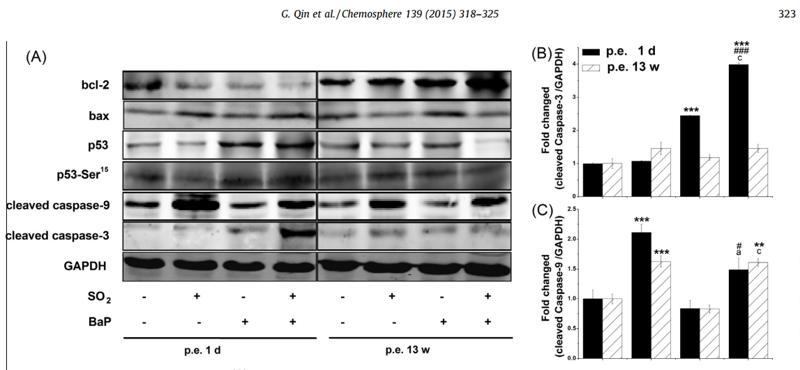
Fig. 4. Effects of SO2 and/or BaP on protein expression in mouse livers. (A), Representative bands of mouse liver proteins after chlorpyrifos exposure. Proteins (50 lg) obtained from mouse liver after post-exposure to SO2 and/or BaP for 1 d or 13 weeks were used to blot. GAPDH was used as a loading control. (B–H), The protein level of cleaved casepase-3 (B), cleaved caspase-9 (C), bcl-2 (D), bax (E), bcl-2/bax ratio (F), p53 (G), and p53 phosphorylation on Ser15 (H) were measured by densitometric analysis and normalized to the loading control (n = 1). Each column and bar represents the mean ± SE of at least six individual sample; ⁄ P < 0.05, ⁄⁄P < 0.01, ⁄⁄⁄P < 0.001 versus control group; #P < 0.05, ##P < 0.01, ###P < 0.001 versus SO2 alone; a P < 0.05, b P < 0.01, c P < 0.001 versus BaP alone.

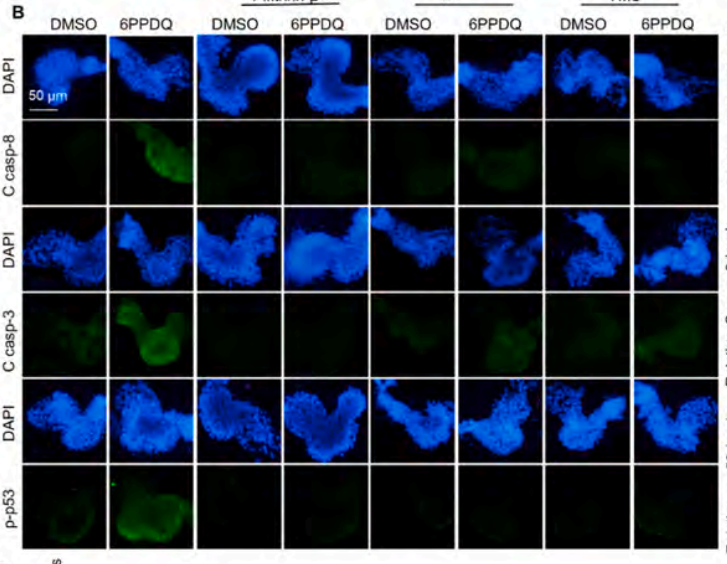
4). AHR/cyp1b1 signaling-mediated extrinsic apoptosis contributes to 6PPDQ-induced cardiac dysfunction in zebrafish embryos. Environmental Pollution, 2024 (PubMed: 38311157) [IF=7.6]

Application: IHC    Species: zebrafish    Sample: zebrafish embryos

Fig. 4. AHR mediates 6PPDQ-induced extrinsic apoptosis in the hearts of zebrafish embryos at 72 hpf. B, E) Immunostaining and quantification results for cleaved caspase-8/3 (c caspa-8/3) and phosphorylated p53 (p-p53).

5). TRIP13 interference inhibits the proliferation and metastasis of thyroid cancer cells through regulating TTC5/p53 pathway and epithelial-mesenchymal transition related genes expression. BIOMEDICINE & PHARMACOTHERAPY, 2019 (PubMed: 31648166) [IF=6.9]

6). Patulin induces ROS-dependent cardiac cell toxicity by inducing DNA damage and activating endoplasmic reticulum stress apoptotic pathway. Ecotoxicology and environmental safety, 2024 (PubMed: 38061079) [IF=6.2]

7). The activated ATM/p53 pathway promotes autophagy in response to oxidative stress-mediated DNA damage induced by Microcystin-LR in male germ cells. Ecotoxicology and Environmental Safety, 2021 (PubMed: 34715501) [IF=6.2]

Application: WB    Species: Mouse    Sample: GC-1 cells

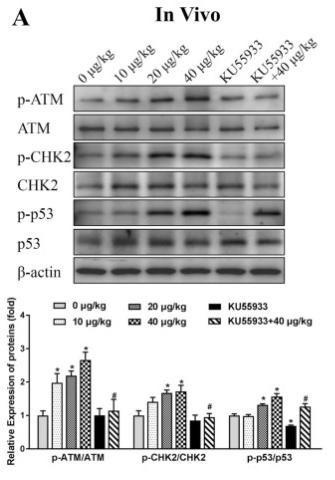
Fig. 4. Changes of ATM and its downstream proteins in GC-1 cells and mouse testis. Expression and analysis of the related proteins in mouse testis (A) and in GC-1 cells (B). *p < 0.05 vs. the control group; #p < 0.05 vs. the corresponding MC-LR exposure group. All data were expressed as  ± SD (n = 3).

8). Single-cell transcriptomics reveals ependymal subtypes related to cytoskeleton dynamics as the core driver of syringomyelia pathological development. iScience, 2023 (PubMed: 37275526) [IF=5.8]

Application: IF/ICC    Species: Human    Sample: EPCs

Figure 5 Dynamic changes in spinal cord ependymal populations post-op for syringomyelia (A–D) We performed further subtype analysis of the EPCs, the UMAP of which revealed two main distinct subtypes. Subtypes were annotated using a combination of canonical markers, transcription factor expression, and GO enrichment analyses of the DEGs (A). Cell cycle assignment showed that cells in Cluster 5 were mainly in the S phase (B). In general, ependymal cells showed high expression of the EPC-specific marker Foxj1 in the dot plot and violin plot (C). Cells are colored by the expression value of canonical marker genes, the motor ciliary ependymal subpopulations (with featured genes of Cfap43 and Sncg), and the glial ependymal subpopulations (with a higher expression of GFAP and Rnd3). Values are log-transformed normalized expression counts (D). (E, F, and H) The differentiation and development trajectory of EPCs was constructed by the diffusion map of pseudotime analysis. Pseudotime trajectory analysis corresponding to the differentiation of EPCs from Cluster 0 to other clusters. Cluster 5 (proliferating) between Cluster 0 in the early state and the other clusters. Cells are colored by cell types, pseudotime, and cell states, as indicated, from the left to the right panels (E). A ridge plot was drawn based on the pseudotime analysis to simulate the trajectory of cell clusters in cell differentiation and the proportions of ependymal-lineage subtypes at each time point from the top to the bottom (F). GO biological process terms associated with the top DEGs among two different cell subtypes (H). (G) Immunofluorescence shows the expression pattern of Sox2, Foxj1, and Nestin during syringomyelia development. EPCs (Foxj1+) in the CC area represented the EPCs with colocalization of Sox2 and Nestin. During the syringomyelia formation with the CC expansion, the syringomyelia was in a moniliform shape, and there were obvious separations between adjacent dilated compartments. The EPCs are irregularly distributed throughout the central canal due to moniliform-like dilatation. The stacked cells remained as EPCs (Foxj1+) in the stenosis, resembling a tunneling effect and showing that the CC was still communicating, but the monolayer of cells was organized in the significant dilatated area and even disrupted in some regions. The cytoskeleton outlined by Netin also became gradually disorganized or even disordered at post-op 6 weeks.

9). Hua-Tan-Sheng-Jing Decoction Treats Obesity With Oligoasthenozoospermia by Up-Regulating the PI3K-AKT and Down-Regulating the JNK MAPK Signaling Pathways: At the Crossroad of Obesity and Oligoasthenozoospermia. Frontiers in Pharmacology, 2022 (PubMed: 35559247) [IF=5.6]

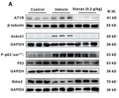
10). Storax Attenuates Cardiac Fibrosis following Acute Myocardial Infarction in Rats via Suppression of AT1R–Ankrd1–P53 Signaling Pathway. INTERNATIONAL JOURNAL OF MOLECULAR SCIENCES, 2022 (PubMed: 36361958) [IF=5.6]

Application: WB    Species: Rat    Sample: cardiac tissue

Figure 8 Storax inhibits the AT1R–Ankrd1–P53 pathway in AMI rats against cardiomyocyte apoptosis. (A) WB bands showing the protein expression levels of AT1R, Ankrd1, P-p53 (ser15), P53 and Mdm2 in cardiac tissue. (B–E) Relative protein for AT1R, Ankrd1, P-p53 (ser15) and Mdm2 were quantified by densitometry based on immunoblot images. Results are presented as mean ± SD (n = 3). One-way ANOVA follow by Bonferroni’s post hoc test: # p < 0.05, ## p < 0.01 vs. vehicle group.

もっと読みます

Restrictive clause


Affinity Biosciences tests all products strictly. Citations are provided as a resource for additional applications that have not been validated by Affinity Biosciences. Please choose the appropriate format for each application and consult Materials and Methods sections for additional details about the use of any product in these publications.

For Research Use Only.
Not for use in diagnostic or therapeutic procedures. Not for resale. Not for distribution without written consent. Affinity Biosciences will not be held responsible for patent infringement or other violations that may occur with the use of our products. Affinity Biosciences, Affinity Biosciences Logo and all other trademarks are the property of Affinity Biosciences LTD.