製品: Cytokeratin 19 Antibody
カタログ: AF0192
タンパク質の説明: Rabbit polyclonal antibody to Cytokeratin 19
アプリケーション: WB IHC IF/ICC IHC-F
反応性: Human, Mouse, Rat
予測: Bovine, Rabbit, Chicken
分子量: 44kDa; 44kD(Calculated).
ユニプロット: P08727
RRID: AB_2833385

類似製品を見る>>

   サイズ 価格 在庫状況
 100ul $280 在庫あり
 200ul $350 在庫あり

リードタイム: 当日配達

For pricing and ordering contact:
お問い合わせ先

製品説明

ソース:
Rabbit
アプリケーション:
WB 1:500-1:3000, IHC 1:50-1:200, IF/ICC 1:100-1:500
*The optimal dilutions should be determined by the end user.
*Tips:

WB: For western blot detection of denatured protein samples. IHC: For immunohistochemical detection of paraffin sections (IHC-p) or frozen sections (IHC-f) of tissue samples. IF/ICC: For immunofluorescence detection of cell samples. ELISA(peptide): For ELISA detection of antigenic peptide.

反応性:
Human,Mouse,Rat
予測:
Bovine(100%), Rabbit(100%), Chicken(100%)
クローナリティ:
Polyclonal
特異性:
Cytokeratin 19 Antibody detects endogenous levels of total Cytokeratin 19.
RRID:
AB_2833385
引用形式: Affinity Biosciences Cat# AF0192, RRID:AB_2833385.
コンジュゲート:
Unconjugated.
精製:
The antiserum was purified by peptide affinity chromatography using SulfoLink™ Coupling Resin (Thermo Fisher Scientific).
保存:
Rabbit IgG in phosphate buffered saline , pH 7.4, 150mM NaCl, 0.02% sodium azide and 50% glycerol. Store at -20 °C. Stable for 12 months from date of receipt.
別名:

折りたたみ/展開

40 kDa keratin intermediate filament; CK 19; CK-19; CK19; Cytokeratin 19; Cytokeratin-19; K19; K1C19_HUMAN; K1CS; Keratin 19; Keratin type I 40 kD; Keratin type I 40kD; Keratin type I cytoskeletal 19; Keratin, type I cytoskeletal 19; Keratin, type I, 40 kd; Keratin-19; KRT19; MGC15366;

免疫原

免疫原:
Uniprot:
遺伝子(ID):
発現特異性:
P08727 K1C19_HUMAN:

Expressed in a defined zone of basal keratinocytes in the deep outer root sheath of hair follicles. Also observed in sweat gland and mammary gland ductal and secretory cells, bile ducts, gastrointestinal tract, bladder urothelium, oral epithelia, esophagus, ectocervical epithelium (at protein level). Expressed in epidermal basal cells, in nipple epidermis and a defined region of the hair follicle. Also seen in a subset of vascular wall cells in both the veins and artery of human umbilical cord, and in umbilical cord vascular smooth muscle. Observed in muscle fibers accumulating in the costameres of myoplasm at the sarcolemma in structures that contain dystrophin and spectrin.

タンパク質の説明:
K19 a type I cytoskeletal keratin. The keratins are intermediate filament proteins responsible for the structural integrity of epithelial cells and are subdivided into cytokeratins and hair keratins. Keratin 19 differs from all other IF proteins in lacking the C-terminal tail domain.
タンパク質配列:
MTSYSYRQSSATSSFGGLGGGSVRFGPGVAFRAPSIHGGSGGRGVSVSSARFVSSSSSGAYGGGYGGVLTASDGLLAGNEKLTMQNLNDRLASYLDKVRALEAANGELEVKIRDWYQKQGPGPSRDYSHYYTTIQDLRDKILGATIENSRIVLQIDNARLAADDFRTKFETEQALRMSVEADINGLRRVLDELTLARTDLEMQIEGLKEELAYLKKNHEEEISTLRGQVGGQVSVEVDSAPGTDLAKILSDMRSQYEVMAEQNRKDAEAWFTSRTEELNREVAGHTEQLQMSRSEVTDLRRTLQGLEIELQSQLSMKAALEDTLAETEARFGAQLAHIQALISGIEAQLGDVRADSERQNQEYQRLMDIKSRLEQEIATYRSLLEGQEDHYNNLSASKVL

種類予測

種類予測:

Score>80(red) has high confidence and is suggested to be used for WB detection. *The prediction model is mainly based on the alignment of immunogen sequences, the results are for reference only, not as the basis of quality assurance.

Species
Results
Score
Bovine
100
Chicken
100
Rabbit
100
Xenopus
75
Pig
0
Horse
0
Sheep
0
Dog
0
Zebrafish
0
Model Confidence:
High(score>80) Medium(80>score>50) Low(score<50) No confidence

PTMs - P08727 基板として

Site PTM Type Enzyme
S3 Phosphorylation
Y4 Phosphorylation
R7 Methylation
S9 Phosphorylation
S10 Phosphorylation
T12 Phosphorylation
S13 Phosphorylation
S14 Phosphorylation
S22 Phosphorylation
R24 Methylation
R32 Methylation
S35 Phosphorylation P31749 (AKT1)
S40 Phosphorylation
R43 Methylation
S46 Phosphorylation
S48 Phosphorylation
S49 Phosphorylation
R51 Methylation
S55 Phosphorylation
S58 Phosphorylation
Y61 Phosphorylation
Y65 Phosphorylation
T70 Phosphorylation
S72 Phosphorylation
K81 Ubiquitination
S93 Phosphorylation
Y94 Phosphorylation
K97 Acetylation
K97 Ubiquitination
K111 Ubiquitination
Y116 Phosphorylation
K118 Methylation
K118 Ubiquitination
Y127 Phosphorylation
S128 Phosphorylation
Y130 Phosphorylation
Y131 Phosphorylation
T132 Phosphorylation
T133 Phosphorylation
K140 Ubiquitination
T145 Phosphorylation
K168 Methylation
K168 Sumoylation
K168 Ubiquitination
S178 Phosphorylation
K208 Sumoylation
K208 Ubiquitination
Y213 Phosphorylation
K215 Sumoylation
K215 Ubiquitination
K216 Ubiquitination
S223 Phosphorylation
T224 Phosphorylation
S234 Phosphorylation
S239 Phosphorylation
K247 Ubiquitination
S250 Phosphorylation
S254 Phosphorylation
Y256 Phosphorylation
K265 Ubiquitination
T272 Phosphorylation
S273 Phosphorylation
T275 Phosphorylation
T286 Phosphorylation
S292 Phosphorylation
S294 Phosphorylation
T302 Phosphorylation
S312 Phosphorylation
S315 Phosphorylation
K317 Ubiquitination
T323 Phosphorylation
T327 Phosphorylation
S343 Phosphorylation
K370 Ubiquitination
S371 Phosphorylation
T379 Phosphorylation
Y380 Phosphorylation
S382 Phosphorylation
Y391 Phosphorylation P12931 (SRC)
S395 Phosphorylation
S397 Phosphorylation
K398 Ubiquitination

研究背景

機能:

Involved in the organization of myofibers. Together with KRT8, helps to link the contractile apparatus to dystrophin at the costameres of striated muscle.

組織特異性:

Expressed in a defined zone of basal keratinocytes in the deep outer root sheath of hair follicles. Also observed in sweat gland and mammary gland ductal and secretory cells, bile ducts, gastrointestinal tract, bladder urothelium, oral epithelia, esophagus, ectocervical epithelium (at protein level). Expressed in epidermal basal cells, in nipple epidermis and a defined region of the hair follicle. Also seen in a subset of vascular wall cells in both the veins and artery of human umbilical cord, and in umbilical cord vascular smooth muscle. Observed in muscle fibers accumulating in the costameres of myoplasm at the sarcolemma in structures that contain dystrophin and spectrin.

サブユニット構造:

Heterotetramer of two type I and two type II keratins. Interacts with PNN and the actin-binding domain of DMD. Interacts with HCV core protein.

(Microbial infection) Interacts with hepatitis C virus/HCV core protein.

タンパク質ファミリー:

This keratin differs from all other IF proteins in lacking the C-terminal tail domain.

Belongs to the intermediate filament family.

研究領域

· Organismal Systems > Endocrine system > Estrogen signaling pathway.   (View pathway)

参考文献

1). BMSC loaded photo-crosslinked hyaluronic acid/collagen hydrogel incorporating FG4592 for enhanced cell proliferation and nucleus pulposus differentiation. International journal of biological macromolecules, 2024 (PubMed: 38834125) [IF=8.2]

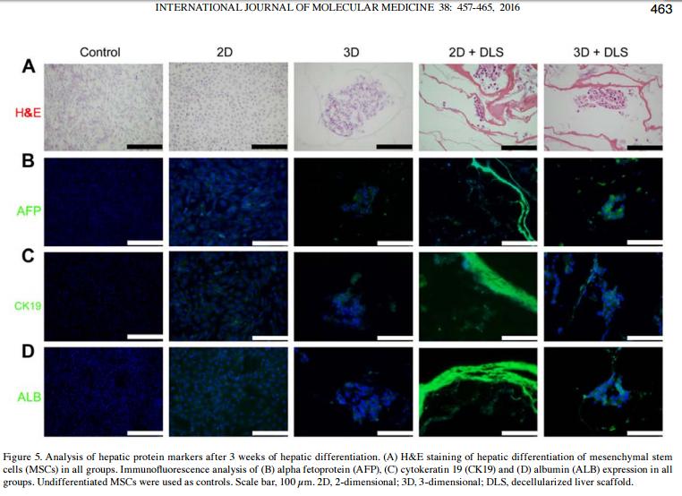
2). Enhanced hepatic differentiation of rat bone marrow-derived mesenchymal stem cells in spheroidal aggregate culture on a decellularized liver scaffold. INTERNATIONAL JOURNAL OF MOLECULAR MEDICINE, 2016 (PubMed: 27314916) [IF=5.4]

Application: IF/ICC    Species: rat    Sample:

Figure 5. Analysis of hepatic protein markers after 3 weeks of hepatic differentiation. (A) H&E staining of hepatic differentiation of mesenchymal stem cells (MSCs) in all groups. Immunofluorescence analysis of (B) alpha fetoprotein (AFP), (C) cytokeratin 19 (CK19) and (D) albumin (ALB) expression in all groups. Undifferentiated MSCs were used as controls. Scale bar, 100 µm. 2D, 2-dimensional; 3D, 3-dimensional; DLS, decellularized liver scaffold.

3). Human umbilical cord mesenchymal stem cells implantation accelerates cutaneous wound healing in diabetic rats via the Wnt signaling pathway. EUROPEAN JOURNAL OF MEDICAL RESEARCH, 2019 (PubMed: 30736851) [IF=4.2]

Application: WB    Species: Human    Sample: hUCMSCs

Fig. 5 Activation of the Wnt signaling pathway enhanced hUCMSCs differentiation. a Western blotting was applied to detect the changes in the levels of β-catenin, c-Myc, p63, CK19, and PCNA proteins. b Statistical analysis of a results. c The differentiation degree of hUCMSCs was evaluated by cell morphology under optical microscope (scale: 100 μm). d Statistical analysis on the ratio of cells with round or small polygonal morphology displayed in c;

4). Hepatic differentiation of mouse bone marrow‑derived mesenchymal stem cells using a novel 3D culture system. Molecular Medicine Reports, 2017 (PubMed: 29152658) [IF=3.4]

Application: IF/ICC    Species: Mouse    Sample:

Figure 4. Analysis of hepatic protein markers in mouse MSCs following 23 days of hepatic differentiation. Green immunofluorescence staining of ALB, AFP and CK19 in undifferentiated MSC control, 2D and 3D groups. Nuclei were stained blue with DAPI. Scale bars, 100 µm. MSCs, mesenchymal stem cells; ALB, albumin; AFP, α-fetoprotein; CK19, cytokeratin-19.

5). Adenomyosis-derived extracellular vesicles endow endometrial epithelial cells with an invasive phenotype through epithelial-mesenchymal transition. Genes & Diseases, 2020 (PubMed: 33335963)

Restrictive clause


Affinity Biosciences tests all products strictly. Citations are provided as a resource for additional applications that have not been validated by Affinity Biosciences. Please choose the appropriate format for each application and consult Materials and Methods sections for additional details about the use of any product in these publications.

For Research Use Only.
Not for use in diagnostic or therapeutic procedures. Not for resale. Not for distribution without written consent. Affinity Biosciences will not be held responsible for patent infringement or other violations that may occur with the use of our products. Affinity Biosciences, Affinity Biosciences Logo and all other trademarks are the property of Affinity Biosciences LTD.