 by IF/ICC. The samples were fixed with PFA and permeabilized in 0.1% Triton X-100, then blocked in 10% serum for 45 minutes at 25°C. Samples were then incubated with primary Ab(#AF3262) and mouse anti-beta tubulin Ab(#T0023) for 1 hour at 37°C. An AlexaFluor594 conjugated goat anti-rabbit IgG Ab(Red) and an AlexaFluor488 conjugated goat anti-mouse IgG Ab(Green) were used as the secondary antibody.
The nuclear counter stain is DAPI (blue).")

 for 24 h, after which whole-cell proteins were extracted for Western blot assay using (a) p-Akt, Akt, (b) bax, bcl-2, cleaved-caspase-3 and cleaved-PARP antibodies. (c) HepG2 cells were pretreated with LY294002 (5 µmol/L) and evodiamine (0, 5, 10 µmol/L) for 24 h. Whole-cell proteins were extracted for Western blot assay using p-Akt and Akt antibodies. (d) HepG2 cells were pretreated with SC79 (4 µg/mL) and evodiamine (0, 5, 10 µmol/L). Whole-cell extracts were processed for Western blot assay using p-Akt, Akt, bax, bcl-2 and cleaved-caspase3 antibodies. (e) Tumor tissues were fixed, sectioned and processed for IHC analysis. The protein expression of cleaved-caspase3 was detected. Data are presented as means ± S.D (n=3).")
 BxPC-3, MIAPaCa-2, and CFPAC-1 cells were treated with vehicle control or variable concentrations of BVD-523 and VS-5584, alone or in combination, for 48 h. Viable cells were measured by MTT assays. Standard isobologram analyses of the antitumor interactions are shown. The IC50 values of each drug are plotted on the axes; the solid line represents the additive effect, while the points represent the concentrations of each drug resulting in 50% inhibition of proliferation. Points falling below the line indicate synergism whereas those above the line indicate antagonism. (B) HPAC cells were treated with vehicle control, VS, BVD-523 (BVD), or VS plus BVD for 48 h. Cells were fxed with 80% ice-cold ethanol and stained with PI for cell cycle analysis. The percentage of cells with sub-G1 DNA content are graphed as means of triplicates ± SEM from one representative experiment. ***indicates p < 0.001; combined treatment compared to individual treatments and vehicle control. Combination index (CI) values were calculated using CompuSyn software. (C) CI vs. Fa plot (combination index vs. fraction affected) for the cell death data is presented. (D) HPAC cells were treated with vehicle control or the indicated drugs for 48 h. Whole cell lysates were subjected to Western blotting and probed with the indicated antibody. The fold changes for the densitometry measurements, normalized to β-actin and then compared to no drug treatment control, are indicated.")
 BxPC‐3 and HPAC cells were treated with vehicle control or different concentrations of CuB for 24 hr. Whole cell lysates were subjected to western blot analysis and probed with antibodies to EGFR, pEGFR, Akt, pAkt (T308), or β‐actin.")
 with or without OA (240 μM) for 24 hr. (a) Protein expression (including p‐AKT, AKT, p‐ACC, ACC, p‐AMPKα, AMPKα,PPARα, and SREBP‐1c) was analyzed by western blot.")


 Immunoblot for LPP3
from the heart tissue of high fat diet-fed
mice at different time points. (b–d)
Immunoblot quantification for LPP3 expression (b) ratio of AKT phosphorylation at
Ser473 and Thr 308/total AKT (c), and AKT
expression (d). Data represent three separate experiments.")
 and PA (330 μmol/L)‐induced HepG2 cells. GEN was added into HepG2 cells prior 1 h to stimulation of OA and PA for 18 h. A‐C, Protein expression of Nrf2, PPARα, PPARγ in nucleus and HO‐1 in cytoplasm was detected by Western blot. (D and E) Protein expression of P‐ACC, ACC, P‐AKT, AKT, P‐AMPKα, AMPKα, P‐AMPKβ and AMPKβ was detected by Western blot. F‐H, Protein expression of PI3K, P‐mTORC, mTORC, P‐S6K, S6K, P‐S6, S6, SREBP‐1c and HMGB1 was detected by Western blot. The similar results were collected from three dependent experiments. All data were expressed by mean ± SEM (n = 5 in each group). ## P < .01 vs Control Group; *P < .05 and **P < .01 vs OA and PA Group")
 and LNCaP (panel B) cells were treated with variable concentrations of CUDC‐907 for 6‐48 h. Whole cell lysates were subjected to Western blotting and probed with the indicated antibodies. The fold changes for the densitometry measurements, normalized to β‐actin and then compared to no drug control, are shown below the corresponding blot. C‐F, 22Rv1 and LNCaP cells were treated with 100 nmol/L CUDC‐907 with or without 500 nmol/L ERK inhibitor SCH772984 for 48 h. Whole cell lysates of 22Rv1 (panel C) and LNCaP (panel E) cells were subjected to Western blotting. The CUDC‐907 treated 22Rv1 (panel D) and LNCaP (panel F) cells were subjected to Annexin V/PI staining and flow cytometry analyses. ***indicates P < .001 compared to no drug treatment control, while ###indicates P < .001 compared to individual drug treatment")
 Western blot analysis of AKT/ERK1/2, G1-phase checkpoint and EMT molecules. (A) The representative western blot images. (B) The relative level of p-AKT (T308)/AKT, p-AKT (S473)/AKT and p-ERK1/2/ERK1/2 as well as p21, p27, cyclin D1, cyclin E1, CDK2, CDK4, E-cadherin, N-cadherin, vimentin, Snail and Slug in A549-SIRT7 (A549-mock served as a control) or H292-shSIRT7 (H292-shcontrol served as a control) NSCLC cells. *P<0.05, Student's t-test, n=6 per group. (C) IHC analysis of AKT/ERK1/2, G1-phase checkpoint and EMT molecules in xenograft tumor tissues. The representative IHC images are presented. SIRT7, sirtuin 7; EMT, epithelial-mesenchymal transition; NSCLC, non-small cell lung cancer; AKT, protein kinase; ERK1/2, extracellular signal-regulated kinase 1/2; CDK, cyclin-dependent kinase; IHC, immunohistochemistry.")
 Western blot analysis of AKT/ERK1/2, G1-phase checkpoint and EMT molecules. (A) The representative western blot images. (B) The relative level of p-AKT (T308)/AKT, p-AKT (S473)/AKT and p-ERK1/2/ERK1/2 as well as p21, p27, cyclin D1, cyclin E1, CDK2, CDK4, E-cadherin, N-cadherin, vimentin, Snail and Slug in A549-SIRT7 (A549-mock served as a control) or H292-shSIRT7 (H292-shcontrol served as a control) NSCLC cells. *P<0.05, Student's t-test, n=6 per group. (C) IHC analysis of AKT/ERK1/2, G1-phase checkpoint and EMT molecules in xenograft tumor tissues. The representative IHC images are presented. SIRT7, sirtuin 7; EMT, epithelial-mesenchymal transition; NSCLC, non-small cell lung cancer; AKT, protein kinase; ERK1/2, extracellular signal-regulated kinase 1/2; CDK, cyclin-dependent kinase; IHC, immunohistochemistry.")

. The relative protein abundances of FoxO (B), P-FoxO (C), AKT (D), and P-AKT (E) in hepatopancreas were measured by Western blot and expressed as relative expression values to those in the LFD group. Values are represented as mean ± S.E., n = 3. Values with different letters are significantly different (p < 0.05).")
 WB analysis of the AKT/ERK1/2-GSK3β-β-catenin/Snail axis and its regulatory targets in TIPE3 knock-down and TIPE3-overexpressing NSCLC cells. Representative WB images are shown. (B) IF analysis of the AKT/ERK1/2-GSK3β-β-catenin/Snail axis in TIPE3 knock-down and TIPE3-overexpressing NSCLC xenograft tumor tissues. Representative IF images of merge (red, Alexa Fluor 647; green, GFP; and blue, DAPI) (×200) are shown. (C) WB analysis of β-catenin, Snail1, and Slug in TIPE3-overexpressing NSCLC cells after treatment with PI3K or MEK inhibitor and TIPE3 knock-down NSCLC cells after treatment with GSK3β or proteasome inhibitor. Representative WB images are shown. (D) WB analysis of β-catenin, Snail1, and Slug in TIPE3 knock-down NSCLC cells after β-Trcp or STUB1 knockdown. Representative WB images are shown. (E,F) Clinical association of TIPE3 with the AKT/ERK1/2-GSK3β-β-catenin/Snail axis in lung cancer. Representative IHC images of Case 27 (+++, high TIPE3 expression) and Case 34 (-, low TIPE3 expression) lung cancer T tissues (×400) (E) are shown. Percentage of lung cancer T tissue specimens showing high or low TIPE3 expression in relation to the expression level of proteins related to the AKT/ERK1/2-GSK3β-β-catenin/Snail axis (F) is shown. *, P<0.05, Pearson’s χ2 test. (G) A schematic model for the mechanism of action of TIPE3 during NSCLC progression. Upregulation of TIPE3 promotes growth and metastasis in NSCLC cells by activating β-catenin, Snail1, and Slug transcriptional signaling via the AKT/ERK1/2-GSK3β axis. NSCLC, non-small cell lung cancer; WB: western blotting; AKT, protein kinase B; ERK1/2, extracellular signal-regulated kinase 1/2; GSK3β, glycogen synthase kinase 3β; IF, immunofluorescence; GFP, green fluorescent protein; DAPI: 4’,6-diamidino-2-phenylindole; PI3K, phosphatidylinositol 3-kinase; MEK: mitogen-activated protein kinase kinase; IHC, immunohistochemistry; T, lung cancer tumor.")
 WB analysis of the AKT/ERK1/2-GSK3β-β-catenin/Snail axis and its regulatory targets in TIPE3 knock-down and TIPE3-overexpressing NSCLC cells. Representative WB images are shown. (B) IF analysis of the AKT/ERK1/2-GSK3β-β-catenin/Snail axis in TIPE3 knock-down and TIPE3-overexpressing NSCLC xenograft tumor tissues. Representative IF images of merge (red, Alexa Fluor 647; green, GFP; and blue, DAPI) (×200) are shown. (C) WB analysis of β-catenin, Snail1, and Slug in TIPE3-overexpressing NSCLC cells after treatment with PI3K or MEK inhibitor and TIPE3 knock-down NSCLC cells after treatment with GSK3β or proteasome inhibitor. Representative WB images are shown. (D) WB analysis of β-catenin, Snail1, and Slug in TIPE3 knock-down NSCLC cells after β-Trcp or STUB1 knockdown. Representative WB images are shown. (E,F) Clinical association of TIPE3 with the AKT/ERK1/2-GSK3β-β-catenin/Snail axis in lung cancer. Representative IHC images of Case 27 (+++, high TIPE3 expression) and Case 34 (-, low TIPE3 expression) lung cancer T tissues (×400) (E) are shown. Percentage of lung cancer T tissue specimens showing high or low TIPE3 expression in relation to the expression level of proteins related to the AKT/ERK1/2-GSK3β-β-catenin/Snail axis (F) is shown. *, P<0.05, Pearson’s χ2 test. (G) A schematic model for the mechanism of action of TIPE3 during NSCLC progression. Upregulation of TIPE3 promotes growth and metastasis in NSCLC cells by activating β-catenin, Snail1, and Slug transcriptional signaling via the AKT/ERK1/2-GSK3β axis. NSCLC, non-small cell lung cancer; WB: western blotting; AKT, protein kinase B; ERK1/2, extracellular signal-regulated kinase 1/2; GSK3β, glycogen synthase kinase 3β; IF, immunofluorescence; GFP, green fluorescent protein; DAPI: 4’,6-diamidino-2-phenylindole; PI3K, phosphatidylinositol 3-kinase; MEK: mitogen-activated protein kinase kinase; IHC, immunohistochemistry; T, lung cancer tumor.")
 Bar plot revealing the relative mRNA expression (left panel). *P")
 Notch1, (B) NICD1, (C) hairy and enhancer of split 1, (D) PTEN, (E-1) p-AKT (Thr308), (E-2) AKT, (E-3) p-AKT (Ser473), (F-1) p-mTOR (Ser2448), (F-2) mTORC1, (G) S6K1 (p70S6Kα), (H) S6K2 (p70S6Kβ) and (I) IL-17A in skin samples of experimental mice. *P")
 CCK-8 cell proliferation assay. (B) TUNEL assay of MKN28 cells treated with 20 mg/L CDDP for 24 h. (C) Cell apoptosis measured by TUNEL assay of MKN28 cells. (D) Cell apoptosis measured by flow cytometry of MKN45 cells. (E) Transwell cell migration assay of gastric cancer (GC) cells. (F) Expression of epithelial–mesenchymal transition (EMT) markers and AKT by western blotting in GC cells. (G) Centromere protein I (CENP-I) promoter methylation level and (H) CENP-I expression level in TP53-mutant and nonmutant groups in GC tissues from UALCAN database. (I) CENP-I promoter methylation levels between gender groups in GC tissues from UALCAN database.")
 inhibits ASFV replication. (A) BMDM cells, PAMs, and MA-104 cells were infected with ASFV (MOI = 1) for 12 and 24 h. The cell lysates were analyzed using immunoblotting with the indicated antibody. β-Actin was used as an internal loading control. The relative expression levels of proteins were determined through densitometric analysis using Image J software (NIH). (B) BMDM cells, PAMs, and MA-104 cells were treated with LY294002 (5 μM) for 4 h and infected with ASFV (MOI = 1) for 12 h. The ASFV copy number was determined using qPCR. Akt and p-Akt expressions were analyzed using western blotting. (C) Before the MA-104 cells were treated with LY294002 (5 μM) for 4 h, they were transfected with Myc-PIK3AP1 (4 μg), followed by infection with ASFV (MOI = 1) for 12 h. The ASFV copy number was determined using qPCR. Expression of Myc-PIK3AP1 was analyzed using western blotting. The statistical significance of the differences between groups was determined using t-test with GraphPad Prism v.8.")

 the schematic diagram of expression levels in p‐mTOR, mTOR, p‐PIK3, PI3K, p‐Akt(473), p‐Aky(308), and Akt, measured by Western blot; (b–e) the semiquantitative analysis of the expression of p‐mTOR (b), p‐PI3K (c), p‐Akt(473) (d), and p‐Akt(308) (e). WT, wild type; KO, knockout; SR, sleep restriction. All groups: n = 3 (three times repeated). Data are the means ± SEM.")
 sEVs can be uptake by AC16 cells; (B) sEVs derived from DM-CAD group diminished phosphorylation of AKT at Ser473 and Thr308 site; (C) Insulin stimulated 2-NBDG glucose uptake were decreased by sEVs-DM-CAD treatment; (D) Volcano blot of fold changes of individual lipid molecules in AC16 cells treated with control-sEVs or DM-CAD-sEVs; (E) A heat map of lipidomic analysis in AC16 cells; (F, G) Relative abundance of n-3 DHA (22:6) enriched TG and PC in AC16 cells. #p < .05. DHA, docosahexaenoic acid; DM, diabetic patients without cardiovascular diseases; DMCAD, diabetic patients with cardiovascular diseases; NS, no significant difference; PC, phosphatidylcholine; sEV, small extracellular vesicle; TG, triacylglycerol.")
 Detection of PI3K/AKT/mTOR signaling pathway-related protein expression in MCF-7 cells was done using Western blot assay; (B) Western blot assay was used to measure the PI3K/AKT/mTOR signaling pathway-related protein expression in MCF-7-TS cells. Data are expressed as mean ± SD, n = 3. Compared with the control group, ▲P")
| 製品: | Phospho-pan-AKT1/2/3 (Thr308) Antibody |
| カタログ: | AF3262 |
| タンパク質の説明: | Rabbit polyclonal antibody to Phospho-pan-AKT1/2/3 (Thr308) |
| アプリケーション: | WB IHC IF/ICC |
| Cited expt.: | WB, IHC, IF/ICC |
| 反応性: | Human, Mouse, Rat |
| 予測: | Pig, Zebrafish, Bovine, Horse, Dog, Chicken, Xenopus |
| 分子量: | 60kDa(Observed); 56kD(Calculated). |
| ユニプロット: | P31749 | P31751 | Q9Y243 |
| RRID: | AB_2834688 |

製品説明
*The optimal dilutions should be determined by the end user. For optimal experimental results, antibody reuse is not recommended.
*Tips:
WB: For western blot detection of denatured protein samples. IHC: For immunohistochemical detection of paraffin sections (IHC-p) or frozen sections (IHC-f) of tissue samples. IF/ICC: For immunofluorescence detection of cell samples. ELISA(peptide): For ELISA detection of antigenic peptide.
引用形式: Affinity Biosciences Cat# AF3262, RRID:AB_2834688.
折りたたみ/展開
AKT 1; AKT; AKT1; AKT1_HUMAN; MGC99656; PKB; PKB-ALPHA; PRKBA; Protein Kinase B Alpha; Protein kinase B; Proto-oncogene c-Akt; RAC Alpha; RAC; RAC-alpha serine/threonine-protein kinase; RAC-PK-alpha; Akt2; AKT2_HUMAN; HIHGHH; murine thymoma viral (v-akt) homolog-2; PKB; PKB beta; PKBB; PKBBETA; PRKBB; Protein kinase Akt 2; Protein kinase Akt-2; Protein kinase B beta; rac protein kinase beta; RAC-BETA; RAC-beta serine/threonine-protein kinase; RAC-PK-beta; v akt murine thymoma viral oncogene homolog 2; Akt3; AKT3 kinase; AKT3_HUMAN; DKFZp434N0250; MPPH; PKB gamma; PKBG; PRKBG; Protein kinase Akt-3; Protein Kinase AKT3; Protein kinase B gamma; RAC gamma; RAC gamma serine/threonine protein kinase; RAC-gamma serine/threonine-protein kinase; RAC-PK-gamma; RACPK Gamma; Serine threonine protein kinase Akt 3; Serine threonine protein kinase Akt3; STK 2; STK-2; STK2; V akt murine thymoma viral oncogene homolog 3 (protein kinase B, gamma); V akt murine thymoma viral oncogene homolog 3; V akt murine thymoma viral oncogene homolog 3 protein kinase B gamma;
免疫原
A synthesized peptide derived from human Akt around the phosphorylation site of Thr308.
Expressed in prostate cancer and levels increase from the normal to the malignant state (at protein level). Expressed in all human cell types so far analyzed. The Tyr-176 phosphorylated form shows a significant increase in expression in breast cancers during the progressive stages i.e. normal to hyperplasia (ADH), ductal carcinoma in situ (DCIS), invasive ductal carcinoma (IDC) and lymph node metastatic (LNMM) stages.
P31751 AKT2_HUMAN:Expressed in all cell types so far analyzed.
Q9Y243 AKT3_HUMAN:In adult tissues, it is highly expressed in brain, lung and kidney, but weakly in heart, testis and liver. In fetal tissues, it is highly expressed in heart, liver and brain and not at all in kidney.
- P31749 AKT1_HUMAN:
- Protein BLAST With
- NCBI/
- ExPASy/
- Uniprot
MSDVAIVKEGWLHKRGEYIKTWRPRYFLLKNDGTFIGYKERPQDVDQREAPLNNFSVAQCQLMKTERPRPNTFIIRCLQWTTVIERTFHVETPEEREEWTTAIQTVADGLKKQEEEEMDFRSGSPSDNSGAEEMEVSLAKPKHRVTMNEFEYLKLLGKGTFGKVILVKEKATGRYYAMKILKKEVIVAKDEVAHTLTENRVLQNSRHPFLTALKYSFQTHDRLCFVMEYANGGELFFHLSRERVFSEDRARFYGAEIVSALDYLHSEKNVVYRDLKLENLMLDKDGHIKITDFGLCKEGIKDGATMKTFCGTPEYLAPEVLEDNDYGRAVDWWGLGVVMYEMMCGRLPFYNQDHEKLFELILMEEIRFPRTLGPEAKSLLSGLLKKDPKQRLGGGSEDAKEIMQHRFFAGIVWQHVYEKKLSPPFKPQVTSETDTRYFDEEFTAQMITITPPDQDDSMECVDSERRPHFPQFSYSASGTA
- P31751 AKT2_HUMAN:
- Protein BLAST With
- NCBI/
- ExPASy/
- Uniprot
MNEVSVIKEGWLHKRGEYIKTWRPRYFLLKSDGSFIGYKERPEAPDQTLPPLNNFSVAECQLMKTERPRPNTFVIRCLQWTTVIERTFHVDSPDEREEWMRAIQMVANSLKQRAPGEDPMDYKCGSPSDSSTTEEMEVAVSKARAKVTMNDFDYLKLLGKGTFGKVILVREKATGRYYAMKILRKEVIIAKDEVAHTVTESRVLQNTRHPFLTALKYAFQTHDRLCFVMEYANGGELFFHLSRERVFTEERARFYGAEIVSALEYLHSRDVVYRDIKLENLMLDKDGHIKITDFGLCKEGISDGATMKTFCGTPEYLAPEVLEDNDYGRAVDWWGLGVVMYEMMCGRLPFYNQDHERLFELILMEEIRFPRTLSPEAKSLLAGLLKKDPKQRLGGGPSDAKEVMEHRFFLSINWQDVVQKKLLPPFKPQVTSEVDTRYFDDEFTAQSITITPPDRYDSLGLLELDQRTHFPQFSYSASIRE
- Q9Y243 AKT3_HUMAN:
- Protein BLAST With
- NCBI/
- ExPASy/
- Uniprot
MSDVTIVKEGWVQKRGEYIKNWRPRYFLLKTDGSFIGYKEKPQDVDLPYPLNNFSVAKCQLMKTERPKPNTFIIRCLQWTTVIERTFHVDTPEEREEWTEAIQAVADRLQRQEEERMNCSPTSQIDNIGEEEMDASTTHHKRKTMNDFDYLKLLGKGTFGKVILVREKASGKYYAMKILKKEVIIAKDEVAHTLTESRVLKNTRHPFLTSLKYSFQTKDRLCFVMEYVNGGELFFHLSRERVFSEDRTRFYGAEIVSALDYLHSGKIVYRDLKLENLMLDKDGHIKITDFGLCKEGITDAATMKTFCGTPEYLAPEVLEDNDYGRAVDWWGLGVVMYEMMCGRLPFYNQDHEKLFELILMEDIKFPRTLSSDAKSLLSGLLIKDPNKRLGGGPDDAKEIMRHSFFSGVNWQDVYDKKLVPPFKPQVTSETDTRYFDEEFTAQTITITPPEKYDEDGMDCMDNERRPHFPQFSYSASGRE
種類予測
Score>80(red) has high confidence and is suggested to be used for WB detection. *The prediction model is mainly based on the alignment of immunogen sequences, the results are for reference only, not as the basis of quality assurance.
High(score>80) Medium(80>score>50) Low(score<50) No confidence
研究背景
AKT1 is one of 3 closely related serine/threonine-protein kinases (AKT1, AKT2 and AKT3) called the AKT kinase, and which regulate many processes including metabolism, proliferation, cell survival, growth and angiogenesis. This is mediated through serine and/or threonine phosphorylation of a range of downstream substrates. Over 100 substrate candidates have been reported so far, but for most of them, no isoform specificity has been reported. AKT is responsible of the regulation of glucose uptake by mediating insulin-induced translocation of the SLC2A4/GLUT4 glucose transporter to the cell surface (By similarity). Phosphorylation of PTPN1 at 'Ser-50' negatively modulates its phosphatase activity preventing dephosphorylation of the insulin receptor and the attenuation of insulin signaling (By similarity). Phosphorylation of TBC1D4 triggers the binding of this effector to inhibitory 14-3-3 proteins, which is required for insulin-stimulated glucose transport. AKT regulates also the storage of glucose in the form of glycogen by phosphorylating GSK3A at 'Ser-21' and GSK3B at 'Ser-9', resulting in inhibition of its kinase activity (By similarity). Phosphorylation of GSK3 isoforms by AKT is also thought to be one mechanism by which cell proliferation is driven (By similarity). AKT regulates also cell survival via the phosphorylation of MAP3K5 (apoptosis signal-related kinase). Phosphorylation of 'Ser-83' decreases MAP3K5 kinase activity stimulated by oxidative stress and thereby prevents apoptosis. AKT mediates insulin-stimulated protein synthesis by phosphorylating TSC2 at 'Ser-939' and 'Thr-1462', thereby activating mTORC1 signaling and leading to both phosphorylation of 4E-BP1 and in activation of RPS6KB1. AKT is involved in the phosphorylation of members of the FOXO factors (Forkhead family of transcription factors), leading to binding of 14-3-3 proteins and cytoplasmic localization. In particular, FOXO1 is phosphorylated at 'Thr-24', 'Ser-256' and 'Ser-319'. FOXO3 and FOXO4 are phosphorylated on equivalent sites. AKT has an important role in the regulation of NF-kappa-B-dependent gene transcription and positively regulates the activity of CREB1 (cyclic AMP (cAMP)-response element binding protein). The phosphorylation of CREB1 induces the binding of accessory proteins that are necessary for the transcription of pro-survival genes such as BCL2 and MCL1. AKT phosphorylates 'Ser-454' on ATP citrate lyase (ACLY), thereby potentially regulating ACLY activity and fatty acid synthesis (By similarity). Activates the 3B isoform of cyclic nucleotide phosphodiesterase (PDE3B) via phosphorylation of 'Ser-273', resulting in reduced cyclic AMP levels and inhibition of lipolysis (By similarity). Phosphorylates PIKFYVE on 'Ser-318', which results in increased PI(3)P-5 activity (By similarity). The Rho GTPase-activating protein DLC1 is another substrate and its phosphorylation is implicated in the regulation cell proliferation and cell growth. AKT plays a role as key modulator of the AKT-mTOR signaling pathway controlling the tempo of the process of newborn neurons integration during adult neurogenesis, including correct neuron positioning, dendritic development and synapse formation (By similarity). Signals downstream of phosphatidylinositol 3-kinase (PI(3)K) to mediate the effects of various growth factors such as platelet-derived growth factor (PDGF), epidermal growth factor (EGF), insulin and insulin-like growth factor I (IGF-I). AKT mediates the antiapoptotic effects of IGF-I (By similarity). Essential for the SPATA13-mediated regulation of cell migration and adhesion assembly and disassembly. May be involved in the regulation of the placental development (By similarity). Phosphorylates STK4/MST1 at 'Thr-120' and 'Thr-387' leading to inhibition of its: kinase activity, nuclear translocation, autophosphorylation and ability to phosphorylate FOXO3. Phosphorylates STK3/MST2 at 'Thr-117' and 'Thr-384' leading to inhibition of its: cleavage, kinase activity, autophosphorylation at Thr-180, binding to RASSF1 and nuclear translocation. Phosphorylates SRPK2 and enhances its kinase activity towards SRSF2 and ACIN1 and promotes its nuclear translocation. Phosphorylates RAF1 at 'Ser-259' and negatively regulates its activity. Phosphorylation of BAD stimulates its pro-apoptotic activity. Phosphorylates KAT6A at 'Thr-369' and this phosphorylation inhibits the interaction of KAT6A with PML and negatively regulates its acetylation activity towards p53/TP53. Phosphorylates palladin (PALLD), modulating cytoskeletal organization and cell motility. Phosphorylates prohibitin (PHB), playing an important role in cell metabolism and proliferation. Phosphorylates CDKN1A, for which phosphorylation at 'Thr-145' induces its release from CDK2 and cytoplasmic relocalization. These recent findings indicate that the AKT1 isoform has a more specific role in cell motility and proliferation. Phosphorylates CLK2 thereby controlling cell survival to ionizing radiation.
O-GlcNAcylation at Thr-305 and Thr-312 inhibits activating phosphorylation at Thr-308 via disrupting the interaction between AKT1 and PDPK1. O-GlcNAcylation at Ser-473 also probably interferes with phosphorylation at this site.
Phosphorylation on Thr-308, Ser-473 and Tyr-474 is required for full activity. Activated TNK2 phosphorylates it on Tyr-176 resulting in its binding to the anionic plasma membrane phospholipid PA. This phosphorylated form localizes to the cell membrane, where it is targeted by PDPK1 and PDPK2 for further phosphorylations on Thr-308 and Ser-473 leading to its activation. Ser-473 phosphorylation by mTORC2 favors Thr-308 phosphorylation by PDPK1. Phosphorylated at Thr-308 and Ser-473 by IKBKE and TBK1. Ser-473 phosphorylation is enhanced by interaction with AGAP2 isoform 2 (PIKE-A). Ser-473 phosphorylation is enhanced in focal cortical dysplasias with Taylor-type balloon cells. Ser-473 phosphorylation is enhanced by signaling through activated FLT3 (By similarity). Ser-473 is dephosphorylated by PHLPP. Dephosphorylated at Thr-308 and Ser-473 by PP2A phosphatase. The phosphorylated form of PPP2R5B is required for bridging AKT1 with PP2A phosphatase. Ser-473 is dephosphorylated by CPPED1, leading to termination of signaling.
Ubiquitinated via 'Lys-48'-linked polyubiquitination by ZNRF1, leading to its degradation by the proteasome (By similarity). Ubiquitinated; undergoes both 'Lys-48'- and 'Lys-63'-linked polyubiquitination. TRAF6-induced 'Lys-63'-linked AKT1 ubiquitination is critical for phosphorylation and activation. When ubiquitinated, it translocates to the plasma membrane, where it becomes phosphorylated. When fully phosphorylated and translocated into the nucleus, undergoes 'Lys-48'-polyubiquitination catalyzed by TTC3, leading to its degradation by the proteasome. Also ubiquitinated by TRIM13 leading to its proteasomal degradation. Phosphorylated, undergoes 'Lys-48'-linked polyubiquitination preferentially at Lys-284 catalyzed by MUL1, leading to its proteasomal degradation.
Acetylated on Lys-14 and Lys-20 by the histone acetyltransferases EP300 and KAT2B. Acetylation results in reduced phosphorylation and inhibition of activity. Deacetylated at Lys-14 and Lys-20 by SIRT1. SIRT1-mediated deacetylation relieves the inhibition.
Cytoplasm. Nucleus. Cell membrane.
Note: Nucleus after activation by integrin-linked protein kinase 1 (ILK1). Nuclear translocation is enhanced by interaction with TCL1A. Phosphorylation on Tyr-176 by TNK2 results in its localization to the cell membrane where it is targeted for further phosphorylations on Thr-308 and Ser-473 leading to its activation and the activated form translocates to the nucleus. Colocalizes with WDFY2 in intracellular vesicles (PubMed:16792529).
Expressed in prostate cancer and levels increase from the normal to the malignant state (at protein level). Expressed in all human cell types so far analyzed. The Tyr-176 phosphorylated form shows a significant increase in expression in breast cancers during the progressive stages i.e. normal to hyperplasia (ADH), ductal carcinoma in situ (DCIS), invasive ductal carcinoma (IDC) and lymph node metastatic (LNMM) stages.
Binding of the PH domain to phosphatidylinositol 3,4,5-trisphosphate (PI(3,4,5)P3) following phosphatidylinositol 3-kinase alpha (PIK3CA) activity results in its targeting to the plasma membrane. The PH domain mediates interaction with TNK2 and Tyr-176 is also essential for this interaction.
The AGC-kinase C-terminal mediates interaction with THEM4.
Belongs to the protein kinase superfamily. AGC Ser/Thr protein kinase family. RAC subfamily.
AKT2 is one of 3 closely related serine/threonine-protein kinases (AKT1, AKT2 and AKT3) called the AKT kinase, and which regulate many processes including metabolism, proliferation, cell survival, growth and angiogenesis. This is mediated through serine and/or threonine phosphorylation of a range of downstream substrates. Over 100 substrate candidates have been reported so far, but for most of them, no isoform specificity has been reported. AKT is responsible of the regulation of glucose uptake by mediating insulin-induced translocation of the SLC2A4/GLUT4 glucose transporter to the cell surface. Phosphorylation of PTPN1 at 'Ser-50' negatively modulates its phosphatase activity preventing dephosphorylation of the insulin receptor and the attenuation of insulin signaling. Phosphorylation of TBC1D4 triggers the binding of this effector to inhibitory 14-3-3 proteins, which is required for insulin-stimulated glucose transport. AKT regulates also the storage of glucose in the form of glycogen by phosphorylating GSK3A at 'Ser-21' and GSK3B at 'Ser-9', resulting in inhibition of its kinase activity. Phosphorylation of GSK3 isoforms by AKT is also thought to be one mechanism by which cell proliferation is driven. AKT regulates also cell survival via the phosphorylation of MAP3K5 (apoptosis signal-related kinase). Phosphorylation of 'Ser-83' decreases MAP3K5 kinase activity stimulated by oxidative stress and thereby prevents apoptosis. AKT mediates insulin-stimulated protein synthesis by phosphorylating TSC2 at 'Ser-939' and 'Thr-1462', thereby activating mTORC1 signaling and leading to both phosphorylation of 4E-BP1 and in activation of RPS6KB1. AKT is involved in the phosphorylation of members of the FOXO factors (Forkhead family of transcription factors), leading to binding of 14-3-3 proteins and cytoplasmic localization. In particular, FOXO1 is phosphorylated at 'Thr-24', 'Ser-256' and 'Ser-319'. FOXO3 and FOXO4 are phosphorylated on equivalent sites. AKT has an important role in the regulation of NF-kappa-B-dependent gene transcription and positively regulates the activity of CREB1 (cyclic AMP (cAMP)-response element binding protein). The phosphorylation of CREB1 induces the binding of accessory proteins that are necessary for the transcription of pro-survival genes such as BCL2 and MCL1. AKT phosphorylates 'Ser-454' on ATP citrate lyase (ACLY), thereby potentially regulating ACLY activity and fatty acid synthesis. Activates the 3B isoform of cyclic nucleotide phosphodiesterase (PDE3B) via phosphorylation of 'Ser-273', resulting in reduced cyclic AMP levels and inhibition of lipolysis. Phosphorylates PIKFYVE on 'Ser-318', which results in increased PI(3)P-5 activity. The Rho GTPase-activating protein DLC1 is another substrate and its phosphorylation is implicated in the regulation cell proliferation and cell growth. AKT plays a role as key modulator of the AKT-mTOR signaling pathway controlling the tempo of the process of newborn neurons integration during adult neurogenesis, including correct neuron positioning, dendritic development and synapse formation. Signals downstream of phosphatidylinositol 3-kinase (PI(3)K) to mediate the effects of various growth factors such as platelet-derived growth factor (PDGF), epidermal growth factor (EGF), insulin and insulin-like growth factor I (IGF-I). AKT mediates the antiapoptotic effects of IGF-I. Essential for the SPATA13-mediated regulation of cell migration and adhesion assembly and disassembly. May be involved in the regulation of the placental development.
One of the few specific substrates of AKT2 identified recently is PITX2. Phosphorylation of PITX2 impairs its association with the CCND1 mRNA-stabilizing complex thus shortening the half-life of CCND1. AKT2 seems also to be the principal isoform responsible of the regulation of glucose uptake. Phosphorylates C2CD5 on 'Ser-197' during insulin-stimulated adipocytes. AKT2 is also specifically involved in skeletal muscle differentiation, one of its substrates in this process being ANKRD2. Down-regulation by RNA interference reduces the expression of the phosphorylated form of BAD, resulting in the induction of caspase-dependent apoptosis. Phosphorylates CLK2 on 'Thr-343'.
Phosphorylation on Thr-309 and Ser-474 is required for full activity.
Ubiquitinated; undergoes both 'Lys-48'- and 'Lys-63'-linked polyubiquitination. TRAF6-induced 'Lys-63'-linked AKT2 ubiquitination. When fully phosphorylated and translocated into the nucleus, undergoes 'Lys-48'-polyubiquitination catalyzed by TTC3, leading to its degradation by the proteasome.
O-GlcNAcylation at Thr-306 and Thr-313 inhibits activating phosphorylation at Thr-309 via disrupting the interaction between AKT and PDK1.
Cytoplasm. Nucleus. Cell membrane>Peripheral membrane protein. Early endosome.
Note: Localizes within both nucleus and cytoplasm of proliferative primary myoblasts and mostly within the nucleus of differentiated primary myoblasts. By virtue of the N-terminal PH domain, is recruited to sites of the plasma membrane containing increased PI(3,4,5)P3 or PI(3,4)P2, cell membrane targeting is also facilitared by interaction with CLIP3. Colocalizes with WDFY2 in early endosomes (By similarity).
Expressed in all cell types so far analyzed.
Binding of the PH domain to phosphatidylinositol 3,4,5-trisphosphate (PI(3,4,5)P3) following phosphatidylinositol 3-kinase alpha (PIK3CA) activity results in its targeting to the plasma membrane.
Belongs to the protein kinase superfamily. AGC Ser/Thr protein kinase family. RAC subfamily.
AKT3 is one of 3 closely related serine/threonine-protein kinases (AKT1, AKT2 and AKT3) called the AKT kinase, and which regulate many processes including metabolism, proliferation, cell survival, growth and angiogenesis. This is mediated through serine and/or threonine phosphorylation of a range of downstream substrates. Over 100 substrate candidates have been reported so far, but for most of them, no isoform specificity has been reported. AKT3 is the least studied AKT isoform. It plays an important role in brain development and is crucial for the viability of malignant glioma cells. AKT3 isoform may also be the key molecule in up-regulation and down-regulation of MMP13 via IL13. Required for the coordination of mitochondrial biogenesis with growth factor-induced increases in cellular energy demands. Down-regulation by RNA interference reduces the expression of the phosphorylated form of BAD, resulting in the induction of caspase-dependent apoptosis.
Phosphorylation on Thr-305 and Ser-472 is required for full activity.
Ubiquitinated. When fully phosphorylated and translocated into the nucleus, undergoes 'Lys-48'-polyubiquitination catalyzed by TTC3, leading to its degradation by the proteasome.
O-GlcNAcylation at Thr-302 and Thr-309 inhibits activating phosphorylation at Thr-305 via disrupting the interaction between AKT and PDK1.
Nucleus. Cytoplasm. Membrane>Peripheral membrane protein.
Note: Membrane-associated after cell stimulation leading to its translocation.
In adult tissues, it is highly expressed in brain, lung and kidney, but weakly in heart, testis and liver. In fetal tissues, it is highly expressed in heart, liver and brain and not at all in kidney.
Binding of the PH domain to the phosphatidylinositol 3-kinase alpha (PI(3)K) results in its targeting to the plasma membrane.
Belongs to the protein kinase superfamily. AGC Ser/Thr protein kinase family. RAC subfamily.
研究領域
· Cellular Processes > Transport and catabolism > Autophagy - animal. (View pathway)
· Cellular Processes > Cell growth and death > Apoptosis. (View pathway)
· Cellular Processes > Cell growth and death > Cellular senescence. (View pathway)
· Cellular Processes > Cellular community - eukaryotes > Focal adhesion. (View pathway)
· Cellular Processes > Cellular community - eukaryotes > Signaling pathways regulating pluripotency of stem cells. (View pathway)
· Environmental Information Processing > Signal transduction > MAPK signaling pathway. (View pathway)
· Environmental Information Processing > Signal transduction > ErbB signaling pathway. (View pathway)
· Environmental Information Processing > Signal transduction > Ras signaling pathway. (View pathway)
· Environmental Information Processing > Signal transduction > Rap1 signaling pathway. (View pathway)
· Environmental Information Processing > Signal transduction > cGMP-PKG signaling pathway. (View pathway)
· Environmental Information Processing > Signal transduction > cAMP signaling pathway. (View pathway)
· Environmental Information Processing > Signal transduction > HIF-1 signaling pathway. (View pathway)
· Environmental Information Processing > Signal transduction > FoxO signaling pathway. (View pathway)
· Environmental Information Processing > Signal transduction > Sphingolipid signaling pathway. (View pathway)
· Environmental Information Processing > Signal transduction > Phospholipase D signaling pathway. (View pathway)
· Environmental Information Processing > Signal transduction > mTOR signaling pathway. (View pathway)
· Environmental Information Processing > Signal transduction > PI3K-Akt signaling pathway. (View pathway)
· Environmental Information Processing > Signal transduction > AMPK signaling pathway. (View pathway)
· Environmental Information Processing > Signal transduction > Apelin signaling pathway. (View pathway)
· Environmental Information Processing > Signal transduction > Jak-STAT signaling pathway. (View pathway)
· Environmental Information Processing > Signal transduction > TNF signaling pathway. (View pathway)
· Human Diseases > Drug resistance: Antineoplastic > EGFR tyrosine kinase inhibitor resistance.
· Human Diseases > Drug resistance: Antineoplastic > Endocrine resistance.
· Human Diseases > Drug resistance: Antineoplastic > Platinum drug resistance.
· Human Diseases > Endocrine and metabolic diseases > Insulin resistance.
· Human Diseases > Endocrine and metabolic diseases > Non-alcoholic fatty liver disease (NAFLD).
· Human Diseases > Infectious diseases: Parasitic > Chagas disease (American trypanosomiasis).
· Human Diseases > Infectious diseases: Parasitic > Toxoplasmosis.
· Human Diseases > Infectious diseases: Bacterial > Tuberculosis.
· Human Diseases > Infectious diseases: Viral > Hepatitis C.
· Human Diseases > Infectious diseases: Viral > Hepatitis B.
· Human Diseases > Infectious diseases: Viral > Measles.
· Human Diseases > Infectious diseases: Viral > Influenza A.
· Human Diseases > Infectious diseases: Viral > Human papillomavirus infection.
· Human Diseases > Infectious diseases: Viral > HTLV-I infection.
· Human Diseases > Infectious diseases: Viral > Epstein-Barr virus infection.
· Human Diseases > Cancers: Overview > Pathways in cancer. (View pathway)
· Human Diseases > Cancers: Overview > Proteoglycans in cancer.
· Human Diseases > Cancers: Specific types > Colorectal cancer. (View pathway)
· Human Diseases > Cancers: Specific types > Renal cell carcinoma. (View pathway)
· Human Diseases > Cancers: Specific types > Pancreatic cancer. (View pathway)
· Human Diseases > Cancers: Specific types > Endometrial cancer. (View pathway)
· Human Diseases > Cancers: Specific types > Glioma. (View pathway)
· Human Diseases > Cancers: Specific types > Prostate cancer. (View pathway)
· Human Diseases > Cancers: Specific types > Melanoma. (View pathway)
· Human Diseases > Cancers: Specific types > Chronic myeloid leukemia. (View pathway)
· Human Diseases > Cancers: Specific types > Acute myeloid leukemia. (View pathway)
· Human Diseases > Cancers: Specific types > Small cell lung cancer. (View pathway)
· Human Diseases > Cancers: Specific types > Non-small cell lung cancer. (View pathway)
· Human Diseases > Cancers: Specific types > Breast cancer. (View pathway)
· Human Diseases > Cancers: Specific types > Hepatocellular carcinoma. (View pathway)
· Human Diseases > Cancers: Specific types > Gastric cancer. (View pathway)
· Human Diseases > Cancers: Overview > Central carbon metabolism in cancer. (View pathway)
· Human Diseases > Cancers: Overview > Choline metabolism in cancer. (View pathway)
· Organismal Systems > Immune system > Chemokine signaling pathway. (View pathway)
· Organismal Systems > Aging > Longevity regulating pathway. (View pathway)
· Organismal Systems > Aging > Longevity regulating pathway - multiple species. (View pathway)
· Organismal Systems > Circulatory system > Adrenergic signaling in cardiomyocytes. (View pathway)
· Organismal Systems > Development > Osteoclast differentiation. (View pathway)
· Organismal Systems > Immune system > Platelet activation. (View pathway)
· Organismal Systems > Immune system > Toll-like receptor signaling pathway. (View pathway)
· Organismal Systems > Immune system > T cell receptor signaling pathway. (View pathway)
· Organismal Systems > Immune system > B cell receptor signaling pathway. (View pathway)
· Organismal Systems > Immune system > Fc epsilon RI signaling pathway. (View pathway)
· Organismal Systems > Immune system > Fc gamma R-mediated phagocytosis. (View pathway)
· Organismal Systems > Nervous system > Neurotrophin signaling pathway. (View pathway)
· Organismal Systems > Nervous system > Cholinergic synapse.
· Organismal Systems > Nervous system > Dopaminergic synapse.
· Organismal Systems > Endocrine system > Insulin signaling pathway. (View pathway)
· Organismal Systems > Endocrine system > Progesterone-mediated oocyte maturation.
· Organismal Systems > Endocrine system > Estrogen signaling pathway. (View pathway)
· Organismal Systems > Endocrine system > Prolactin signaling pathway. (View pathway)
· Organismal Systems > Endocrine system > Thyroid hormone signaling pathway. (View pathway)
· Organismal Systems > Endocrine system > Adipocytokine signaling pathway.
· Organismal Systems > Endocrine system > Glucagon signaling pathway.
· Organismal Systems > Endocrine system > Regulation of lipolysis in adipocytes.
· Organismal Systems > Endocrine system > Relaxin signaling pathway.
· Organismal Systems > Digestive system > Carbohydrate digestion and absorption.
参考文献

Application: WB Species: Mouse Sample: PDAC cells
Application: WB Species: mouse Sample: AML cell
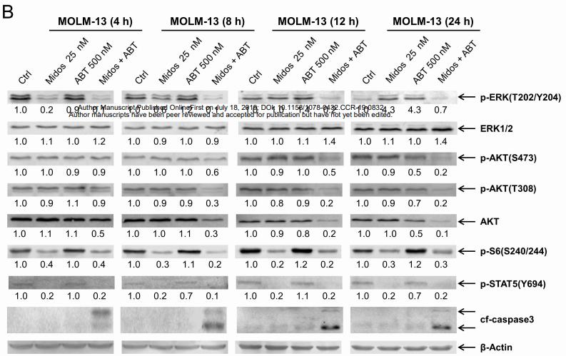
Application: WB Species: human Sample: MOLM-13 cells

Application: WB Species: human Sample: SGC-7901 cell

Application: IHC Species: Mouse Sample:

Application: WB Species: Mouse Sample:

Application: IHC Species: Mouse Sample: lung tissues
Application: WB Species: mouse Sample: Liver
Application: IF/ICC Species: mouse Sample: HepG2 cells
Restrictive clause
Affinity Biosciences tests all products strictly. Citations are provided as a resource for additional applications that have not been validated by Affinity Biosciences. Please choose the appropriate format for each application and consult Materials and Methods sections for additional details about the use of any product in these publications.
For Research Use Only.
Not for use in diagnostic or therapeutic procedures. Not for resale. Not for distribution without written consent. Affinity Biosciences will not be held responsible for patent infringement or other violations that may occur with the use of our products. Affinity Biosciences, Affinity Biosciences Logo and all other trademarks are the property of Affinity Biosciences LTD.