製品: PCNA Antibody
カタログ: AF0239
タンパク質の説明: Rabbit polyclonal antibody to PCNA
アプリケーション: WB IHC IF/ICC
反応性: Human, Mouse, Rat
予測: Pig, Zebrafish, Bovine, Horse, Sheep, Rabbit, Dog, Chicken
分子量: 36kDa; 29kD(Calculated).
ユニプロット: P12004
RRID: AB_2833414

類似製品を見る>>

   サイズ 価格 在庫状況
 50ul $150 在庫あり
 100ul $250 在庫あり
 200ul $350 在庫あり
 1ml $1200 在庫あり

リードタイム: 当日配達

For pricing and ordering contact:
お問い合わせ先

製品説明

ソース:
Rabbit
アプリケーション:
WB 1:500-1:3000, IHC 1:50-1:200, IF/ICC 1:100
*The optimal dilutions should be determined by the end user.
*Tips:

WB: For western blot detection of denatured protein samples. IHC: For immunohistochemical detection of paraffin sections (IHC-p) or frozen sections (IHC-f) of tissue samples. IF/ICC: For immunofluorescence detection of cell samples. ELISA(peptide): For ELISA detection of antigenic peptide.

反応性:
Human,Mouse,Rat
予測:
Pig(100%), Zebrafish(88%), Bovine(100%), Horse(100%), Sheep(100%), Rabbit(100%), Dog(100%), Chicken(100%)
クローナリティ:
Polyclonal
特異性:
PCNA antibody detects endogenous levels of total PCNA.
RRID:
AB_2833414
引用形式: Affinity Biosciences Cat# AF0239, RRID:AB_2833414.
コンジュゲート:
Unconjugated.
精製:
The antiserum was purified by peptide affinity chromatography using SulfoLink™ Coupling Resin (Thermo Fisher Scientific).
保存:
Rabbit IgG in phosphate buffered saline , pH 7.4, 150mM NaCl, 0.02% sodium azide and 50% glycerol. Store at -20 °C. Stable for 12 months from date of receipt.
別名:

折りたたみ/展開

ATLD2; cb16; Cyclin; DNA polymerase delta auxiliary protein; etID36690.10; fa28e03; fb36g03; HGCN8729; MGC8367; Mutagen-sensitive 209 protein; OTTHUMP00000030189; OTTHUMP00000030190; PCNA; Pcna/cyclin; PCNA_HUMAN; PCNAR; Polymerase delta accessory protein; Proliferating cell nuclear antigen; wu:fa28e03; wu:fb36g03;

免疫原

免疫原:
Uniprot:
遺伝子(ID):
タンパク質の説明:
PCNA This protein is an auxiliary protein of DNA polymerase delta and is involved in the control of eukaryotic DNA replication by increasing the polymerase's processibility during elongation of the leading strand. Belongs to the PCNA family. Homotrimer. Forms a complex with activator 1 heteropentamer in the presence of ATP.
タンパク質配列:
MFEARLVQGSILKKVLEALKDLINEACWDISSSGVNLQSMDSSHVSLVQLTLRSEGFDTYRCDRNLAMGVNLTSMSKILKCAGNEDIITLRAEDNADTLALVFEAPNQEKVSDYEMKLMDLDVEQLGIPEQEYSCVVKMPSGEFARICRDLSHIGDAVVISCAKDGVKFSASGELGNGNIKLSQTSNVDKEEEAVTIEMNEPVQLTFALRYLNFFTKATPLSSTVTLSMSADVPLVVEYKIADMGHLKYYLAPKIEDEEGS

種類予測

種類予測:

Score>80(red) has high confidence and is suggested to be used for WB detection. *The prediction model is mainly based on the alignment of immunogen sequences, the results are for reference only, not as the basis of quality assurance.

Species
Results
Score
Pig
100
Horse
100
Bovine
100
Sheep
100
Dog
100
Chicken
100
Rabbit
100
Zebrafish
88
Xenopus
63
Model Confidence:
High(score>80) Medium(80>score>50) Low(score<50) No confidence

PTMs - P12004 基板として

Site PTM Type Enzyme
R5 Methylation
S10 Phosphorylation
K13 Acetylation
K13 Ubiquitination
K14 Acetylation
K14 Ubiquitination
Y60 Phosphorylation
R61 Methylation
K77 Acetylation
K77 Ubiquitination
K80 Acetylation
K80 Ubiquitination
C81 S-Nitrosylation
K110 Ubiquitination
K117 Ubiquitination
Y133 Phosphorylation
S134 Phosphorylation
C135 S-Nitrosylation
K138 Ubiquitination
R146 Methylation
S152 Phosphorylation
S161 Phosphorylation
C162 S-Nitrosylation
K164 Acetylation
K164 Sumoylation
K164 Ubiquitination
K168 Ubiquitination
K181 Ubiquitination
T185 Phosphorylation
K190 Acetylation
R210 Methylation
Y211 Phosphorylation P00533 (EGFR)
T216 Phosphorylation
K217 Acetylation
S228 Phosphorylation
K248 Acetylation
K248 Ubiquitination
Y250 Phosphorylation
K254 Sumoylation
K254 Ubiquitination
S261 Phosphorylation

研究背景

機能:

Auxiliary protein of DNA polymerase delta and is involved in the control of eukaryotic DNA replication by increasing the polymerase's processibility during elongation of the leading strand. Induces a robust stimulatory effect on the 3'-5' exonuclease and 3'-phosphodiesterase, but not apurinic-apyrimidinic (AP) endonuclease, APEX2 activities. Has to be loaded onto DNA in order to be able to stimulate APEX2. Plays a key role in DNA damage response (DDR) by being conveniently positioned at the replication fork to coordinate DNA replication with DNA repair and DNA damage tolerance pathways. Acts as a loading platform to recruit DDR proteins that allow completion of DNA replication after DNA damage and promote postreplication repair: Monoubiquitinated PCNA leads to recruitment of translesion (TLS) polymerases, while 'Lys-63'-linked polyubiquitination of PCNA is involved in error-free pathway and employs recombination mechanisms to synthesize across the lesion.

PTMs:

Phosphorylated. Phosphorylation at Tyr-211 by EGFR stabilizes chromatin-associated PCNA.

Acetylated by CREBBP and p300/EP300; preferentially acetylated by CREBBP on Lys-80, Lys-13 and Lys-14 and on Lys-77 by p300/EP300 upon loading on chromatin in response to UV irradiation. Lysine acetylation disrupts association with chromatin, hence promoting PCNA ubiquitination and proteasomal degradation in response to UV damage in a CREBBP- and EP300-dependent manner. Acetylation disrupts interaction with NUDT15 and promotes degradation.

Ubiquitinated. Following DNA damage, can be either monoubiquitinated to stimulate direct bypass of DNA lesions by specialized DNA polymerases or polyubiquitinated to promote recombination-dependent DNA synthesis across DNA lesions by template switching mechanisms. Following induction of replication stress, monoubiquitinated by the UBE2B-RAD18 complex on Lys-164, leading to recruit translesion (TLS) polymerases, which are able to synthesize across DNA lesions in a potentially error-prone manner. An error-free pathway also exists and requires non-canonical polyubiquitination on Lys-164 through 'Lys-63' linkage of ubiquitin moieties by the E2 complex UBE2N-UBE2V2 and the E3 ligases, HLTF, RNF8 and SHPRH. This error-free pathway, also known as template switching, employs recombination mechanisms to synthesize across the lesion, using as a template the undamaged, newly synthesized strand of the sister chromatid. Monoubiquitination at Lys-164 also takes place in undamaged proliferating cells, and is mediated by the DCX(DTL) complex, leading to enhance PCNA-dependent translesion DNA synthesis. Sumoylated during S phase.

Methylated on glutamate residues by ARMT1/C6orf211.

細胞の位置付け:

Nucleus.
Note: Colocalizes with CREBBP, EP300 and POLD1 to sites of DNA damage (PubMed:24939902). Forms nuclear foci representing sites of ongoing DNA replication and vary in morphology and number during S phase. Together with APEX2, is redistributed in discrete nuclear foci in presence of oxidative DNA damaging agents.

Extracellular region or secreted Cytosol Plasma membrane Cytoskeleton Lysosome Endosome Peroxisome ER Golgi apparatus Nucleus Mitochondrion Manual annotation Automatic computational assertionSubcellular location
サブユニット構造:

Homotrimer. Interacts with p300/EP300; the interaction occurs on chromatin in UV-irradiated damaged cells. Interacts with CREBBP (via transactivation domain and C-terminus); the interaction occurs on chromatin in UV-irradiated damaged cells. Directly interacts with POLD1, POLD3 and POLD4 subunits of the DNA polymerase delta complex, POLD3 being the major interacting partner; the interaction with POLD3 is inhibited by CDKN1A/p21(CIP1). Forms a complex with activator 1 heteropentamer in the presence of ATP. Interacts with EXO1, POLH, POLK, DNMT1, ERCC5, FEN1, CDC6 and POLDIP2. Interacts with APEX2; this interaction is triggered by reactive oxygen species and increased by misincorporation of uracil in nuclear DNA. Forms a ternary complex with DNTTIP2 and core histone. Interacts with KCTD10 and PPP1R15A (By similarity). Directly interacts with BAZ1B. Interacts with HLTF and SHPRH. Interacts with NUDT15; this interaction is disrupted in response to UV irradiation and acetylation. Interacts with CDKN1A/p21(CIP1) and CDT1; interacts via their PIP-box which also recruits the DCX(DTL) complex. The interaction with CDKN1A inhibits POLD3 binding. Interacts with DDX11. Interacts with EGFR; positively regulates PCNA. Interacts with PARPBP. Interacts (when ubiquitinated) with SPRTN; leading to enhance RAD18-mediated PCNA ubiquitination. Interacts (when polyubiquitinated) with ZRANB3. Interacts with SMARCAD1. Interacts with CDKN1C. Interacts with PCLAF (via PIP-box). Interacts with RTEL1 (via PIP-box); the interaction is direct and essential for the suppression of telomere fragility. Interacts with FAM111A (via PIP-box); the interaction is direct and required for PCNA loading on chromatin binding. Interacts with LIG1. Interacts with SETMAR. Interacts with ANKRD17. Interacts with FBXO18/FBH1 (via PIP-box); the interaction recruits the DCX(DTL) complex and promotes ubiquitination and degradation of FBXO18/FBH1. Interacts with POLN. Interacts with SDE2 (via PIP-box); the interaction is direct and prevents ultraviolet light induced monoubiquitination. Component of the replisome complex composed of at least DONSON, MCM2, MCM7, PCNA and TICRR; interaction at least with PCNA occurs during DNA replication. Interacts with MAPK15; the interaction is chromatin binding dependent and prevents MDM2-mediated PCNA destruction by inhibiting the association of PCNA with MDM2. Interacts with PARP10 (via PIP-box). Interacts with DDI2. Interacts with HMCES (via PIP-box).

(Microbial infection) Interacts with herpes virus 8 protein LANA1.

タンパク質ファミリー:

Belongs to the PCNA family.

研究領域

· Cellular Processes > Cell growth and death > Cell cycle.   (View pathway)

· Cellular Processes > Cellular community - eukaryotes > Tight junction.   (View pathway)

· Genetic Information Processing > Replication and repair > DNA replication.

· Genetic Information Processing > Replication and repair > Base excision repair.

· Genetic Information Processing > Replication and repair > Nucleotide excision repair.

· Genetic Information Processing > Replication and repair > Mismatch repair.

· Human Diseases > Infectious diseases: Viral > Hepatitis B.

· Human Diseases > Infectious diseases: Viral > HTLV-I infection.

参考文献

1). A novel UV-curable extravascular stent to prevent restenosis of venous grafts. Composites Part B: Engineering, 2021 [IF=13.1]

Application: WB    Species: Rat    Sample:

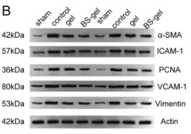
Fig. 6. A: PCNA immunofluorescence staining of venous graft vessels in rats. B, C: Western blot analysis of α-SMA, PCNA, ICAM-1, VCAM-1, and vimentin in rat vein graft vessels. D: ICAM-1 immunofluorescence staining of venous graft vessels in rats. E: qPCR of α-SMA, PCNA, ICAM-1, VCAM-1, and vimentin in rat vein graft vessels. Data are presented as the mean ± SD; *: compared with control group, p < 0.05. #: compared with sham group,

Application: IF/ICC    Species: Rat    Sample:

Fig. 6. A: PCNA immunofluorescence staining of venous graft vessels in rats. B, C: Western blot analysis of α-SMA, PCNA, ICAM-1, VCAM-1, and vimentin in rat vein graft vessels. D: ICAM-1 immunofluorescence staining of venous graft vessels in rats. E: qPCR of α-SMA, PCNA, ICAM-1, VCAM-1, and vimentin in rat vein graft vessels. Data are presented as the mean ± SD; *: compared with control group, p < 0.05. #: compared with sham group, p < 0.05.

2). Graphene foam/hydrogel scaffolds for regeneration of peripheral nerve using ADSCs in a diabetic mouse model. Nano Research, 2021 [IF=9.9]

3). Quercetin inhibited mesangial cell proliferation of early diabetic nephropathy through the Hippo pathway. PHARMACOLOGICAL RESEARCH, 2019 (PubMed: 31220559) [IF=9.3]

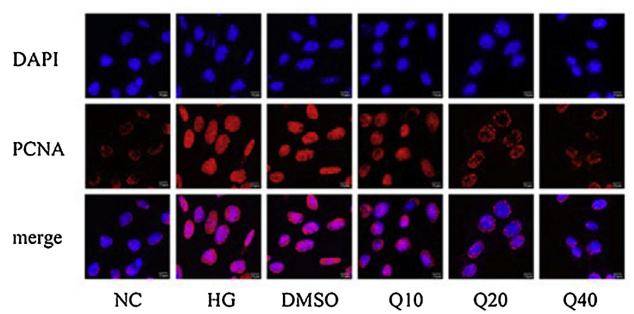
Application: IF/ICC    Species: mouse    Sample: glomerular mesangial cells

Fig.1.|Treatment with quercetin ameliorated the proliferation of high glucose-cultured glomerular mesangial cells. (A and B)Distribution and expression of PCNA and ki67 through immunofluorescence.

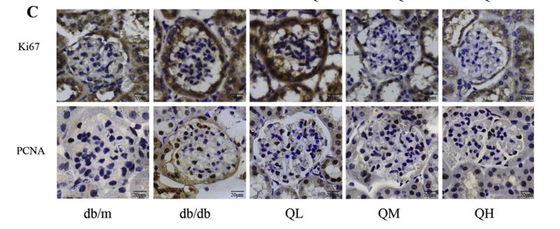
Application: IHC    Species: mouse    Sample: renal cortex

Fig. 5.| Quercetin ameliorated the glomerular mesangial cell proliferation of diabetic mice. (A) Renal ultra-structural changes of diabetic mice by transmission electron microscope. (B) Glomerular mesangial cell proliferation activity detected by 5-ethynyl-20-deoxyuridine incorporation assay. Scale bar = 20 μm. (C)Distribution and expression of ki67 and PCNA in renal cortex of diabetic mice through immunohistochemistry. Scale bar = 20 μm. 4.

4). Berberine inhibits high fat diet-associated colorectal cancer through modulation of the gut microbiota-mediated lysophosphatidylcholine. International Journal of Biological Sciences, 2023 (PubMed: 37151876) [IF=9.2]

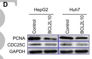
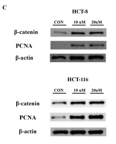
Application: WB    Species: Human    Sample: HCT 8 cells and HCT 116 cells

Figure 5 The BBR-associated differential metabolite LPC contributes to cell proliferation and cell junction impairment. (A) Cell growth curves of the CRC cell lines HCT116, HCT-8, SW480, and the normal intestinal epithelial cell line IEC-6, with or without LPC treatment. (B) HCT 116 cells and HCT 8 cells with or without LPC treatment were analyzed with flow cytometry. (C) Expression of β-catenin and PCNA in HCT 8 cells and HCT 116 cells with or without LPC treatment. (D) Expression of IL-6 and p-STAT3 in Raw 264.7 cells with or without LPC treatment. (E) Expression of occludin and E-cadherin in HCT-8 cells and HCT-116 cells co-cultured with Raw264.7 cells with or without LPC treatment.

5). Metformin: A promising clinical therapeutical approach for BPH treatment via inhibiting dysregulated steroid hormones-induced prostatic epithelial cells proliferation. Journal of Pharmaceutical Analysis, 2023 (PubMed: 38352949) [IF=8.8]

Application: IHC    Species: Human    Sample:

Fig. 1 Disruption of sex steroid hormones in clinical samples of benign prostate hyperplasia (BPH) patients. (A) Hematoxylin and eosin (H&E) staining of prostate sections from BPH patients (n = 6). (B) The relative protein levels of proliferating cell nuclear antigen (PCNA) in clinical prostate tissue by immunohistochemistry (IHC) staining and western blot analysis (n = 6). (C) The relative protein levels of Ki67 in clinical prostate tissue by IHC staining (n = 6). (D) The relative protein levels of cell cycle protein 1 (CyclinD1) in clinical prostate tissue by western blot analysis (n = 6). (E) Hierarchical clustered heat map of steroid hormones in the serum of normal men and BPH patients (n = 100). (F) 3D scatter plot of steroid hormones in the serum of normal men and BPH patients. (G) Variable importance for the projection (VIP) value of steroid hormones in the serum of normal men and BPH patients. (H) Hierarchical clustered heat maps of steroid hormones in the prostate tissue of normal men (n = 8) and BPH patients (n = 19). (I) 3D scatter plot of steroid hormones in the prostate tissue of normal men and BPH patients. (J) VIP value of steroid hormones in the prostate of normal men and BPH patients. Arrows indicate anatomical regions: prostatic epithelium or the acini (yellow arrow) and prostatic stroma (blue arrow). Data are mean ± standard error of mean (SEM). ∗∗P < 0.01, compared to N as indicated. A: androsterone; A4: androstenedione; DHEA: dehydroepiandrosterone; DHT: dihydrotestosterone; E1: estrone; E2: estradiol; T: testosterone.

Application: WB    Species: Human    Sample:

Fig. 1 Disruption of sex steroid hormones in clinical samples of benign prostate hyperplasia (BPH) patients. (A) Hematoxylin and eosin (H&E) staining of prostate sections from BPH patients (n = 6). (B) The relative protein levels of proliferating cell nuclear antigen (PCNA) in clinical prostate tissue by immunohistochemistry (IHC) staining and western blot analysis (n = 6). (C) The relative protein levels of Ki67 in clinical prostate tissue by IHC staining (n = 6). (D) The relative protein levels of cell cycle protein 1 (CyclinD1) in clinical prostate tissue by western blot analysis (n = 6). (E) Hierarchical clustered heat map of steroid hormones in the serum of normal men and BPH patients (n = 100). (F) 3D scatter plot of steroid hormones in the serum of normal men and BPH patients. (G) Variable importance for the projection (VIP) value of steroid hormones in the serum of normal men and BPH patients. (H) Hierarchical clustered heat maps of steroid hormones in the prostate tissue of normal men (n = 8) and BPH patients (n = 19). (I) 3D scatter plot of steroid hormones in the prostate tissue of normal men and BPH patients. (J) VIP value of steroid hormones in the prostate of normal men and BPH patients. Arrows indicate anatomical regions: prostatic epithelium or the acini (yellow arrow) and prostatic stroma (blue arrow). Data are mean ± standard error of mean (SEM). ∗∗P < 0.01, compared to N as indicated. A: androsterone; A4: androstenedione; DHEA: dehydroepiandrosterone; DHT: dihydrotestosterone; E1: estrone; E2: estradiol; T: testosterone.

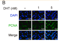
Application: IF/ICC    Species: Human    Sample:

Fig. 4 Effects of metformin (Met) on dihydrotestosterone (DHT)-induced proliferation in vitro. (A) The relative protein levels of androgen receptor (AR) in benign prostate hyperplasia (BPH)-1 cells cultured with DHT. (B) The relative protein levels of proliferating cell nuclear antigen (PCNA) in BPH-1 cells cultured with DHT by immunofluorescence (IF) and western blot analysis. (C) The relative protein levels of Ki67 in BPH-1 cells cultured with DHT by IF. (D) BPH-1 cell proliferation activity detected by 5-Ethynyl-20-deoxyuridine (EdU) assay. (E) Cell viability in DHT treated BPH-1 cells. (F) The relative protein levels of cell cycle protein 1 (CyclinD1) in BPH-1 cells cultured with DHT. (G) The relative protein levels of phosphorylation-AMP-activated protein kinase (p-AMPK) in BPH-1 cells cultured with DHT and Met. (H) The relative protein levels of AR in BPH-1 cells cultured with DHT and Met. (I) The relative protein levels of PCNA in BPH-1 cells co-cultured with DHT and Met by IF and western blot analysis. (J) The relative protein levels of Ki67 in BPH-1 cells cultured with DHT and Met by IF. (K) BPH-1 cell proliferation activity detected by EdU assay. (L) Cell viability in DHT and Met cultured BPH-1 cells. (M) The relative protein levels of CyclinD1 in BPH-1 cells cultured with DHT and Met. Data are mean ± standard error of mean (SEM) for cell groups of three. ∗P < 0.05, ∗∗P < 0.01, compared to normal control (NC) as indicated; #P < 0.05, ##P < 0.01, compared to DHT as indicated. t-AMPK: total-AMP-activated protein kinase.

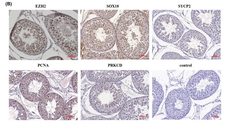
6). Long noncoding RNAs regulated spermatogenesis in varicocele‐induced spermatogenic dysfunction. CELL PROLIFERATION, 2022 (PubMed: 35297519) [IF=8.5]

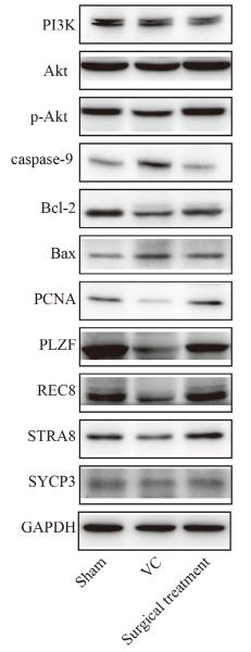
Application: WB    Species: rat    Sample: Testicular

FIGURE 13| Validation of regulated signalling pathways, spermatogenic cell apoptosis and proliferation, and meiotic spermatocytes by Western blot. Representative Western blot images of PI3K, Akt, p-Akt, caspase-9, Bcl-2, Bax, PCNA, PLZF, REC8, STRA8, and SYCP3.

7). ASIC1a promotes hepatic stellate cell activation through the exosomal miR-301a-3p/BTG1 pathway. International Journal of Biological Macromolecules, 2022 (PubMed: 35561854) [IF=8.2]

8). Ethyl ferulate suppresses post-myocardial infarction myocardial fibrosis by inhibiting transforming growth factor receptor 1. Phytomedicine, 2023 (PubMed: 37801895) [IF=7.9]

9). Caffeic acid mitigates myocardial fibrosis and improves heart function in post-myocardial infarction by inhibiting transforming growth factor-β receptor 1 signaling pathways. Biomedicine & pharmacotherapy = Biomedecine & pharmacotherapie, 2024 (PubMed: 38906025) [IF=7.5]

10). Human umbilical cord-derived mesenchymal stem cells improve the function of liver in rats with acute-on-chronic liver failure via downregulating Notch and Stat1/Stat3 signaling. Stem Cell Research & Therapy, 2021 (PubMed: 34256837) [IF=7.5]

Application: IHC    Species: Rat    Sample: liver tissue

Fig. 7 The effects of hUC-MSC transplantation on hepatocyte regeneration in ACLF rats. Liver sections from ACLF rats 12 and 24 h post-hUC-MSC transplantation or 0.9% sodium chloride injection as a control were used for immunohistochemical staining of AFP, CK18, HGF, and PCNA and microscopic examination (n = 4/group). Representative photographs are shown (a). Positive staining was quantified and is presented as the mean ± SD (b, c). d Serum levels of HGF were detected by ELISA (4h, n = 3/group; 12h and 24h, n = 4/group). Data are presented as the mean ± SD. *P < 0.05, **P < 0.01, ***P < 0.001

もっと読みます

Restrictive clause


Affinity Biosciences tests all products strictly. Citations are provided as a resource for additional applications that have not been validated by Affinity Biosciences. Please choose the appropriate format for each application and consult Materials and Methods sections for additional details about the use of any product in these publications.

For Research Use Only.
Not for use in diagnostic or therapeutic procedures. Not for resale. Not for distribution without written consent. Affinity Biosciences will not be held responsible for patent infringement or other violations that may occur with the use of our products. Affinity Biosciences, Affinity Biosciences Logo and all other trademarks are the property of Affinity Biosciences LTD.