製品: Phospho-FAK (Tyr397) Antibody
カタログ: AF3398
タンパク質の説明: Rabbit polyclonal antibody to Phospho-FAK (Tyr397)
アプリケーション: WB IHC IF/ICC
反応性: Human, Mouse, Rat
予測: Pig, Horse, Sheep, Rabbit, Dog, Chicken, Xenopus
分子量: 119kDa; 119kD(Calculated).
ユニプロット: Q05397
RRID: AB_2834829

類似製品を見る>>

   サイズ 価格 在庫状況
 100ul $280 在庫あり
 200ul $350 在庫あり

リードタイム: 当日配達

For pricing and ordering contact:
お問い合わせ先

製品説明

ソース:
Rabbit
アプリケーション:
WB 1:500-1:2000, IHC 1:100-1:500, IF/ICC 1:100-1:500
*The optimal dilutions should be determined by the end user.
*Tips:

WB: For western blot detection of denatured protein samples. IHC: For immunohistochemical detection of paraffin sections (IHC-p) or frozen sections (IHC-f) of tissue samples. IF/ICC: For immunofluorescence detection of cell samples. ELISA(peptide): For ELISA detection of antigenic peptide.

反応性:
Human,Mouse,Rat
予測:
Pig(100%), Horse(100%), Sheep(100%), Rabbit(100%), Dog(100%), Chicken(100%), Xenopus(100%)
クローナリティ:
Polyclonal
特異性:
Phospho-FAK (Tyr397) Antibody detects endogenous levels of FAK only when phosphorylated at Tyrosine 397.
RRID:
AB_2834829
引用形式: Affinity Biosciences Cat# AF3398, RRID:AB_2834829.
コンジュゲート:
Unconjugated.
精製:
The antibody is from purified rabbit serum by affinity purification via sequential chromatography on phospho-peptide and non-phospho-peptide affinity columns.
保存:
Rabbit IgG in phosphate buffered saline , pH 7.4, 150mM NaCl, 0.02% sodium azide and 50% glycerol. Store at -20 °C. Stable for 12 months from date of receipt.
別名:

折りたたみ/展開

FADK 1; FADK; FAK related non kinase polypeptide; FAK1; FAK1_HUMAN; Focal adhesion kinase 1; Focal adhesion Kinase; Focal adhesion kinase isoform FAK Del33; Focal adhesion kinase related nonkinase; FRNK; p125FAK; pp125FAK; PPP1R71; Protein phosphatase 1 regulatory subunit 71; Protein tyrosine kinase 2; Protein-tyrosine kinase 2; Ptk2; PTK2 protein tyrosine kinase 2;

免疫原

免疫原:
Uniprot:
遺伝子(ID):
発現特異性:
Q05397 FAK1_HUMAN:

Detected in B and T-lymphocytes. Isoform 1 and isoform 6 are detected in lung fibroblasts (at protein level). Ubiquitous.

タンパク質の説明:
This gene encodes a cytoplasmic protein tyrosine kinase which is found concentrated in the focal adhesions that form between cells growing in the presence of extracellular matrix constituents.
タンパク質配列:
MAAAYLDPNLNHTPNSSTKTHLGTGMERSPGAMERVLKVFHYFESNSEPTTWASIIRHGDATDVRGIIQKIVDSHKVKHVACYGFRLSHLRSEEVHWLHVDMGVSSVREKYELAHPPEEWKYELRIRYLPKGFLNQFTEDKPTLNFFYQQVKSDYMLEIADQVDQEIALKLGCLEIRRSYWEMRGNALEKKSNYEVLEKDVGLKRFFPKSLLDSVKAKTLRKLIQQTFRQFANLNREESILKFFEILSPVYRFDKECFKCALGSSWIISVELAIGPEEGISYLTDKGCNPTHLADFTQVQTIQYSNSEDKDRKGMLQLKIAGAPEPLTVTAPSLTIAENMADLIDGYCRLVNGTSQSFIIRPQKEGERALPSIPKLANSEKQGMRTHAVSVSETDDYAEIIDEEDTYTMPSTRDYEIQRERIELGRCIGEGQFGDVHQGIYMSPENPALAVAIKTCKNCTSDSVREKFLQEALTMRQFDHPHIVKLIGVITENPVWIIMELCTLGELRSFLQVRKYSLDLASLILYAYQLSTALAYLESKRFVHRDIAARNVLVSSNDCVKLGDFGLSRYMEDSTYYKASKGKLPIKWMAPESINFRRFTSASDVWMFGVCMWEILMHGVKPFQGVKNNDVIGRIENGERLPMPPNCPPTLYSLMTKCWAYDPSRRPRFTELKAQLSTILEEEKAQQEERMRMESRRQATVSWDSGGSDEAPPKPSRPGYPSPRSSEGFYPSPQHMVQTNHYQVSGYPGSHGITAMAGSIYPGQASLLDQTDSWNHRPQEIAMWQPNVEDSTVLDLRGIGQVLPTHLMEERLIRQQQEMEEDQRWLEKEERFLKPDVRLSRGSIDREDGSLQGPIGNQHIYQPVGKPDPAAPPKKPPRPGAPGHLGSLASLSSPADSYNEGVKLQPQEISPPPTANLDRSNDKVYENVTGLVKAVIEMSSKIQPAPPEEYVPMVKEVGLALRTLLATVDETIPLLPASTHREIEMAQKLLNSDLGELINKMKLAQQYVMTSLQQEYKKQMLTAAHALAVDAKNLLDVIDQARLKMLGQTRPH

種類予測

種類予測:

Score>80(red) has high confidence and is suggested to be used for WB detection. *The prediction model is mainly based on the alignment of immunogen sequences, the results are for reference only, not as the basis of quality assurance.

Species
Results
Score
Pig
100
Horse
100
Sheep
100
Dog
100
Xenopus
100
Chicken
100
Rabbit
100
Bovine
0
Zebrafish
0
Model Confidence:
High(score>80) Medium(80>score>50) Low(score<50) No confidence

PTMs - Q05397 基板として

Site PTM Type Enzyme
A2 Acetylation
Y5 Phosphorylation P09619 (PDGFRB) , P08581 (MET) , P00533 (EGFR)
T13 Phosphorylation
S16 Phosphorylation
T20 Phosphorylation
T24 Phosphorylation
S29 Phosphorylation
R65 Methylation
K70 Ubiquitination
Y148 Phosphorylation
Y155 Phosphorylation
Y194 Phosphorylation P09619 (PDGFRB) , P12931 (SRC) , P00533 (EGFR) , P51813 (BMX) , P08581 (MET)
K199 Ubiquitination
K216 Ubiquitination
S248 Phosphorylation
Y347 Phosphorylation
K364 Ubiquitination
S372 Phosphorylation
T386 Phosphorylation
S390 Phosphorylation
S392 Phosphorylation
T394 Phosphorylation
Y397 Phosphorylation P12931 (SRC) , Q05397 (PTK2) , P08581 (MET) , P46108 (CRK) , A0A059VC25 (FAK) , P09769 (FGR)
T406 Phosphorylation
Y407 Phosphorylation P08581 (MET) , Q05397 (PTK2) , Q14289 (PTK2B) , P12931 (SRC)
T408 Phosphorylation
Y441 Phosphorylation P51813 (BMX) , P12931 (SRC)
S443 Phosphorylation
T460 Phosphorylation
S461 Phosphorylation
S463 Phosphorylation
K467 Acetylation
T474 Phosphorylation
T503 Phosphorylation
S517 Phosphorylation
S568 Phosphorylation
Y570 Phosphorylation
S574 Phosphorylation
T575 Phosphorylation
Y576 Phosphorylation P08581 (MET) , P06241 (FYN) , P07949 (RET) , Q05397 (PTK2) , A0A059VC25 (FAK) , P51813 (BMX) , P07947 (YES1) , P09769 (FGR) , P12931 (SRC)
Y577 Phosphorylation P12931 (SRC) , A0A059VC25 (FAK) , P51813 (BMX) , P07949 (RET) , P08581 (MET) , Q05397 (PTK2)
S580 Phosphorylation
S677 Phosphorylation
K684 Ubiquitination
S695 Phosphorylation P31749 (AKT1)
T700 Phosphorylation P31749 (AKT1)
S702 Phosphorylation
S705 Phosphorylation
S708 Phosphorylation
S716 Phosphorylation
Y720 Phosphorylation
S722 Phosphorylation P49840 (GSK3A)
S726 Phosphorylation
S732 Phosphorylation Q13464 (ROCK1) , Q00535 (CDK5)
Y742 Phosphorylation P12931 (SRC) , P51813 (BMX)
S759 Phosphorylation
Y761 Phosphorylation
T805 Phosphorylation
S840 Phosphorylation
S843 Phosphorylation Q9UQM7 (CAMK2A)
S850 Phosphorylation
Y861 Phosphorylation Q05397 (PTK2) , P07949 (RET) , P08581 (MET) , Q13882 (PTK6) , P16591 (FER) , P12931 (SRC) , P51813 (BMX) , P07947 (YES1)
S887 Phosphorylation
S892 Phosphorylation
S893 Phosphorylation
Y898 Phosphorylation P51813 (BMX) , P12931 (SRC)
S910 Phosphorylation Q00535 (CDK5) , P27361 (MAPK3) , P28482 (MAPK1)
T914 Phosphorylation
S920 Phosphorylation
K923 Ubiquitination
Y925 Phosphorylation P12931 (SRC) , P16591 (FER) , P09769 (FGR) , P51813 (BMX) , P07947 (YES1) , P07949 (RET) , P08581 (MET)
T929 Phosphorylation
T963 Phosphorylation
T967 Phosphorylation
T971 Phosphorylation
K1000 Ubiquitination
Y1007 Phosphorylation P12931 (SRC) , P51813 (BMX)
K1017 Acetylation

PTMs - Q05397 酵素として

Substrate Site Source
O00401 (WASL) Y256 Uniprot
O43707 (ACTN4) Y4 Uniprot
O43707 (ACTN4) Y31 Uniprot
O43707 (ACTN4) Y265 Uniprot
O75962-1 (TRIO) Y2796 Uniprot
P07949 (RET) Y905 Uniprot
P12814-1 (ACTN1) Y12 Uniprot
P23634-6 (ATP2B4) Y1176 Uniprot
P26010 (ITGB7) Y753 Uniprot
P26010 (ITGB7) Y758 Uniprot
P29353 (SHC1) Y315 Uniprot
P49023 (PXN) Y31 Uniprot
P49023 (PXN) Y118 Uniprot
P51813 (BMX) Y40 Uniprot
P56945 (BCAR1) Y410 Uniprot
P56945-1 (BCAR1) Y664 Uniprot
P56945-1 (BCAR1) Y666 Uniprot
P63000 (RAC1) Y64 Uniprot
Q05397-1 (PTK2) S390 Uniprot
Q05397 (PTK2) Y397 Uniprot
Q05397 (PTK2) Y407 Uniprot
Q05397 (PTK2) Y576 Uniprot
Q05397 (PTK2) Y577 Uniprot
Q05397-1 (PTK2) S850 Uniprot
Q05397 (PTK2) Y861 Uniprot
Q05397-1 (PTK2) Y925 Uniprot
Q14451 (GRB7) Y188 Uniprot
Q14451 (GRB7) Y338 Uniprot
Q658W2 (DKFZp666O0110) S412 Uniprot
Q658W2 (DKFZp666O0110) Y419 Uniprot
Q658W2 (DKFZp666O0110) S872 Uniprot
Q658W2 (DKFZp666O0110) Y947 Uniprot
Q99961 (SH3GL1) Y315 Uniprot
Q9H5V8 (CDCP1) Y734 Uniprot
Q9H9S0 (NANOG) Y35 Uniprot
Q9H9S0 (NANOG) Y174 Uniprot
Q9Y2X7 (GIT1) Y321 Uniprot

研究背景

機能:

Non-receptor protein-tyrosine kinase that plays an essential role in regulating cell migration, adhesion, spreading, reorganization of the actin cytoskeleton, formation and disassembly of focal adhesions and cell protrusions, cell cycle progression, cell proliferation and apoptosis. Required for early embryonic development and placenta development. Required for embryonic angiogenesis, normal cardiomyocyte migration and proliferation, and normal heart development. Regulates axon growth and neuronal cell migration, axon branching and synapse formation; required for normal development of the nervous system. Plays a role in osteogenesis and differentiation of osteoblasts. Functions in integrin signal transduction, but also in signaling downstream of numerous growth factor receptors, G-protein coupled receptors (GPCR), EPHA2, netrin receptors and LDL receptors. Forms multisubunit signaling complexes with SRC and SRC family members upon activation; this leads to the phosphorylation of additional tyrosine residues, creating binding sites for scaffold proteins, effectors and substrates. Regulates numerous signaling pathways. Promotes activation of phosphatidylinositol 3-kinase and the AKT1 signaling cascade. Promotes activation of MAPK1/ERK2, MAPK3/ERK1 and the MAP kinase signaling cascade. Promotes localized and transient activation of guanine nucleotide exchange factors (GEFs) and GTPase-activating proteins (GAPs), and thereby modulates the activity of Rho family GTPases. Signaling via CAS family members mediates activation of RAC1. Recruits the ubiquitin ligase MDM2 to P53/TP53 in the nucleus, and thereby regulates P53/TP53 activity, P53/TP53 ubiquitination and proteasomal degradation. Phosphorylates SRC; this increases SRC kinase activity. Phosphorylates ACTN1, ARHGEF7, GRB7, RET and WASL. Promotes phosphorylation of PXN and STAT1; most likely PXN and STAT1 are phosphorylated by a SRC family kinase that is recruited to autophosphorylated PTK2/FAK1, rather than by PTK2/FAK1 itself. Promotes phosphorylation of BCAR1; GIT2 and SHC1; this requires both SRC and PTK2/FAK1. Promotes phosphorylation of BMX and PIK3R1. Isoform 6 (FRNK) does not contain a kinase domain and inhibits PTK2/FAK1 phosphorylation and signaling. Its enhanced expression can attenuate the nuclear accumulation of LPXN and limit its ability to enhance serum response factor (SRF)-dependent gene transcription.

PTMs:

Phosphorylated on tyrosine residues upon activation, e.g. upon integrin signaling. Tyr-397 is the major autophosphorylation site, but other kinases can also phosphorylate this residue. Phosphorylation at Tyr-397 promotes interaction with SRC and SRC family members, leading to phosphorylation at Tyr-576, Tyr-577 and at additional tyrosine residues. FGR promotes phosphorylation at Tyr-397 and Tyr-576. FER promotes phosphorylation at Tyr-577, Tyr-861 and Tyr-925, even when cells are not adherent. Tyr-397, Tyr-576 and Ser-722 are phosphorylated only when cells are adherent. Phosphorylation at Tyr-397 is important for interaction with BMX, PIK3R1 and SHC1. Phosphorylation at Tyr-925 is important for interaction with GRB2. Dephosphorylated by PTPN11; PTPN11 is recruited to PTK2 via EPHA2 (tyrosine phosphorylated). Microtubule-induced dephosphorylation at Tyr-397 is crucial for the induction of focal adhesion disassembly; this dephosphorylation could be catalyzed by PTPN11 and regulated by ZFYVE21. Phosphorylation on tyrosine residues is enhanced by NTN1 (By similarity).

Sumoylated; this enhances autophosphorylation.

細胞の位置付け:

Cell junction>Focal adhesion. Cell membrane>Peripheral membrane protein>Cytoplasmic side. Cytoplasm>Cell cortex. Cytoplasm>Cytoskeleton. Cytoplasm>Cytoskeleton>Microtubule organizing center>Centrosome. Nucleus.
Note: Constituent of focal adhesions. Detected at microtubules.

Extracellular region or secreted Cytosol Plasma membrane Cytoskeleton Lysosome Endosome Peroxisome ER Golgi apparatus Nucleus Mitochondrion Manual annotation Automatic computational assertionSubcellular location
組織特異性:

Detected in B and T-lymphocytes. Isoform 1 and isoform 6 are detected in lung fibroblasts (at protein level). Ubiquitous.

サブユニット構造:

Interacts (via first Pro-rich region) with CAS family members (via SH3 domain), including BCAR1, BCAR3, CASS4 and NEDD9. Interacts with GIT1. Interacts with SORBS1. Interacts with ARHGEF28. Interacts with SHB. Interacts with PXN and TLN1. Interacts with STAT1. Interacts with DCC. Interacts with WASL. Interacts with ARHGEF7. Interacts with GRB2 and GRB7 (By similarity). Component of a complex that contains at least FER, CTTN and PTK2/FAK1. Interacts with BMX. Interacts with TGFB1I1. Interacts with STEAP4. Interacts with ZFYVE21. Interacts with ESR1. Interacts with PIK3R1 or PIK3R2. Interacts with SRC, FGR, FLT4 and RET. Interacts with EPHA2 in resting cells; activation of EPHA2 recruits PTPN11, leading to dephosphorylation of PTK2/FAK1 and dissociation of the complex. Interacts with EPHA1 (kinase activity-dependent). Interacts with CD4; this interaction requires the presence of HIV-1 gp120. Interacts with PIAS1. Interacts with ARHGAP26 and SHC1. Interacts with RB1CC1; this inhibits PTK2/FAK1 activity and activation of downstream signaling pathways. Interacts with P53/TP53 and MDM2. Interacts with LPXN (via LD motif 3). Interacts with MISP. Interacts with CIB1 isoform 2. Interacts with CD36. Interacts with EMP2; regulates PTK2 activation and localization. Interacts with DSCAM (By similarity).

タンパク質ファミリー:

The Pro-rich regions interact with the SH3 domain of CAS family members, such as BCAR1 and NEDD9, and with the GTPase activating protein ARHGAP26.

The carboxy-terminal region is the site of focal adhesion targeting (FAT) sequence which mediates the localization of FAK1 to focal adhesions.

Belongs to the protein kinase superfamily. Tyr protein kinase family. FAK subfamily.

研究領域

· Cellular Processes > Cellular community - eukaryotes > Focal adhesion.   (View pathway)

· Cellular Processes > Cell motility > Regulation of actin cytoskeleton.   (View pathway)

· Environmental Information Processing > Signal transduction > ErbB signaling pathway.   (View pathway)

· Environmental Information Processing > Signal transduction > PI3K-Akt signaling pathway.   (View pathway)

· Human Diseases > Drug resistance: Antineoplastic > Endocrine resistance.

· Human Diseases > Infectious diseases: Bacterial > Bacterial invasion of epithelial cells.

· Human Diseases > Infectious diseases: Parasitic > Amoebiasis.

· Human Diseases > Infectious diseases: Viral > Human papillomavirus infection.

· Human Diseases > Cancers: Overview > Pathways in cancer.   (View pathway)

· Human Diseases > Cancers: Overview > Transcriptional misregulation in cancer.

· Human Diseases > Cancers: Overview > Proteoglycans in cancer.

· Human Diseases > Cancers: Specific types > Small cell lung cancer.   (View pathway)

· Organismal Systems > Immune system > Chemokine signaling pathway.   (View pathway)

· Organismal Systems > Development > Axon guidance.   (View pathway)

· Organismal Systems > Immune system > Leukocyte transendothelial migration.   (View pathway)

参考文献

1). A tropomyosin-like Meretrix meretrix Linnaeus polypeptide inhibits the proliferation and metastasis of glioma cells via microtubule polymerization and FAK/Akt/MMPs signaling. International Journal of Biological Macromolecules, 2020 (PubMed: 31866539) [IF=7.7]

Application: WB    Species: Human    Sample: U87 cells

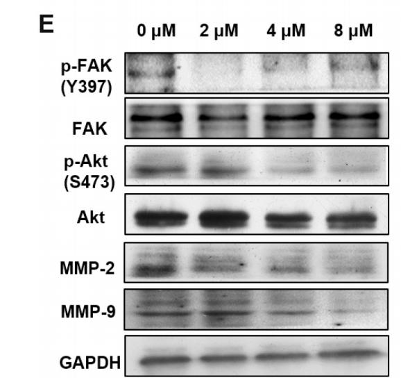
Fig. 5. re-MM15 inhibited the metastasis of U87 cells through downregulating FAK/Akt/MMPs signaling. (A and B) Suppression of U87 cell migration by re-MM15. Wound closures of U87 cells were photographed at 0 h, 12 h and 24 h following treatment with different concentrations of re-MM15 (scale bar: 100 μm). Error bars represented the mean ± SEM of triplicate independent experiments. (C–D) Inhibition of U87 cell invasion by re-MM15. After treated with indicated concentrations of re-MM15, U87 cells were seeded in Matrigel-coated transwell chambers and the invaded cells were stained and calculated. Shown are representative images of invaded cells (scale bar: 100 μm). The bar graphs showed mean ± SEM of the numbers of invaded cells from three independent assays. (E–F) Downregulation of FAK/Akt/MMPs signaling by re-MM15. The expression of p-FAK, FAK, p-Akt, Akt, MMP-2, MMP- 9 were detected by Western blot. Band density was quantified and normalized with GAPDH. The results shown are mean ± SEM (n = 3). *P b 0.05, **P b 0.01 versus control group.

2). Anti-proliferation and anti-migration effects of Yishen Tongbi decoction in experimental rheumatoid arthritis by suppressing SLC3A2/integrin β3 signaling pathways. Phytomedicine, 2023 (PubMed: 36990010) [IF=6.7]

3). Down-regulating Myoferlin inhibits the vasculogenic mimicry of melanoma via decreasing MMP-2 and inducing mesenchymal-to-epithelial transition. Journal of Cellular and Molecular Medicine, 2018 (PubMed: 29164766) [IF=5.3]

4). Phellinus linteus activates Treg cells via FAK to promote M2 macrophage polarization in hepatocellular carcinoma. Cancer immunology, immunotherapy : CII, 2024 (PubMed: 38240856) [IF=4.6]

Application: WB    Species: Mouse    Sample:

Fig. 3 FAK promotes the polarization of macrophages in the Treg-macrophage co-culture system through PI3K/AKT/JAK/STAT3 and p38/JNK/ERK signal pathway A, B The expression of the PI3K, p-PI3K, AKT, p-AKT, p-FAK, FAK, JAK2, STAT3, p-STAT3 and p38, p-p38/JNK, p-JNK/ERK1/2, p-ERK1/2 in Treg-macrophage co-culture system were observed by the western blot, n = 3 in each group. #P 

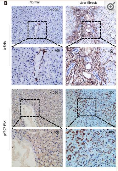
5). Focal adhesion kinase-related non-kinase ameliorates liver fibrosis by inhibiting aerobic glycolysis via the FAK/Ras/c-myc/ENO1 pathway. WORLD JOURNAL OF GASTROENTEROLOGY, 2022 (PubMed: 35125823) [IF=4.3]

Application: IHC    Species: Human    Sample: fibrotic liver tissue

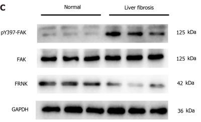
Figure 1 Tyrosine phosphorylation at position 397 of FAK is upregulated, while FRNK expression is downregulated in human fibrotic liver tissue. A: H&E, Masson’s trichrome and Sirius Red staining were performed after liver biopsy to assess the tissues of normal subjects and patients with liver fibrosis under a light microscope at 200× magnification; B: Immunohistochemistry showed changes in the expression of α-smooth muscle actin (α-SMA) and tyrosine phosphorylation at position 397 of FAK(pY397-FAK) in the livers of normal subjects compared with patients with liver fibrosis under a light microscope at 200× or 400× magnification; C and D: Protein expression in biopsy tissues was analyzed using Western blotting. Representative results from three independent replicate assays are shown. aP < 0.05. Data are presented as the mean ± SD. FAK: Focal adhesion kinase; FRNK: Focal adhesion kinase-related non-kinase; GAPDH: Glyceraldehyde 3-phosphate dehydrogenase.

Application: WB    Species: Human    Sample: fibrotic liver tissue

Figure 1 Tyrosine phosphorylation at position 397 of FAK is upregulated, while FRNK expression is downregulated in human fibrotic liver tissue. A: H&E, Masson’s trichrome and Sirius Red staining were performed after liver biopsy to assess the tissues of normal subjects and patients with liver fibrosis under a light microscope at 200× magnification; B: Immunohistochemistry showed changes in the expression of α-smooth muscle actin (α-SMA) and tyrosine phosphorylation at position 397 of FAK(pY397-FAK) in the livers of normal subjects compared with patients with liver fibrosis under a light microscope at 200× or 400× magnification; C and D: Protein expression in biopsy tissues was analyzed using Western blotting. Representative results from three independent replicate assays are shown. aP < 0.05. Data are presented as the mean ± SD. FAK: Focal adhesion kinase; FRNK: Focal adhesion kinase-related non-kinase; GAPDH: Glyceraldehyde 3-phosphate dehydrogenase.

6). FOXF1 inhibits invasion and metastasis of lung adenocarcinoma cells and enhances anti-tumor immunity via MFAP4/FAK signal axis. Scientific reports, 2024 (PubMed: 39271782) [IF=3.8]

7). KRT7 promotes epithelial‑mesenchymal transition in ovarian cancer via the TGF‑β/Smad2/3 signaling pathway. ONCOLOGY REPORTS, 2021 (PubMed: 33416175) [IF=3.8]

Application: WB    Species: Human    Sample: HEY cells

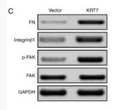
Figure 5. - KRT7 expression affects the integrin-β1-FAK signaling and TGF-β signaling pathways. (A and B) Expression of proliferation- and migration-associated genes (PCNA, MMP9 and TIMP-1) were evaluated using western blotting in HEY cells. (C and D) Western blotting of proteins involved in integrin-β1-FAK signaling pathway in the KRT7-overexpressing HEY cells. (E) Expression of MMPs after knockdown of KRT7 in OVCAR433 cells. (F and G) Expression of the TGF-β signaling pathway-related proteins was evaluated by western blotting in KRT7-overexpressing HEY cells and KRT7-knockdown OVCAR433 cells. All experiments were performed at least three times. Results are presented as the mean ± standard deviation. **P<0.01. FAK, focal adhesion kinase; PCNA, proliferating cell nuclear antigen; FN, fibronectin; TIMP-1, TIMP metallopeptidase inhibitor 1; p-, phosphorylated; MMP, matrix metalloproteinase; KRT7, keratin 7; sh, short hairpin RNA; NC, negative control.

8). Niban apoptosis regulator 1 promotes gemcitabine resistance by activating the focal adhesion kinase signaling pathway in bladder cancer. Journal of Cancer, 2022 (PubMed: 35281857) [IF=3.3]

Application: WB    Species: Human    Sample: bladder cancer cells

Figure 7 High NIBAN1 expression correlates with highly enriched focal adhesion signaling in bladder cancer. A. Gene Set Enrichment Analysis of The Cancer Genome Atlas bladder cancer dataset revealed a highly enriched gene signature of focal adhesion pathway in NIBAN1-high bladder cancer. B. GEM-resistant cells T24GR exhibited highly activated FAK and its downstream SRC/AKT signaling compared to GEM-sensitive T24 and 5637 cells. β-Actin was used as a loading control. C. Differentially expressed genes in T24GR cells with or without NIBAN1 knockdown. D. Pathway enrichment analysis was conducted on downregulated genes (n = 173) using GO, KEGG, and Reactome Gene Sets annotations.

9). Ovostatin 2 knockdown significantly inhibits the growth, migration, and tumorigenicity of cutaneous malignant melanoma cells. PLoS One, 2018 (PubMed: 29684087) [IF=2.9]

Application: WB    Species: mouse    Sample: A375 cells

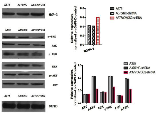
Fig 5. |Western blot results. (a) The levels of cyclin A, cyclin B, cyclin D1, and CDK2 were reduced in A375 cells transfected with OVOS2-shRNA; (b) The downregulated expression of N-cadherin accompanied with the upregulated expression of E-cadherin and β-catenin were observed in A375 cells transfected with OVOS2-shRNA; (c) The expression of p-FAK, p-AKT, and p-ERK were reduced in A375 cells transfected with OVOS2-shRNA; (d) The increased production of MMP-2 was observed in A375 transfected with OVOS2-shRNA; (e) GAPDH was used as the reference.

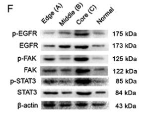
10). Distribution pattern of invasion‑related bio‑markers in head Marjolin's ulcer. Experimental and Therapeutic Medicine, 2020 (PubMed: 32855703) [IF=2.4]

Application: WB    Species: human    Sample: MU tissue

Figure 5.| Expression levels of tumor invasion-associated cell signaling pathways molecules at different sites of Marjolin's ulcer. Relative mRNA expression levels of (A) FAK, (B) STAT3, (C) EGFR, (D) CXCL9 and (E) RANKL.Values are expressed as the mean ± standard error of the mean (n=3). Western blot was used to determine the protein level of phosphorylated STAT3, FAK and EGFR (F) and statistical analysis has been performed (G-I).

もっと読みます

Restrictive clause


Affinity Biosciences tests all products strictly. Citations are provided as a resource for additional applications that have not been validated by Affinity Biosciences. Please choose the appropriate format for each application and consult Materials and Methods sections for additional details about the use of any product in these publications.

For Research Use Only.
Not for use in diagnostic or therapeutic procedures. Not for resale. Not for distribution without written consent. Affinity Biosciences will not be held responsible for patent infringement or other violations that may occur with the use of our products. Affinity Biosciences, Affinity Biosciences Logo and all other trademarks are the property of Affinity Biosciences LTD.