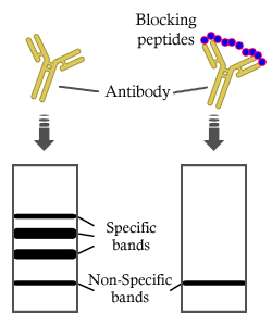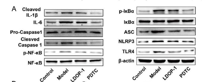
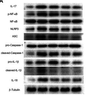
製品: Cleaved-Caspase 1 (Ala317), p10 Antibody
カタログ: AF4022
タンパク質の説明: Rabbit polyclonal antibody to Cleaved-Caspase 1 (Ala317), p10
アプリケーション: WB IHC IF/ICC
Cited expt.: WB, IHC, IF/ICC
反応性: Human, Mouse, Rat
予測: Pig, Bovine, Horse, Rabbit, Dog
分子量: 10kD,45kD; 45kD(Calculated).
ユニプロット: P29466
RRID: AB_2845464

類似製品を見る>>

   サイズ 価格 在庫状況
 50ul $250 在庫あり
 100ul $350 在庫あり
 200ul $450 在庫あり

リードタイム: 当日配達

For pricing and ordering contact:
お問い合わせ先

製品説明

ソース:
Rabbit
アプリケーション:
WB 1:500-1:2000, IHC 1:50-1:200, IF/ICC 1:100-1:500
*The optimal dilutions should be determined by the end user. For optimal experimental results, antibody reuse is not recommended.
*Tips:

WB: For western blot detection of denatured protein samples. IHC: For immunohistochemical detection of paraffin sections (IHC-p) or frozen sections (IHC-f) of tissue samples. IF/ICC: For immunofluorescence detection of cell samples. ELISA(peptide): For ELISA detection of antigenic peptide.

反応性:
Human,Mouse,Rat
予測:
Pig(100%), Bovine(100%), Horse(88%), Rabbit(100%), Dog(88%)
クローナリティ:
Polyclonal
特異性:
Cleaved-Caspase-1 (Ala317,p10) Antibody detects endogenous levels of fragment of activated Caspase-1 resulting from cleavage adjacent to Ala317.
RRID:
AB_2845464
引用形式: Affinity Biosciences Cat# AF4022, RRID:AB_2845464.
コンジュゲート:
Unconjugated.
精製:
The antiserum was purified by peptide affinity chromatography using SulfoLink™ Coupling Resin (Thermo Fisher Scientific).
保存:
Rabbit IgG in phosphate buffered saline , pH 7.4, 150mM NaCl, 0.02% sodium azide and 50% glycerol. Store at -20 °C. Stable for 12 months from date of receipt.
別名:

折りたたみ/展開

CASP-1; CASP1; CASP1_HUMAN; Caspase 1; Caspase-1 subunit p10; ICE; IL-1 beta-converting enzyme; IL-1BC; IL1 beta converting enzyme; IL1B convertase; Interleukin 1 beta convertase; Interleukin 1B converting enzyme; Interleukin-1 beta convertase; Interleukin-1 beta-converting enzyme; p45;

免疫原

免疫原:

The antiserum was produced against synthesized peptide derived from human Caspase 1.

Uniprot:
遺伝子(ID):
発現特異性:
P29466 CASP1_HUMAN:

Expressed in larger amounts in spleen and lung. Detected in liver, heart, small intestine, colon, thymus, prostate, skeletal muscle, peripheral blood leukocytes, kidney and testis. No expression in the brain.

タンパク質配列:
MADKVLKEKRKLFIRSMGEGTINGLLDELLQTRVLNKEEMEKVKRENATVMDKTRALIDSVIPKGAQACQICITYICEEDSYLAGTLGLSADQTSGNYLNMQDSQGVLSSFPAPQAVQDNPAMPTSSGSEGNVKLCSLEEAQRIWKQKSAEIYPIMDKSSRTRLALIICNEEFDSIPRRTGAEVDITGMTMLLQNLGYSVDVKKNLTASDMTTELEAFAHRPEHKTSDSTFLVFMSHGIREGICGKKHSEQVPDILQLNAIFNMLNTKNCPSLKDKPKVIIIQACRGDSPGVVWFKDSVGVSGNLSLPTTEEFEDDAIKKAHIEKDFIAFCSSTPDNVSWRHPTMGSVFIGRLIEHMQEYACSCDVEEIFRKVRFSFEQPDGRAQMPTTERVTLTRCFYLFPGH

種類予測

種類予測:

Score>80(red) has high confidence and is suggested to be used for WB detection. *The prediction model is mainly based on the alignment of immunogen sequences, the results are for reference only, not as the basis of quality assurance.

Species
Results
Score
Pig
100
Bovine
100
Rabbit
100
Horse
88
Dog
88
Sheep
0
Xenopus
0
Zebrafish
0
Chicken
0
Model Confidence:
High(score>80) Medium(80>score>50) Low(score<50) No confidence

研究背景

機能:

Thiol protease that cleaves IL-1 beta between an Asp and an Ala, releasing the mature cytokine which is involved in a variety of inflammatory processes. Important for defense against pathogens. Cleaves and activates sterol regulatory element binding proteins (SREBPs). Can also promote apoptosis. Upon inflammasome activation, during DNA virus infection but not RNA virus challenge, controls antiviral immunity through the cleavage of CGAS, rendering it inactive. In apoptotic cells, cleaves SPHK2 which is released from cells and remains enzymatically active extracellularly.

PTMs:

The two subunits are derived from the precursor sequence by an autocatalytic mechanism.

細胞の位置付け:

Cytoplasm. Cell membrane.

Extracellular region or secreted Cytosol Plasma membrane Cytoskeleton Lysosome Endosome Peroxisome ER Golgi apparatus Nucleus Mitochondrion Manual annotation Automatic computational assertionSubcellular location
組織特異性:

Expressed in larger amounts in spleen and lung. Detected in liver, heart, small intestine, colon, thymus, prostate, skeletal muscle, peripheral blood leukocytes, kidney and testis. No expression in the brain.

タンパク質ファミリー:

Belongs to the peptidase C14A family.

研究領域

· Cellular Processes > Cell growth and death > Necroptosis.   (View pathway)

· Human Diseases > Neurodegenerative diseases > Amyotrophic lateral sclerosis (ALS).

· Human Diseases > Infectious diseases: Bacterial > Salmonella infection.

· Human Diseases > Infectious diseases: Bacterial > Pertussis.

· Human Diseases > Infectious diseases: Bacterial > Legionellosis.

· Human Diseases > Infectious diseases: Viral > Influenza A.

· Organismal Systems > Immune system > NOD-like receptor signaling pathway.   (View pathway)

· Organismal Systems > Immune system > Cytosolic DNA-sensing pathway.   (View pathway)

参考文献
1). An artificial metabzyme for tumour-cell-specific metabolic therapy. Nature nanotechnology, 2024 (PubMed: 39103450) [IF=38.1]

2). NAD+-Boosters Improve Mitochondria Quality Control In Parkinson's Disease Models Via Mitochondrial UPR. Advanced science (Weinheim, Baden-Wurttemberg, Germany), 2025 (PubMed: 40685704) [IF=15.1]

3). p66Shc Contributes to Liver Fibrosis through the Regulation of Mitochondrial Reactive Oxygen Species. Theranostics, 2019 (PubMed: 30867846) [IF=12.4]

Application: WB    Species: mouse    Sample: Liver

Figure 2.| p66Shc silencing attenuates liver fibrosis in mice. p66Shc silencing was induced via lentivirus delivered to C57BL/6 mice exposed to CCl4 (2 ml/kg). (A) Liver p66Shc mRNA expression, n=6. (B) Liver p66Shc, SOD2, UCP1, Col1a1, α-SMA protein, n=3. (C) H2O2 content, n=8. (D) SOD activity, n=8. (E) Cytochrome c expression in the cytoplasm and mitochondria, n=3. (F) H&E and Masson staining. Scale bar, 200 μm. (G) Ishak score of Masson staining. (H) Serum ALT and AST levels, n=8. (I) Liver NLRP3 inflammasome protein expression, n=3.

4). Inflammation-driven biomimetic nano-polyphenol drug delivery system alleviates severe acute pancreatitis by inhibiting macrophage PANoptosis and pancreatic enzymes oversecretion. Journal of advanced research, 2025 (PubMed: 40210149) [IF=11.4]

Application: WB    Species: Mouse    Sample: BMDMs

Fig. 4. PTX formulations inhibited macrophage PANoptosis. (A) Gene clustering analysis of PTX, FePTX, FePTX@CM-treated BMDMs. (B-C) GSEA analysis of transcriptomics results of BMDMs after LPS intervention. (D-E) Inflammatory-related pathway analysis in transcriptomics results of BMDMs after different PTX formulation interventions. (F) Heatmap of transcriptional profiling of inflammatory factors in BMDMs after PTX formulation interventions. (G) Results of qPCR sequencing (n = 4). (H) Cell viability of BMDMs with different treatments (n = 4). (I) PI staining images of BMDMs after LPS and FePTX@CM NPs treatment and its analysis (n = 4). Scale bar = 100 μm. (J) WB detection of the PANoptosis biomarkers in BMDMs upon treatment with FePTX@CM NPs. (K) TEM images of BMDMs after different treatments. Scale bar = 5 μm. (L) Co-IP analysis of PANoptosomes biomarkers in RAW264.7 after various interventions (n = 4). *P < 0.05, **P < 0.01, ***P < 0.001, N.s. = no significance. Data = mean ± SD.

5). Water-extracted Lonicera japonica polysaccharide attenuates allergic rhinitis by regulating NLRP3-IL-17 signaling axis. Carbohydrate Polymers, 2022 (PubMed: 36184153) [IF=10.7]

6). An elastase-responsive "plug-and-play" quercetin-loaded engineered extracellular vesicle platform anchored with anti-PRDX1 antibody for severe acute pancreatitis therapy. Journal of controlled release : official journal of the Controlled Release Society, 2026 (PubMed: 41621758) [IF=10.5]

7). AIM2 Inflammasome Mediates Hallmark Neuropathological Alterations and Cognitive Impairment in a Mouse Model of Vascular Dementia. Molecular Psychiatry, 2020 (PubMed: 33299135) [IF=9.6]

8). NLRP3-GABA signaling pathway contributes to the pathogenesis of impulsive-like behaviors and cognitive deficits in aged mice. Journal of neuroinflammation, 2023 (PubMed: 37434240) [IF=9.3]

Application: IF/ICC    Species: Mouse    Sample: hippocampus

Fig. 6 Cognitive impairment may be related to pyroptosis-associated inflammatory responses in astrocytes. A Representative photomicrographs of NLRP3-, IL-18-, IL-1β- and cleaved caspase-1-positive cells in the CA1 of the hippocampus at 12 h and 10 days after surgical exposure. Scale bar = 50 μm. B Image of CA1 in hippocampus. C–J Co-stained area of NLRP3-, IL-18-, IL-1β-, cleaved caspase-1- and GFAP-positive cells in the CA1 of the hippocampus. K Representative western blot of NLRP3, IL-1β, IL-18 and cleaved caspase-1 at 12 h after surgical exposure. L–O The optical density values of NLRP3, IL-1β, IL-18, and cleaved caspase-1 at 12 h after surgical exposure. P Representative western blot of NLRP3, IL-1β, IL-18 and cleaved caspase-1 at 10 days after surgical exposure. Q–T The optical density values of NLRP3, IL-1β, IL-18 and cleaved caspase-1 at 10 days after surgical exposure. Data are presented as the mean ± SD (n = 6 mice/group or n = 3 mice/group). Data were analyzed by one-way ANOVA with Tukey’s multiple comparison test or Kruskal–Wallis and Dunn’s multiple comparison test. ***P 

9). JC124 confers multimodal neuroprotection in epilepsy by suppressing NLRP3 inflammasome activation: evidence from animal and human neuronal models. Cell communication and signaling : CCS, 2025 (PubMed: 40629339) [IF=8.4]

Application: WB    Species: mouse    Sample:

Fig. 3 From: JC124 confers multimodal neuroprotection in epilepsy by suppressing NLRP3 inflammasome activation: evidence from animal and human neuronal models JC124 treatment inhibits NLRP3 inflammasome activation in KA-induced epileptic mice. A Representative WB bands. In KA-induced epileptic mice, treatment with JC124 significantly reduced the relative intensities of (B) NLRP3/GAPDH, (C) ASC/GAPDH, (D) pro-Caspase-1/GAPDH, and (E) Caspase-1 p10/GAPDH in the hippocampus (n = 3). F–H Representative immunohistochemical images of NLRP3 (brown), ASC (brown), and Caspase-1 p10 (brown) in the hippocampal CA1 region. Scale bar, 50 μm. The black arrows indicate typical positively stained cells. KA-induced epileptic mice treated with JC124 showed a significant decrease in the mean optical densities of the (I) NLRP3, (J) ASC, and (K) Caspase-1 p10 (n = 5). L-N Representative immunofluorescence images of NLRP3 (green)/GFAP (red), NLRP3 (green)/Iba1 (red), NLRP3 (green)/NeuN (red), ASC (green)/GFAP (red), ASC (green)/Iba1 (red), ASC (green)/NeuN (red), Caspase-1 p10 (green)/GFAP (red), Caspase-1 p10 (green)/Iba1 (red), and Caspase-1 p10 (green)/NeuN (red) in the hippocampal CA1 region (n = 3). Scale bar, 50 μm. The white arrows indicate typical co-labeled cells. Data are represented as mean ± S.E.M. *p < 0.05, **p < 0.01, ***p < 0.001

Application: IF/ICC    Species: mouse    Sample:

Fig. 3 From: JC124 confers multimodal neuroprotection in epilepsy by suppressing NLRP3 inflammasome activation: evidence from animal and human neuronal models JC124 treatment inhibits NLRP3 inflammasome activation in KA-induced epileptic mice. A Representative WB bands. In KA-induced epileptic mice, treatment with JC124 significantly reduced the relative intensities of (B) NLRP3/GAPDH, (C) ASC/GAPDH, (D) pro-Caspase-1/GAPDH, and (E) Caspase-1 p10/GAPDH in the hippocampus (n = 3). F–H Representative immunohistochemical images of NLRP3 (brown), ASC (brown), and Caspase-1 p10 (brown) in the hippocampal CA1 region. Scale bar, 50 μm. The black arrows indicate typical positively stained cells. KA-induced epileptic mice treated with JC124 showed a significant decrease in the mean optical densities of the (I) NLRP3, (J) ASC, and (K) Caspase-1 p10 (n = 5). L-N Representative immunofluorescence images of NLRP3 (green)/GFAP (red), NLRP3 (green)/Iba1 (red), NLRP3 (green)/NeuN (red), ASC (green)/GFAP (red), ASC (green)/Iba1 (red), ASC (green)/NeuN (red), Caspase-1 p10 (green)/GFAP (red), Caspase-1 p10 (green)/Iba1 (red), and Caspase-1 p10 (green)/NeuN (red) in the hippocampal CA1 region (n = 3). Scale bar, 50 μm. The white arrows indicate typical co-labeled cells. Data are represented as mean ± S.E.M. *p < 0.05, **p < 0.01, ***p < 0.001

10). Mesenchymal stem cells protect against TBI-induced pyroptosis in vivo and in vitro through TSG-6. Cell Communication and Signaling, 2022 (PubMed: 35982465) [IF=8.4]

もっと読みます

Restrictive clause


Affinity Biosciences tests all products strictly. Citations are provided as a resource for additional applications that have not been validated by Affinity Biosciences. Please choose the appropriate format for each application and consult Materials and Methods sections for additional details about the use of any product in these publications.

For Research Use Only.
Not for use in diagnostic or therapeutic procedures. Not for resale. Not for distribution without written consent. Affinity Biosciences will not be held responsible for patent infringement or other violations that may occur with the use of our products. Affinity Biosciences, Affinity Biosciences Logo and all other trademarks are the property of Affinity Biosciences LTD.