製品: HIF1 alpha Mouse monoclonal Antibody
カタログ: BF8002
タンパク質の説明: Mouse monoclonal antibody to HIF1 alpha
アプリケーション: WB IHC IF/ICC
反応性: Mouse
分子量: 120kDa; 93kD(Calculated).
ユニプロット: Q16665
RRID: AB_2846221

類似製品を見る>>

   サイズ 価格 在庫状況
 50ul $250 在庫あり
 100ul $350 在庫あり
 200ul $450 在庫あり

リードタイム: 当日配達

For pricing and ordering contact:
お問い合わせ先

製品説明

ソース:
Mouse
アプリケーション:
WB 1:500-1:3000, IHC 1:50-1:200, IF/ICC 1:100-1:500
*The optimal dilutions should be determined by the end user.
*Tips:

WB: For western blot detection of denatured protein samples. IHC: For immunohistochemical detection of paraffin sections (IHC-p) or frozen sections (IHC-f) of tissue samples. IF/ICC: For immunofluorescence detection of cell samples. ELISA(peptide): For ELISA detection of antigenic peptide.

反応性:
Mouse
クローナリティ:
Monoclonal [AFfirm8002(AFB17813)]
特異性:
HIF1 alpha mouse monoclonal antibody detects endogenous levels of HIF1a.
RRID:
AB_2846221
引用形式: Affinity Biosciences Cat# BF8002, RRID:AB_2846221.
コンジュゲート:
Unconjugated.
精製:
The ascites was purified by peptide affinity chromatography using SulfoLink™ Coupling Resin (Thermo Fisher Scientific).
保存:
Mouse IgG in phosphate buffered saline , pH 7.4, 150mM NaCl, 0.02% sodium azide and 50% glycerol.Stable for 15 months from date of receipt. Store at -20 °C. Stable for 12 months from date of receipt.
別名:

折りたたみ/展開

ARNT interacting protein; ARNT-interacting protein; Basic helix loop helix PAS protein MOP1; Basic-helix-loop-helix-PAS protein MOP1; bHLHe78; Class E basic helix-loop-helix protein 78; HIF 1A; HIF 1alpha; HIF-1-alpha; HIF1 A; HIF1 Alpha; HIF1; HIF1-alpha; HIF1A; HIF1A_HUMAN; Hypoxia inducible factor 1 alpha; Hypoxia inducible factor 1 alpha isoform I.3; Hypoxia inducible factor 1 alpha subunit; Hypoxia inducible factor 1 alpha subunit basic helix loop helix transcription factor; Hypoxia inducible factor 1, alpha subunit (basic helix loop helix transcription factor); Hypoxia inducible factor1alpha; Hypoxia-inducible factor 1-alpha; Member of PAS protein 1; Member of PAS superfamily 1; Member of the PAS Superfamily 1; MOP 1; MOP1; PAS domain-containing protein 8; PASD 8; PASD8;

免疫原

免疫原:

A synthesized peptide derived from human HIF1a.

Uniprot:
遺伝子(ID):
発現特異性:
Q16665 HIF1A_HUMAN:

Expressed in most tissues with highest levels in kidney and heart. Overexpressed in the majority of common human cancers and their metastases, due to the presence of intratumoral hypoxia and as a result of mutations in genes encoding oncoproteins and tumor suppressors. A higher level expression seen in pituitary tumors as compared to the pituitary gland.

タンパク質の説明:
Cell growth and viability is compromised by oxygen deprivation (hypoxia). Hypoxia-inducible factors, including HIF-1α, Arnt 1 (also designated HIF-1β), EPAS-1 (also designated HIF-2α) and HIF-3α, induce glycolysis, erythropoiesis and angiogenesis in order to restore oxygen homeostasis. Hypoxia-inducible factors are members of the Per-Arnt-Sim (PAS) domain transcription factor family.
タンパク質配列:
MEGAGGANDKKKISSERRKEKSRDAARSRRSKESEVFYELAHQLPLPHNVSSHLDKASVMRLTISYLRVRKLLDAGDLDIEDDMKAQMNCFYLKALDGFVMVLTDDGDMIYISDNVNKYMGLTQFELTGHSVFDFTHPCDHEEMREMLTHRNGLVKKGKEQNTQRSFFLRMKCTLTSRGRTMNIKSATWKVLHCTGHIHVYDTNSNQPQCGYKKPPMTCLVLICEPIPHPSNIEIPLDSKTFLSRHSLDMKFSYCDERITELMGYEPEELLGRSIYEYYHALDSDHLTKTHHDMFTKGQVTTGQYRMLAKRGGYVWVETQATVIYNTKNSQPQCIVCVNYVVSGIIQHDLIFSLQQTECVLKPVESSDMKMTQLFTKVESEDTSSLFDKLKKEPDALTLLAPAAGDTIISLDFGSNDTETDDQQLEEVPLYNDVMLPSPNEKLQNINLAMSPLPTAETPKPLRSSADPALNQEVALKLEPNPESLELSFTMPQIQDQTPSPSDGSTRQSSPEPNSPSEYCFYVDSDMVNEFKLELVEKLFAEDTEAKNPFSTQDTDLDLEMLAPYIPMDDDFQLRSFDQLSPLESSSASPESASPQSTVTVFQQTQIQEPTANATTTTATTDELKTVTKDRMEDIKILIASPSPTHIHKETTSATSSPYRDTQSRTASPNRAGKGVIEQTEKSHPRSPNVLSVALSQRTTVPEEELNPKILALQNAQRKRKMEHDGSLFQAVGIGTLLQQPDDHAATTSLSWKRVKGCKSSEQNGMEQKTIILIPSDLACRLLGQSMDESGLPQLTSYDCEVNAPIQGSRNLLQGEELLRALDQVN

PTMs - Q16665 基板として

Site PTM Type Enzyme
K10 Acetylation
K10 Sumoylation
K11 Acetylation
K12 Acetylation
K19 Acetylation
K21 Acetylation
S31 Phosphorylation
K32 Ubiquitination
T63 Phosphorylation
Y66 Phosphorylation
K71 Ubiquitination
K85 Ubiquitination
S113 Phosphorylation
K172 Ubiquitination
K185 Sumoylation
K185 Ubiquitination
S247 Phosphorylation P48730 (CSNK1D)
K251 Ubiquitination
Y276 Phosphorylation
K289 Ubiquitination
K297 Ubiquitination
K377 Ubiquitination
K389 Acetylation
K389 Ubiquitination
K391 Methylation
K391 Sumoylation
K391 Ubiquitination
S451 Phosphorylation
T455 Phosphorylation
T458 Phosphorylation
K460 Ubiquitination
S465 Phosphorylation
K477 Sumoylation
K477 Ubiquitination
C520 S-Nitrosylation
K532 Acetylation
K532 Ubiquitination
K538 Ubiquitination
K547 Ubiquitination
S551 Phosphorylation
T555 Phosphorylation
Y565 Phosphorylation
S576 Phosphorylation Q9H4B4 (PLK3)
S589 Phosphorylation
K636 Ubiquitination
S641 Phosphorylation P28482 (MAPK1) , P27361 (MAPK3)
S643 Phosphorylation P28482 (MAPK1) , P27361 (MAPK3)
K649 Ubiquitination
T651 Phosphorylation
T652 Phosphorylation
S653 Phosphorylation
S656 Phosphorylation
S657 Phosphorylation Q9H4B4 (PLK3)
S668 Phosphorylation P06493 (CDK1)
K674 Acetylation
K674 Ubiquitination
K682 Ubiquitination
S683 Phosphorylation
S687 Phosphorylation
S692 Phosphorylation
S696 Phosphorylation Q13315 (ATM)
T700 Phosphorylation
K709 Acetylation
K709 Ubiquitination
K721 Ubiquitination
S727 Phosphorylation
S760 Phosphorylation
S761 Phosphorylation
K769 Ubiquitination
T796 Phosphorylation
S797 Phosphorylation Q5S007 (LRRK2)
C800 S-Nitrosylation
S809 Phosphorylation

研究背景

機能:

Functions as a master transcriptional regulator of the adaptive response to hypoxia. Under hypoxic conditions, activates the transcription of over 40 genes, including erythropoietin, glucose transporters, glycolytic enzymes, vascular endothelial growth factor, HILPDA, and other genes whose protein products increase oxygen delivery or facilitate metabolic adaptation to hypoxia. Plays an essential role in embryonic vascularization, tumor angiogenesis and pathophysiology of ischemic disease. Heterodimerizes with ARNT; heterodimer binds to core DNA sequence 5'-TACGTG-3' within the hypoxia response element (HRE) of target gene promoters (By similarity). Activation requires recruitment of transcriptional coactivators such as CREBBP and EP300. Activity is enhanced by interaction with both, NCOA1 or NCOA2. Interaction with redox regulatory protein APEX seems to activate CTAD and potentiates activation by NCOA1 and CREBBP. Involved in the axonal distribution and transport of mitochondria in neurons during hypoxia.

PTMs:

S-nitrosylation of Cys-800 may be responsible for increased recruitment of p300 coactivator necessary for transcriptional activity of HIF-1 complex.

Requires phosphorylation for DNA-binding. Phosphorylation at Ser-247 by CSNK1D/CK1 represses kinase activity and impairs ARNT binding. Phosphorylation by GSK3-beta and PLK3 promote degradation by the proteasome.

Sumoylated; with SUMO1 under hypoxia. Sumoylation is enhanced through interaction with RWDD3. Both sumoylation and desumoylation seem to be involved in the regulation of its stability during hypoxia. Sumoylation can promote either its stabilization or its VHL-dependent degradation by promoting hydroxyproline-independent HIF1A-VHL complex binding, thus leading to HIF1A ubiquitination and proteasomal degradation. Desumoylation by SENP1 increases its stability amd transcriptional activity. There is a disaccord between various publications on the effect of sumoylation and desumoylation on its stability and transcriptional activity.

Acetylation of Lys-532 by ARD1 increases interaction with VHL and stimulates subsequent proteasomal degradation. Deacetylation of Lys-709 by SIRT2 increases its interaction with and hydroxylation by EGLN1 thereby inactivating HIF1A activity by inducing its proteasomal degradation.

Polyubiquitinated; in normoxia, following hydroxylation and interaction with VHL. Lys-532 appears to be the principal site of ubiquitination. Clioquinol, the Cu/Zn-chelator, inhibits ubiquitination through preventing hydroxylation at Asn-803. Ubiquitinated by a CUL2-based E3 ligase.

In normoxia, is hydroxylated on Pro-402 and Pro-564 in the oxygen-dependent degradation domain (ODD) by EGLN1/PHD2 and EGLN2/PHD1. EGLN3/PHD3 has also been shown to hydroxylate Pro-564. The hydroxylated prolines promote interaction with VHL, initiating rapid ubiquitination and subsequent proteasomal degradation. Deubiquitinated by USP20. Under hypoxia, proline hydroxylation is impaired and ubiquitination is attenuated, resulting in stabilization. In normoxia, is hydroxylated on Asn-803 by HIF1AN, thus abrogating interaction with CREBBP and EP300 and preventing transcriptional activation. This hydroxylation is inhibited by the Cu/Zn-chelator, Clioquinol. Repressed by iron ion, via Fe(2+) prolyl hydroxylase (PHD) enzymes-mediated hydroxylation and subsequent proteasomal degradation.

The iron and 2-oxoglutarate dependent 3-hydroxylation of asparagine is (S) stereospecific within HIF CTAD domains.

細胞の位置付け:

Cytoplasm. Nucleus. Nucleus speckle.
Note: Colocalizes with HIF3A in the nucleus and speckles (By similarity). Cytoplasmic in normoxia, nuclear translocation in response to hypoxia (PubMed:9822602).

Extracellular region or secreted Cytosol Plasma membrane Cytoskeleton Lysosome Endosome Peroxisome ER Golgi apparatus Nucleus Mitochondrion Manual annotation Automatic computational assertionSubcellular location
組織特異性:

Expressed in most tissues with highest levels in kidney and heart. Overexpressed in the majority of common human cancers and their metastases, due to the presence of intratumoral hypoxia and as a result of mutations in genes encoding oncoproteins and tumor suppressors. A higher level expression seen in pituitary tumors as compared to the pituitary gland.

サブユニット構造:

Interacts with the ARNT; forms a heterodimer that binds core DNA sequence 5'-TACGTG-3' within the hypoxia response element (HRE) of target gene promoters. Interacts with COPS5; the interaction increases the transcriptional activity of HIF1A through increased stability (By similarity). Interacts with EP300 (via TAZ-type 1 domains); the interaction is stimulated in response to hypoxia and inhibited by CITED2. Interacts with CREBBP (via TAZ-type 1 domains). Interacts with NCOA1, NCOA2, APEX and HSP90. Interacts (hydroxylated within the ODD domain) with VHLL (via beta domain); the interaction, leads to polyubiquitination and subsequent HIF1A proteasomal degradation. During hypoxia, sumoylated HIF1A also binds VHL; the interaction promotes the ubiquitination of HIF1A. Interacts with SENP1; the interaction desumoylates HIF1A resulting in stabilization and activation of transcription. Interacts (Via the ODD domain) with ARD1A; the interaction appears not to acetylate HIF1A nor have any affect on protein stability, during hypoxia. Interacts with RWDD3; the interaction enhances HIF1A sumoylation. Interacts with TSGA10 (By similarity). Interacts with HIF3A (By similarity). Interacts with RORA (via the DNA binding domain); the interaction enhances HIF1A transcription under hypoxia through increasing protein stability. Interaction with PSMA7 inhibits the transactivation activity of HIF1A under both normoxic and hypoxia-mimicking conditions. Interacts with USP20. Interacts with RACK1; promotes HIF1A ubiquitination and proteasome-mediated degradation. Interacts (via N-terminus) with USP19. Interacts with SIRT2. Interacts (deacetylated form) with EGLN1. Interacts with CBFA2T3. Interacts with HSP90AA1 and HSP90AB1.

タンパク質ファミリー:

Contains two independent C-terminal transactivation domains, NTAD and CTAD, which function synergistically. Their transcriptional activity is repressed by an intervening inhibitory domain (ID).

研究領域

· Cellular Processes > Transport and catabolism > Autophagy - animal.   (View pathway)

· Environmental Information Processing > Signal transduction > HIF-1 signaling pathway.   (View pathway)

· Human Diseases > Cancers: Overview > Pathways in cancer.   (View pathway)

· Human Diseases > Cancers: Overview > Proteoglycans in cancer.

· Human Diseases > Cancers: Specific types > Renal cell carcinoma.   (View pathway)

· Human Diseases > Cancers: Overview > Central carbon metabolism in cancer.   (View pathway)

· Human Diseases > Cancers: Overview > Choline metabolism in cancer.   (View pathway)

· Organismal Systems > Immune system > Th17 cell differentiation.   (View pathway)

· Organismal Systems > Endocrine system > Thyroid hormone signaling pathway.   (View pathway)

参考文献

1). ETV2 regulating PHD2-HIF-1α axis controls metabolism reprogramming promotes vascularized bone regeneration. Bioactive Materials, 2024 [IF=18.9]

Application: WB    Species: Mouse    Sample:

Fig. 2. ETV2 induces HIF-1α stabilization and nuclear accumulation by transcriptional inhibition of PHD2 and ERK1/2 phosphorylation (A) KEGG functional enrichment analysis of RNA sequence (B) A heatmap displays genes associated with osteogenesis and HIF-1 signaling (C, D) The protein and mRNA expression of HIF-1α and PHDs following ETV2 overexpression (E) Schematic of putative ETV2 binding elements on PHD2 promoter region (F) Dual luciferase reporter gene assay of ETV2 and PHD2 (G) Cytoplasmic HIF-1α, total and phosphorylated ERK1/2, and intracellular HIF-1α expression post ETV2 overexpression (H) Representative immunofluorescent images of intracellular phosphorylated ERK1/2. The Cy3 channel fluorescence represents lentiviral transfection. Scale bar: 50 μm (I) Representative immunofluorescent images of intranuclear HIF-1α. The Cy3 channel fluorescence represents lentiviral transfection. The orange arrows indicate the fluorescence of HIF-1α in the nucleus, while the white arrows point to the absence of HIF-1α fluorescence in the nucleus. Scale bar: 50 μm (NS, no significant difference, NC, negative control; OE, overexpression. Data are presented as the mean of >3 independent experiments ±SD. *P < 0.05, **P < 0.01, and ***P < 0.001).

Application: IF/ICC    Species: Mouse    Sample:

Fig. 2. ETV2 induces HIF-1α stabilization and nuclear accumulation by transcriptional inhibition of PHD2 and ERK1/2 phosphorylation (A) KEGG functional enrichment analysis of RNA sequence (B) A heatmap displays genes associated with osteogenesis and HIF-1 signaling (C, D) The protein and mRNA expression of HIF-1α and PHDs following ETV2 overexpression (E) Schematic of putative ETV2 binding elements on PHD2 promoter region (F) Dual luciferase reporter gene assay of ETV2 and PHD2 (G) Cytoplasmic HIF-1α, total and phosphorylated ERK1/2, and intracellular HIF-1α expression post ETV2 overexpression (H) Representative immunofluorescent images of intracellular phosphorylated ERK1/2. The Cy3 channel fluorescence represents lentiviral transfection. Scale bar: 50 μm (I) Representative immunofluorescent images of intranuclear HIF-1α. The Cy3 channel fluorescence represents lentiviral transfection. The orange arrows indicate the fluorescence of HIF-1α in the nucleus, while the white arrows point to the absence of HIF-1α fluorescence in the nucleus. Scale bar: 50 μm (NS, no significant difference, NC, negative control; OE, overexpression. Data are presented as the mean of >3 independent experiments ±SD. *P < 0.05, **P < 0.01, and ***P < 0.001).

2). Psychologic Stress Drives Progression of Malignant Tumors via DRD2/HIF1α Signaling. CANCER RESEARCH, 2021 (PubMed: 34321238) [IF=11.2]

Application: WB    Species: Mice    Sample: bladder tissues

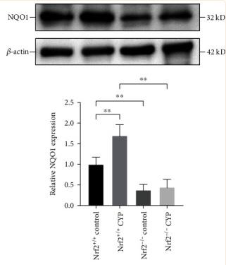
Figure 6 Nrf2 might protect bladder injury by activating its downstream antioxidant genes. (a) The protein expression of HO-1 in the bladder of the four groups. (b) The protein expression of NQO1 in the bladder of the four groups. Relative mRNA expression levels of HO-1 (c) and NQO1 (d) in the bladder of the four groups. ∗P < 0.05, ∗∗P < 0.01. Data are presented as mean ± SD.

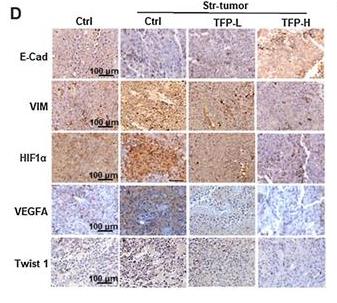
Application: IHC    Species: mouse    Sample: tumor cells

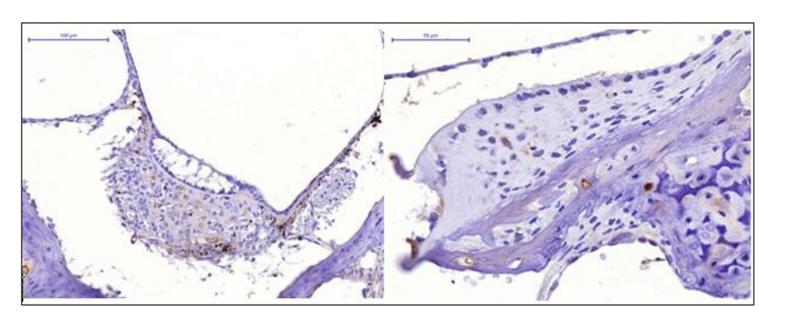
Fig. 6. |TFP inhibits EMT of melanoma cells.D–F, IHC analysis of E-cadherin, vimentin, HIF1α, VEGFA, and Twist1 in melanoma tumor tissues of mice under stress stimulation. Representative images and staining scores are shown. Scale bar, 100 μm.

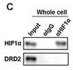
Application: WB    Species: mouse    Sample:

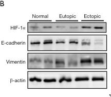
Fig. 4 |DRD2 overexpression inhibited the ubiquitin-dependent degradation of HIF1α. A, Venn diagram of proteins that interacted with DRD2 and HIF1α, as predicted by FPclass website. B, Molecular docking results of DRD2 and VHL. C, Co-IP results of DRD2 and HIF1α.

3). FGF21 ameliorates septic liver injury by restraining proinflammatory macrophages activation through the autophagy/HIF-1α axis. Journal of advanced research, 2024 (PubMed: 38599281) [IF=10.7]

4). Spatiotemporal regulation of injectable heterogeneous silk gel scaffolds for accelerating guided vertebral repair. Advanced Healthcare Materials, 2023 (PubMed: 36465008) [IF=10.0]

5). Inositol hexaphosphate enhances chemotherapy by reversing senescence induced by persistently activated PERK and diphthamide modification of eEF2. Cancer letters, 2024 (PubMed: 38097134) [IF=9.7]

6). Low-dose X-ray irradiation combined with FAK inhibitors improves the immune microenvironment and confers sensitivity to radiotherapy in pancreatic cancer. BIOMEDICINE & PHARMACOTHERAPY, 2022 (PubMed: 35594704) [IF=7.5]

7). Autophagy inhibition and ferroptosis activation during atherosclerosis: Hypoxia-inducible factor 1α inhibitor PX-478 alleviates atherosclerosis by inducing autophagy and suppressing ferroptosis in macrophages. Biomedicine & Pharmacotherapy, 2023 (PubMed: 36948130) [IF=7.5]

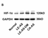
Application: WB    Species: Mouse    Sample:

Fig. 5. The expression HIF-1α protein was enhanced in macrophages of human and mouse atherosclerotic lesions. (A): Dual immunofluorescence staining for HIF-1α (red), CD68 (green) and DAPI (blue) indicated the co-expression of HIF-1α and CD68 (a macrophage marker) in human normal artery free of AS and atherosclerotic lesions from LE and CA with or without DM. (B-C): Compared with histologically normal artery, the protein levels of HIF-1α were upregulated in human atherosclerotic lesions from LE and CA with or without DM by western blot analysis. (D): Dual immunofluorescence staining for HIF-1α (red), CD68 (green) and DAPI (blue) in ApoE-/- mice fed with a HFD or normal diet demonstrated that the HIF-1α expression was present in macrophages, as indicated by HIF-1α immunoreactivity colocalized with CD68. (E-F): Compared with ApoE-/- control mice, the protein levels of HIF-1α were upregulated in HFD-fed ApoE-/- mice by western blot analysis. *P 

8). Aluminum induces neuroinflammation via P2X7 receptor activating NLRP3 inflammasome pathway. Ecotoxicology and Environmental Safety, 2023 (PubMed: 36508838) [IF=6.8]

9). Culin5 aggravates hypoxic pulmonary hypertension by activating TRAF6/NF-κB/HIF-1α/VEGF. iScience, 2023 (PubMed: 37965157) [IF=5.8]

Application: WB    Species: Mouse    Sample:

Figure 7 Upregulated expression of TRAF6/NF-κB/HIF-1α/VEGF pathway in HPH that aggravated by Cul 5 further (A) The effect of hypoxia treatment for 4 weeks on Cul5/TRAF6/NF-κB/HIF-1α/VEGF pathway expression in lung tissues of mice, and reverse effect of pevonedistat. (a) Western blot analysis of TRAF6/NF-κB/HIF-1α/VEGF pathway expression in N4W and H4W group and statistical analysis of the proteins relative expression normalized to GAPDH. (b) Western blot analysis of TRAF6/NF-κB/HIF-1α/VEGF pathway expression in N4W + DMSO, N4W + pevonedistat, H4W+ DMSO, and H4W + pevonedistat group, and statistical analysis of the proteins relative expression normalized to GAPDH. (B) The effect of hypoxia and/or Cul 5 treatment on TRAF6/NF-κB/HIF-1α/VEGF pathway expression in PAECs. Western blot analysis of TRAF6/NF-κB/HIF-1α/VEGF pathway expression in endothelial cells exposed to normoxia and hypoxia with or without Cul 5, and statistical analysis of the proteins relative expression normalized to GAPDH. ∗p < 0.05, ∗∗p < 0.01, ∗∗∗p < 0.001. Student’s t test was used for statistical analysis in Figure 7A(a). Data are represented as the means ± SD, one-way ANOVA was used for statistical analysis in Figure 7A(b) and 7B.

10). Metformin alleviates the cognitive impairment caused by aluminum by improving energy metabolism disorders in mice. BIOCHEMICAL PHARMACOLOGY, 2022 (PubMed: 35700760) [IF=5.8]

もっと読みます

Restrictive clause


Affinity Biosciences tests all products strictly. Citations are provided as a resource for additional applications that have not been validated by Affinity Biosciences. Please choose the appropriate format for each application and consult Materials and Methods sections for additional details about the use of any product in these publications.

For Research Use Only.
Not for use in diagnostic or therapeutic procedures. Not for resale. Not for distribution without written consent. Affinity Biosciences will not be held responsible for patent infringement or other violations that may occur with the use of our products. Affinity Biosciences, Affinity Biosciences Logo and all other trademarks are the property of Affinity Biosciences LTD.