製品: Collagen X Antibody
カタログ: DF13214
タンパク質の説明: Rabbit polyclonal antibody to Collagen X
アプリケーション: WB IHC
Cited expt.: WB, IHC
反応性: Human, Mouse, Rat
予測: Pig, Bovine, Horse, Sheep, Rabbit, Chicken
分子量: 66kDa; 66kD(Calculated).
ユニプロット: Q03692
RRID: AB_2846233

類似製品を見る>>

   サイズ 価格 在庫状況
 100ul $280 在庫あり
 200ul $350 在庫あり

リードタイム: 当日配達

For pricing and ordering contact:
お問い合わせ先

製品説明

ソース:
Rabbit
アプリケーション:
WB 1:500-1:2000, IHC 1:50-1:200
*The optimal dilutions should be determined by the end user. For optimal experimental results, antibody reuse is not recommended.
*Tips:

WB: For western blot detection of denatured protein samples. IHC: For immunohistochemical detection of paraffin sections (IHC-p) or frozen sections (IHC-f) of tissue samples. IF/ICC: For immunofluorescence detection of cell samples. ELISA(peptide): For ELISA detection of antigenic peptide.

反応性:
Human,Mouse,Rat
予測:
Pig(100%), Bovine(88%), Horse(100%), Sheep(100%), Rabbit(100%), Chicken(100%)
クローナリティ:
Polyclonal
特異性:
Collagen X Antibody detects endogenous levels of total Collagen X.
RRID:
AB_2846233
引用形式: Affinity Biosciences Cat# DF13214, RRID:AB_2846233.
コンジュゲート:
Unconjugated.
精製:
The antiserum was purified by peptide affinity chromatography using SulfoLink™ Coupling Resin (Thermo Fisher Scientific).
保存:
Rabbit IgG in phosphate buffered saline , pH 7.4, 150mM NaCl, 0.02% sodium azide and 50% glycerol. Store at -20 °C. Stable for 12 months from date of receipt.
別名:

折りたたみ/展開

COAA1_HUMAN; Col10a 1; COL10A1; Collagen alpha 1(X) chain; Collagen alpha-1(X) chain; Collagen type X alpha 1 (Schmid metaphyseal chondrodysplasia); Collagen type X alpha 1; Collagen X alpha 1 polypeptide; CollagenX; fa66d11; fb10c08; OTTHUMP00000040411; Procollagen type X alpha 1; Schmid metaphyseal chondrodysplasia; wu:fa66d11; wu:fb10c08;

免疫原

免疫原:

A synthesized peptide derived from human Collagen X, corresponding to a region within C-terminal amino acids.

Uniprot:
遺伝子(ID):
タンパク質配列:
MLPQIPFLLLVSLNLVHGVFYAERYQMPTGIKGPLPNTKTQFFIPYTIKSKGIAVRGEQGTPGPPGPAGPRGHPGPSGPPGKPGYGSPGLQGEPGLPGPPGPSAVGKPGVPGLPGKPGERGPYGPKGDVGPAGLPGPRGPPGPPGIPGPAGISVPGKPGQQGPTGAPGPRGFPGEKGAPGVPGMNGQKGEMGYGAPGRPGERGLPGPQGPTGPSGPPGVGKRGENGVPGQPGIKGDRGFPGEMGPIGPPGPQGPPGERGPEGIGKPGAAGAPGQPGIPGTKGLPGAPGIAGPPGPPGFGKPGLPGLKGERGPAGLPGGPGAKGEQGPAGLPGKPGLTGPPGNMGPQGPKGIPGSHGLPGPKGETGPAGPAGYPGAKGERGSPGSDGKPGYPGKPGLDGPKGNPGLPGPKGDPGVGGPPGLPGPVGPAGAKGMPGHNGEAGPRGAPGIPGTRGPIGPPGIPGFPGSKGDPGSPGPPGPAGIATKGLNGPTGPPGPPGPRGHSGEPGLPGPPGPPGPPGQAVMPEGFIKAGQRPSLSGTPLVSANQGVTGMPVSAFTVILSKAYPAIGTPIPFDKILYNRQQHYDPRTGIFTCQIPGIYYFSYHVHVKGTHVWVGLYKNGTPVMYTYDEYTKGYLDQASGSAIIDLTENDQVWLQLPNAESNGLYSSEYVHSSFSGFLVAPM

種類予測

種類予測:

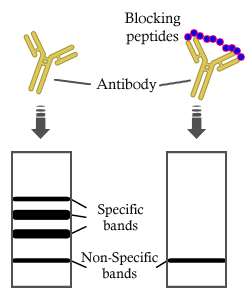
Score>80(red) has high confidence and is suggested to be used for WB detection. *The prediction model is mainly based on the alignment of immunogen sequences, the results are for reference only, not as the basis of quality assurance.

Species
Results
Score
Pig
100
Horse
100
Sheep
100
Chicken
100
Rabbit
100
Bovine
88
Xenopus
75
Dog
0
Zebrafish
0
Model Confidence:
High(score>80) Medium(80>score>50) Low(score<50) No confidence

研究背景

機能:

Type X collagen is a product of hypertrophic chondrocytes and has been localized to presumptive mineralization zones of hyaline cartilage.

PTMs:

Prolines at the third position of the tripeptide repeating unit (G-X-Y) are hydroxylated in some or all of the chains.

細胞の位置付け:

Secreted>Extracellular space>Extracellular matrix.

Extracellular region or secreted Cytosol Plasma membrane Cytoskeleton Lysosome Endosome Peroxisome ER Golgi apparatus Nucleus Mitochondrion Manual annotation Automatic computational assertionSubcellular location

研究領域

· Organismal Systems > Digestive system > Protein digestion and absorption.

参考文献

1). HAMA-SBMA hydrogel with anti-inflammatory properties delivers cartilage organoids, boosting cartilage regeneration. Journal of nanobiotechnology, 2025 (PubMed: 40448111) [IF=10.2]

Application: IF/ICC    Species: Rat    Sample:

Fig. 1 CCO exhibits characteristics of articular cartilage and delays the process of ossification. (A) SO, staining of the CEP. CEP had a quasi-spherical shape. (B) DAPI staining. CEP decellularization of nuclei. (C) DNA content, GAG content, and Total collagen content measurement. CEP was decellularized, preserving a significant amount of GAG and collagen. (D) Cell viability and cytotoxicity assay using Calcein/PI at D1, D3, and D7, with live cells shown in green and dead cells in red. CEP exhibits good biocompatibility. (E) CCK-8 assay at D1, D3, and D7. CEP can promote the proliferation of BMSCs. (F) HE and AB staining of CO and CCO on days 7, 14, and 21. CCO can generate a greater amount of cartilage matrix. (G) Immunofluorescence images for COL2, ACAN in CO and CCO at days 14, with blue representing DAPI and red representing the target proteins. The cartilage markers COL2 and ACAN are expressed at higher levels in CCO. (H)Immunohistochemical images for PRG4 in CO and CCO at days 14 and Immunofluorescence images for COLX in CO and CCO at days 21 with blue representing DAPI and red representing the target proteins. CCO exhibits high expressions of the articular cartilage-specific marker PRG4 and low expressions of the hypertrophic cartilage marker COLX. (I) The mRNA expression of cartilage genes SOX9, COL2, and ACAN, as well as the hypertrophic cartilage gene COLX, was assessed in CO and CCO at 0, 7, 14, and 21 days. Both CCO and CO reached peak cartilage expression in 14 days, after which the expression of hypertrophy-related genes increased. However, CCO demonstrated superior cartilage formation compared to CO, with a delayed hypertrophic process. All experiments were repeated three times. ns p > 0.05, *0.01 

2). Injectable decellularized Wharton's jelly hydrogel containing CD56+ umbilical cord mesenchymal stem cell-derived exosomes for meniscus tear healing and cartilage protection. Materials today. Bio, 2024 (PubMed: 39347017) [IF=8.7]

Application: WB    Species: human    Sample: hBMSC
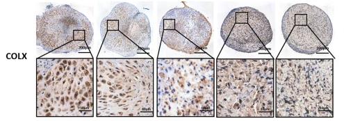
Fig. 5. Evaluation of hBMSC differentiation induced by DWJH/CD56+Exos and sustained release of CD56+Exos in DWJH/CD56+Exos. (A) Histochemical identification using Alcian Blue staining was performed to assess chondrogenic induction of hBMSC by DWJH/CD56+Exos. Scale bar: 100 μm. (E) Statistical analysis was carried out based on the results obtained in A. (B) Immunofluorescence staining for chondrogenic marker SOX9 and (F) quantification of positive cells. (n = 3). Scale bar: 50 μm. (C) Western blot of SOX9 in hBMSC treated with DWJH, CD56+Exos, or DWJH/CD56+Exos. (F) Quantification of SOX9, COL2, and COLX relative protein expression between different groups in control, DWJH, CD56+Exos, and DWJH/CD56+Exos groups. n = 3. ∗P < 0.05, ∗∗P < 0.01, ∗∗∗P < 0.001. (For interpretation of the references to colour in this figure legend, the reader is referred to the Web version of this article.)

3). Co-culture pellet of human Wharton’s jelly mesenchymal stem cells and rat costal chondrocytes as a candidate for articular cartilage regeneration: in vitro and in vivo study. Stem Cell Research & Therapy, 2022 (PubMed: 35907866) [IF=7.5]

Application: IHC    Species: Rat    Sample:

Fig. 3Histological and immunochemistry staining of pellets with semiquantitative analysis. A H–E and Safranin-O staining of pellets and partial enlargement in five groups. B Immunochemistry staining of COLII and COLX of pellets and partial enlargement in five groups. C–E Semiquantitative analysis of AOI of each staining (n = 4). Significant difference symbols: *p < 0.05, **p < 0.01 compared to CCs group, #p < 0.05, ##p < 0.01 compared to hWJMSCs group

4). Receptor tyrosine kinase C-kit promotes a destructive phenotype of FLS in osteoarthritis via intracellular EMT signaling. Molecular medicine (Cambridge, Mass.), 2023 (PubMed: 36959556) [IF=6.0]

Application: WB    Species: Rat    Sample:

Fig. 4 C-kit drives FLS EMT signaling (A) Validation of several shC-kit efficiency by Western blot (n = 3). (B-C) The expression of EMT makers (B) and Transwell invasive assay (C) in OA FLS with pCDNA-Ctrl, pCDNA-C-kit, shCtrl, or shC-kit transfection (n = 3, bar = 100 μm). (D) Morphological observation of DAPI (blue) and vimentin staining (green). Pseudopodia are marked by white arrowheads (bar = 20 μm). (E) The expression of inflammation-related markers (IL-6, IL-8, and MMP13) in FLSs (n = 3). (F) Illustration of co-culture of lenti-virus infected OA-FLS and chondrocytes. (G-H) After co-culture for 7 days, assessment of MMP13 (G) and Runx2 (H) of the chondrocytes (n = 5, bar = 100 μm)

5). Anti-hypertrophic effect of synovium-derived stromal cells on costal chondrocytes promotes cartilage repairs. Journal of Orthopaedic Translation, 2022 (PubMed: 34934627) [IF=5.9]

Application: IHC    Species: Rat    Sample: SDSCs
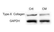
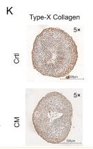
Figure 2 Paracrine effect of SDSCs toward CCs in indirect coculture. A) Schematic of conditioned medium coculture experimental outline. In light blue: CCs; In yellow: SDSCs; In dark blue: CCs pellet. B) Macroscopic appearance of pellets. C) RT-PCR analysis for chondrogenesis and hypertrophic-related gene expression after 3 weeks of culture. D) H&E staining. E) Alcian blue staining. F, G) Safranin-O staining. H) Biochemical evaluation of GAG content and GAG/DNA ratio of pellets. I, J) Immunohistochemistry analysis of type II collagen. K, L) Immunohistochemistry analysis of type X collagen. M) Western blot analysis of type X collagen. Crtl: Control group. CM: SDSCs-conditioned medium group. (For interpretation of the references to color/colour in this figure legend, the reader is referred to the Web version of this article.)

Application: WB    Species: Rat    Sample: SDSCs
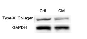
Figure 2 Paracrine effect of SDSCs toward CCs in indirect coculture. A) Schematic of conditioned medium coculture experimental outline. In light blue: CCs; In yellow: SDSCs; In dark blue: CCs pellet. B) Macroscopic appearance of pellets. C) RT-PCR analysis for chondrogenesis and hypertrophic-related gene expression after 3 weeks of culture. D) H&E staining. E) Alcian blue staining. F, G) Safranin-O staining. H) Biochemical evaluation of GAG content and GAG/DNA ratio of pellets. I, J) Immunohistochemistry analysis of type II collagen. K, L) Immunohistochemistry analysis of type X collagen. M) Western blot analysis of type X collagen. Crtl: Control group. CM: SDSCs-conditioned medium group. (For interpretation of the references to color/colour in this figure legend, the reader is referred to the Web version of this article.)

Application: WB    Species: rat    Sample: Costal chondrocytes
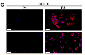
Figure 2.| Paracrine effect of SDSCs toward CCs in indirect coculture.M) Western blot analysis of type X collagen. Crtl: Control group. CM: SDSCs-conditioned medium group. (For interpretation of the references to color/colour in this figure legend, the reader is referred to the Web version of this article.)

6). Enhanced articular cartilage regeneration using costal chondrocyte-derived scaffold-free tissue engineered constructs with ascorbic acid treatment. Journal of orthopaedic translation, 2024 (PubMed: 38559899) [IF=5.9]

7). YAP maintains cartilage stem/progenitor cell homeostasis in osteoarthritis. Journal of orthopaedic translation, 2024 (PubMed: 38817242) [IF=5.9]

8). Near-infrared light-controlled kartogenin delivery of multifunctional Prussian blue nanocomposites for cartilage defect repair. Nanoscale, 2023 (PubMed: 37129436) [IF=5.8]

9). Asiatic acid attenuates hypertrophic and fibrotic differentiation of articular chondrocytes via AMPK/PI3K/AKT signaling pathway. ARTHRITIS RESEARCH & THERAPY, 2020 (PubMed: 32398124) [IF=4.9]

10). Cartilage Regeneration Characteristics of Human and Goat Auricular Chondrocytes. Frontiers in Bioengineering and Biotechnology, 2021 (PubMed: 34993186) [IF=4.3]

Application: IF/ICC    Species: human and goat    Sample:

FIGURE 3 Phenotypic expression of AUCs during in vitro expansion. (A) Alcian staining, and (B) COL II immunofluorescence staining of human and goats at P1 and P3. Gene quantitative analyses of (C) SOX9, (D) Aggrecan, and (E) Col II in human and goat at P1 and P3. (F–I) Analysis of protein and gene expression levels of COL I and COL X in human and goat at P1 and P3. Data were analyzed using t tests (the relative mRNA levels were compared within the species, instead of comparing between the species). Columns with different letters indicate statistical significance (p < 0.05). Abbreviations: AUCs, auricular chondrocytes; Col II, type II collage; Col I, type I collage; Col X, type X collage. Scale bar = 50 μm.

もっと読みます

Restrictive clause


Affinity Biosciences tests all products strictly. Citations are provided as a resource for additional applications that have not been validated by Affinity Biosciences. Please choose the appropriate format for each application and consult Materials and Methods sections for additional details about the use of any product in these publications.

For Research Use Only.
Not for use in diagnostic or therapeutic procedures. Not for resale. Not for distribution without written consent. Affinity Biosciences will not be held responsible for patent infringement or other violations that may occur with the use of our products. Affinity Biosciences, Affinity Biosciences Logo and all other trademarks are the property of Affinity Biosciences LTD.