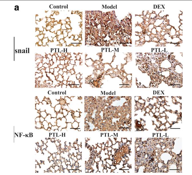
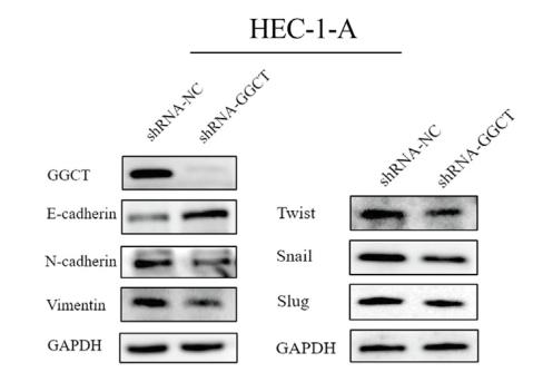
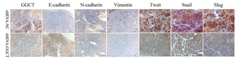
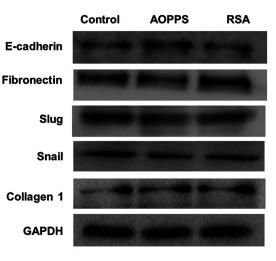
製品: SNAIL Antibody
カタログ: AF6032
タンパク質の説明: Rabbit polyclonal antibody to SNAIL
アプリケーション: WB IHC IF/ICC
Cited expt.: WB, IHC, IF/ICC
反応性: Human, Mouse, Rat
予測: Pig, Bovine, Horse, Rabbit, Dog, Chicken, Xenopus
分子量: 29kDa; 29kD(Calculated).
ユニプロット: O95863
RRID: AB_2834965

類似製品を見る>>

   サイズ 価格 在庫状況
 100ul $280 在庫あり
 200ul $350 在庫あり

リードタイム: 当日配達

For pricing and ordering contact:
お問い合わせ先

製品説明

ソース:
Rabbit
アプリケーション:
WB 1:500-1:2000, IHC 1:50-1:200, IF/ICC 1:100-1:500
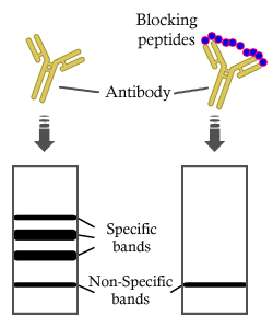
*The optimal dilutions should be determined by the end user. For optimal experimental results, antibody reuse is not recommended.
*Tips:

WB: For western blot detection of denatured protein samples. IHC: For immunohistochemical detection of paraffin sections (IHC-p) or frozen sections (IHC-f) of tissue samples. IF/ICC: For immunofluorescence detection of cell samples. ELISA(peptide): For ELISA detection of antigenic peptide.

反応性:
Human,Mouse,Rat
予測:
Pig(86%), Bovine(86%), Horse(100%), Rabbit(100%), Dog(86%), Chicken(86%), Xenopus(86%)
クローナリティ:
Polyclonal
特異性:
SNAIL Antibody detects endogenous levels of total SNAIL.
RRID:
AB_2834965
引用形式: Affinity Biosciences Cat# AF6032, RRID:AB_2834965.
コンジュゲート:
Unconjugated.
精製:
The antiserum was purified by peptide affinity chromatography using SulfoLink™ Coupling Resin (Thermo Fisher Scientific).
保存:
Rabbit IgG in phosphate buffered saline , pH 7.4, 150mM NaCl, 0.02% sodium azide and 50% glycerol. Store at -20 °C. Stable for 12 months from date of receipt.
別名:

折りたたみ/展開

dJ710H13.1; Protein sna; Protein snail homolog 1; Protein snail homolog; SLUGH2; SNA; Sna protein; SNAH; SNAI; snai1; SNAI1_HUMAN; Snail 1 homolog; Snail 1 zinc finger protein; SNAIL; Snail homolog 1 (Drosophila); SNAIL, Drosophila, homolog of, 1; SNAIL1; Zinc finger protein SNAI1;

免疫原

免疫原:

A synthesized peptide derived from human SNAIL, corresponding to a region within C-terminal amino acids.

Uniprot:
遺伝子(ID):
発現特異性:
O95863 SNAI1_HUMAN:

Expressed in a variety of tissues with the highest expression in kidney. Expressed in mesenchymal and epithelial cell lines.

タンパク質の説明:
This protein has many roles during postimplantation development. It is involved in embryonic mesoderm formation and its maintenance and may also be involved in chondrogenesis and in epithelial-mesenchymal inductive interactions.
タンパク質配列:
MPRSFLVRKPSDPNRKPNYSELQDSNPEFTFQQPYDQAHLLAAIPPPEILNPTASLPMLIWDSVLAPQAQPIAWASLRLQESPRVAELTSLSDEDSGKGSQPPSPPSPAPSSFSSTSVSSLEAEAYAAFPGLGQVPKQLAQLSEAKDLQARKAFNCKYCNKEYLSLGALKMHIRSHTLPCVCGTCGKAFSRPWLLQGHVRTHTGEKPFSCPHCSRAFADRSNLRAHLQTHSDVKKYQCQACARTFSRMSLLHKHQESGCSGCPR

種類予測

種類予測:

Score>80(red) has high confidence and is suggested to be used for WB detection. *The prediction model is mainly based on the alignment of immunogen sequences, the results are for reference only, not as the basis of quality assurance.

Species
Results
Score
Rabbit
100
Horse
100
Pig
86
Bovine
86
Dog
86
Xenopus
86
Chicken
86
Sheep
0
Zebrafish
0
Model Confidence:
High(score>80) Medium(80>score>50) Low(score<50) No confidence

研究背景

機能:

Involved in induction of the epithelial to mesenchymal transition (EMT), formation and maintenance of embryonic mesoderm, growth arrest, survival and cell migration. Binds to 3 E-boxes of the E-cadherin/CDH1 gene promoter and to the promoters of CLDN7 and KRT8 and, in association with histone demethylase KDM1A which it recruits to the promoters, causes a decrease in dimethylated H3K4 levels and represses transcription. The N-terminal SNAG domain competes with histone H3 for the same binding site on the histone demethylase complex formed by KDM1A and RCOR1, and thereby inhibits demethylation of histone H3 at 'Lys-4' (in vitro). During EMT, involved with LOXL2 in negatively regulating pericentromeric heterochromatin transcription (By similarity). SNAI1 recruits LOXL2 to pericentromeric regions to oxidize histone H3 and repress transcription which leads to release of heterochromatin component CBX5/HP1A, enabling chromatin reorganization and acquisition of mesenchymal traits (By similarity). Associates with EGR1 and SP1 to mediate tetradecanoyl phorbol acetate (TPA)-induced up-regulation of CDKN2B, possibly by binding to the CDKN2B promoter region 5'-TCACA-3. In addition, may also activate the CDKN2B promoter by itself.

PTMs:

Phosphorylated by GSK3B. Once phosphorylated, it becomes a target for BTRC ubiquitination. Phosphorylation by CSNK1E, probably at Ser-104, provides the priming site for the subsequent phosphorylation by GSK3B, probably at Ser-100 and Ser-96. Phosphorylation by PAK1 may modulate its transcriptional activity by promoting increased accumulation in the nucleus. Phosphorylation at Ser-11 and Ser-92 positively regulates its functions in induction of EMT and cell survival, respectively. Phosphorylation by LATS2, upon mitotic stress, oncogenic stress or Hippo pathway activation, occurs in the nucleus and promotes nuclear retention and stabilization of total cellular protein level.

Ubiquitinated on Lys-98, Lys-137 and Lys-146 by FBXL14 and BTRC leading to degradation. BTRC-triggered ubiquitination requires previous GSK3B-mediated SNAI1 phosphorylation. Ubiquitination induced upon interaction with NOTCH1 or TP53/p53 is mediated by MDM2.

O-GlcNAcylation at Ser-112 is enhanced in hyperglycaemic conditions, it opposes phosphorylation by GSK3B, and stabilizes the protein.

ADP-ribosylation by PARP1 increases protein half-life and may be involved in TGFB-induced SNAI1 up-regulation.

細胞の位置付け:

Nucleus. Cytoplasm.
Note: Once phosphorylated (probably on Ser-107, Ser-111, Ser-115 and Ser-119) it is exported from the nucleus to the cytoplasm where subsequent phosphorylation of the destruction motif and ubiquitination involving BTRC occurs.

Extracellular region or secreted Cytosol Plasma membrane Cytoskeleton Lysosome Endosome Peroxisome ER Golgi apparatus Nucleus Mitochondrion Manual annotation Automatic computational assertionSubcellular location
組織特異性:

Expressed in a variety of tissues with the highest expression in kidney. Expressed in mesenchymal and epithelial cell lines.

タンパク質ファミリー:

Belongs to the snail C2H2-type zinc-finger protein family.

研究領域

· Cellular Processes > Cellular community - eukaryotes > Adherens junction.   (View pathway)

参考文献
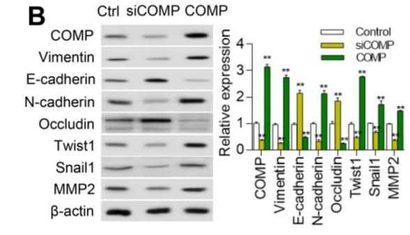
1). Cartilage Oligomeric Matrix Protein promotes epithelial-mesenchymal transition by interacting with Transgelin in Colorectal Cancer. Theranostics, 2020 (PubMed: 32754278) [IF=12.4]

Application: WB    Species: Human    Sample: HCT116 cells
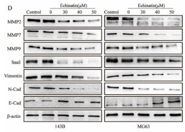
Figure 4. COMP promotes EMT in colorectal cancer. (A) Cell phenotypic changes in cells treated with COMP siRNA and COMP overexpression. (B) WB analysis of COMP and EMT-related markers in HCT116 cells under COMP knockdown or overexpression. (C) Cell wound scratch assay of HCT116, HCT-8, and SW620 cells treated with COMP siRNA or COMP overexpression vectors. (D) Transwell assay of HCT116, HCT-8, and SW620 cells treated with COMP siRNA or COMP overexpression vectors. (E) Immunofluorescence assay of HCT116, HCT-8, and SW620 cells treated with COMP siRNA or COMP overexpression vectors. Image J software was used to analyze the relative intensity of E-cadherin and Vimentin. (F) HCT116 and HCT-8 cells with knocked down or overexpressed COMP were transplanted on nude mice. Tumor volumes were measured every 4 days. (G) Tumor weight in the control, COMP knockdown, and COMP overexpression groups. (H) The in situ spleen model of the colorectal cancer cell line SW620 showed that overexpression of COMP promoted liver metastasis of colorectal cancer, while downregulation of COMP inhibited liver metastasis of colorectal cancer. (I) IHC staining to identify EMT biomarkers and COMP-related proteins in the control, COMP knockdown, and COMP overexpression groups. COMP knockdown displayed strong E-cadherin staining and reduced Vimentin, Snail1, Twist1, and MMP-2 staining. The expression levels of other proteins were identical to those observed in WB experiments. (J) Staining indices of COMP, E-cadherin, Vimentin, Snail1, Twist1, and MMP2. The error bars in all graphs represented SD, and each experiment was repeated three times. * and ** stand for P<0.05 and P<0.01, respectively.

2). Echinatin inhibits the growth and metastasis of human osteosarcoma cells through Wnt/β-catenin and p38 signaling pathways. Pharmacological Research, 2023 (PubMed: 37023991) [IF=9.1]

Application: WB    Species: Human    Sample: OS cells
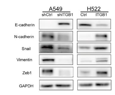
Fig. 4. Ecn inhibits the migration and invasion of OS cells. (A) The effect of Ecn on the migration of OS cells (Wound healing assay, 100 ×). (B) The effect of Ecn on the migration of OS cells (Transwell assay, 100 ×). (C) The effect of Ecn on the invasion of OS cells (Matrigel-coated Transwell assay, 100 ×). (D) The effect of Ecn on the protein level of MMP2, MMP7, MMP9, Snail, Vimentin, N-Cadherin and E-Cadherin in OS cells (Western blot). ##P 

3). ITGB1 enhances the Radioresistance of human Non-small Cell Lung Cancer Cells by modulating the DNA damage response and YAP1-induced Epithelial-mesenchymal Transition. International Journal of Biological Sciences, 2023 (PubMed: 33613118) [IF=8.2]

Application: WB    Species: human    Sample: NSCLC cells
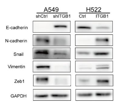
Figure 8. |ITGB1 could promote radioresisntance of NSCLC cells by regulating EMT. A-C. Protein levels of E-cadherin, N-cadherin, Snail, vimentin, and Zeb1 were detected by western blotting of cells from shITGB1 and ITGB1 overexpression groups.

Application: WB    Species: Human    Sample: NSCLC cells

Figure 8 ITGB1 could promote radioresisntance of NSCLC cells by regulating EMT. A-C. Protein levels of E-cadherin, N-cadherin, Snail, vimentin, and Zeb1 were detected by western blotting of cells from shITGB1 and ITGB1 overexpression groups.

4). Targeting TNFAIP2 with NIR-II CRISPR-Cas9 nanosystem to overcome cisplatin resistance in laryngeal cancer. NPJ precision oncology, 2025 (PubMed: 40731142) [IF=7.9]

Application: WB    Species: human    Sample: Tu177/CDDPTNFAIP2-/- cells

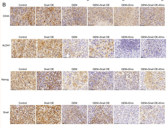
Fig. 5: Effect of NRF2 overexpression on the biological characteristics of Tu177/CDDPTNFAIP2-/- cells. A Quantitative PCR analysis shows the differences in NRF2 mRNA levels between Tu177/CDDP cells and their variants. B Immunofluorescence staining results display NRF2 protein expression in different cell lines, with red indicating NRF2 and blue indicating DAPI-stained nuclei, Bar = 25 μm. C Images of the sphere formation assay (top) and the corresponding statistical graphs of sphere numbers and diameters (bottom), comparing the sphere-forming abilities of the three cell lines, Bar = 100 μm. D, E Western blot analysis showing differences in EMT-related protein expression among the cell lines. F Transwell invasion assay (top, Bar = 50 μm) and wound healing assay (bottom, Bar = 100 μm) results demonstrate the invasion and migration abilities of the different cell lines. The number of invasive cells and the percentage of unhealed areas are compared among the three treatments; ① indicates Tu177/CDDP cells, ② indicates Tu177/CDDPTNFAIP2-/- cells, and ③ indicates Tu177/CDDPTNFAIP2-/-+oe-NRF2 cells. * represents p 

5). CircEGFR reduces the sensitivity of pirarubicin and regulates the malignant progression of triple-negative breast cancer via the miR-1299/EGFR axis. International journal of biological macromolecules, 2023 (PubMed: 37302631) [IF=7.7]

6). CircRNA-3302 promotes endothelial-to-mesenchymal transition via sponging miR-135b-5p to enhance KIT expression in Kawasaki disease. Cell Death Discovery, 2022 (PubMed: 35768408) [IF=7.0]

7). Network pharmacology reveals that Yanghe Decoction inhibits osteosarcoma progression via ROS-induced mitochondrial dysfunction and enhances cisplatin sensitivity. Genes & diseases, 2025 (PubMed: 41716635) [IF=6.9]

8). Subtle structural alteration in indisulam switches the molecular mechanisms for the inhibitory effect on the migration of gastric cancer cells. Biomedicine & pharmacotherapy = Biomedecine & pharmacotherapie, 2024 (PubMed: 38359488) [IF=6.9]

Application: WB    Species: Human    Sample: AGS and MGC803 cells
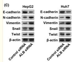
Fig. 2. Compounds SR‐3‐65 and WXM‐1‐170 regulated the protein level of EMT biomarkers and transcription factors in AGS and MGC803 cells. (A, B) SR-3–65 and WXM-1–170 altered the protein level of EMT biomarkers. AGS (A) and MGC803 (B) cells were treated with DMSO, indisulam, SR-3–65, or WXM-1–170 (10 µM) for 72 h. Cell lysates were immunoblotted. Mean ± SD (n = 3). One-way ANOVA, Dunnett’s post hoc analysis, *: P < 0.05, **: P < 0.01, ***: P < 0.001, ns: not significant. (C, D) SR-3–65 and WXM-1–170 downregulated the protein level of EMT-related transcription factors. AGS (C) and MGC803 (D) cells were treated with DMSO, indisulam, SR-3–65, or WXM-1–170 (10 µM) for 72 h. Cell lysates were immunoblotted. Mean ± SD (n = 3). One-way ANOVA, Dunnett’s post hoc analysis, *: P < 0.05, **: P < 0.01, ***: P < 0.001, ****: P < 0.0001, ns: not significant.

9). d-Borneol enhances cisplatin sensitivity via autophagy dependent EMT signaling and NCOA4-mediated ferritinophagy. PHYTOMEDICINE, 2022 (PubMed: 36030746) [IF=6.7]

10). Molecular mechanism of albumin in suppressing invasion and metastasis of hepatocellular carcinoma. LIVER INTERNATIONAL, 2022 (PubMed: 34854209) [IF=6.0]

Application: WB    Species: Human    Sample: HepG2 and Huh7 cells

FIGURE 7 A, Representative images of the western blot results for uPAR, MMP2 and MMP9 in ALB knockdown HepG2 and Huh7 cells; B, Zymography analysis illustrates MMP2 and MMP9 activity in ALB knockdown HepG2 and Huh7 cells; C, Quantitative analysis results and representative images of the western blot results for the EMT‐associated markers, E‐cadherin, N‐cadherin, vimentin, Snail and Twist by western blot in ALB knockdown HepG2 and Huh7 cells; D, Quantification shows a significantly higher uPAR in HCC group with ALB <3.5 g/dL compared to ALB ≥3.5 g/dL (*P < .05); E, Scatterplot showing the correlation between plasma levels of ALB and uPAR. The vertical position represents the expression levels of uPAR (lg pg/mL)

もっと読みます

Restrictive clause


Affinity Biosciences tests all products strictly. Citations are provided as a resource for additional applications that have not been validated by Affinity Biosciences. Please choose the appropriate format for each application and consult Materials and Methods sections for additional details about the use of any product in these publications.

For Research Use Only.
Not for use in diagnostic or therapeutic procedures. Not for resale. Not for distribution without written consent. Affinity Biosciences will not be held responsible for patent infringement or other violations that may occur with the use of our products. Affinity Biosciences, Affinity Biosciences Logo and all other trademarks are the property of Affinity Biosciences LTD.