製品: p70 S6 Kinase Antibody
カタログ: AF6226
タンパク質の説明: Rabbit polyclonal antibody to p70 S6 Kinase
アプリケーション: WB IHC IF/ICC
反応性: Human, Mouse, Rat
予測: Bovine, Horse, Sheep, Rabbit, Dog, Chicken, Xenopus
分子量: 70kDa; 59kD(Calculated).
ユニプロット: P23443
RRID: AB_2835100

類似製品を見る>>

   サイズ 価格 在庫状況
 100ul $280 在庫あり
 200ul $350 在庫あり

リードタイム: 当日配達

For pricing and ordering contact:
お問い合わせ先

製品説明

ソース:
Rabbit
アプリケーション:
WB 1:500-1:2000, IHC 1:50-1:200, IF/ICC 1:100-1:500
*The optimal dilutions should be determined by the end user.
*Tips:

WB: For western blot detection of denatured protein samples. IHC: For immunohistochemical detection of paraffin sections (IHC-p) or frozen sections (IHC-f) of tissue samples. IF/ICC: For immunofluorescence detection of cell samples. ELISA(peptide): For ELISA detection of antigenic peptide.

反応性:
Human,Mouse,Rat
予測:
Bovine(100%), Horse(100%), Sheep(100%), Rabbit(100%), Dog(100%), Chicken(100%), Xenopus(100%)
クローナリティ:
Polyclonal
特異性:
p70 S6 Kinase Antibody detects endogenous levels of total p70 S6 Kinase.
RRID:
AB_2835100
引用形式: Affinity Biosciences Cat# AF6226, RRID:AB_2835100.
コンジュゲート:
Unconjugated.
精製:
The antiserum was purified by peptide affinity chromatography using SulfoLink™ Coupling Resin (Thermo Fisher Scientific).
保存:
Rabbit IgG in phosphate buffered saline , pH 7.4, 150mM NaCl, 0.02% sodium azide and 50% glycerol. Store at -20 °C. Stable for 12 months from date of receipt.
別名:

折りたたみ/展開

70 kDa ribosomal protein S6 kinase 1; KS6B1_HUMAN; p70 alpha; P70 beta 1; p70 ribosomal S6 kinase alpha; p70 ribosomal S6 kinase beta 1; p70 S6 kinase alpha; P70 S6 Kinase; p70 S6 kinase, alpha 1; p70 S6 kinase, alpha 2; p70 S6K; p70 S6K-alpha; p70 S6KA; p70(S6K) alpha; p70(S6K)-alpha; p70-alpha; p70-S6K 1; p70-S6K; P70S6K; P70S6K1; p70S6Kb; PS6K; Ribosomal protein S6 kinase 70kDa polypeptide 1; Ribosomal protein S6 kinase beta 1; Ribosomal protein S6 kinase beta-1; Ribosomal protein S6 kinase I; RPS6KB1; S6K; S6K-beta-1; S6K1; Serine/threonine kinase 14 alpha; Serine/threonine-protein kinase 14A; STK14A;

免疫原

免疫原:
Uniprot:
遺伝子(ID):
発現特異性:
P23443 KS6B1_HUMAN:

Widely expressed.

タンパク質の説明:
This gene encodes a member of the RSK (ribosomal S6 kinase) family of serine/threonine kinases. This kinase contains 2 non-identical kinase catalytic domains and phosphorylates several residues of the S6 ribosomal protein.
タンパク質配列:
MRRRRRRDGFYPAPDFRDREAEDMAGVFDIDLDQPEDAGSEDELEEGGQLNESMDHGGVGPYELGMEHCEKFEISETSVNRGPEKIRPECFELLRVLGKGGYGKVFQVRKVTGANTGKIFAMKVLKKAMIVRNAKDTAHTKAERNILEEVKHPFIVDLIYAFQTGGKLYLILEYLSGGELFMQLEREGIFMEDTACFYLAEISMALGHLHQKGIIYRDLKPENIMLNHQGHVKLTDFGLCKESIHDGTVTHTFCGTIEYMAPEILMRSGHNRAVDWWSLGALMYDMLTGAPPFTGENRKKTIDKILKCKLNLPPYLTQEARDLLKKLLKRNAASRLGAGPGDAGEVQAHPFFRHINWEELLARKVEPPFKPLLQSEEDVSQFDSKFTRQTPVDSPDDSTLSESANQVFLGFTYVAPSVLESVKEKFSFEPKIRSPRRFIGSPRTPVSPVKFSPGDFWGRGASASTANPQTPVEYPMETSGIEQMDVTMSGEASAPLPIRQPNSGPYKKQAFPMISKRPEHLRMNL

種類予測

種類予測:

Score>80(red) has high confidence and is suggested to be used for WB detection. *The prediction model is mainly based on the alignment of immunogen sequences, the results are for reference only, not as the basis of quality assurance.

Species
Results
Score
Horse
100
Bovine
100
Sheep
100
Dog
100
Xenopus
100
Chicken
100
Rabbit
100
Pig
0
Zebrafish
0
Model Confidence:
High(score>80) Medium(80>score>50) Low(score<50) No confidence

PTMs - P23443 基板として

Site PTM Type Enzyme
S40 Phosphorylation
S53 Phosphorylation Q9HC98 (NEK6)
K85 Ubiquitination
K99 Ubiquitination
K104 Ubiquitination
K118 Ubiquitination
S243 Phosphorylation
T248 Phosphorylation
T250 Phosphorylation
T252 Phosphorylation P42336 (PIK3CA) , O15530 (PDPK1)
T256 Phosphorylation
S278 Phosphorylation
K304 Acetylation
K364 Ubiquitination
K370 Ubiquitination
S371 Phosphorylation
T389 Phosphorylation
T390 Phosphorylation P42345 (MTOR)
S394 Phosphorylation P06493 (CDK1) , P42345 (MTOR)
T399 Phosphorylation
S403 Phosphorylation Q9HC98 (NEK6)
T412 Phosphorylation O94768 (STK17B) , O15530 (PDPK1) , Q8TDX7 (NEK7) , Q9HC98 (NEK6) , P42336 (PIK3CA) , P42345 (MTOR) , P23443 (RPS6KB1)
Y413 Phosphorylation
S417 Phosphorylation
S421 Phosphorylation
K425 Ubiquitination
S427 Phosphorylation
S434 Phosphorylation P06493 (CDK1) , P42345 (MTOR) , P45984 (MAPK9) , P27361 (MAPK3) , P45983 (MAPK8) , P28482 (MAPK1)
S441 Phosphorylation
T444 Phosphorylation P06493 (CDK1) , P28482 (MAPK1)
S447 Phosphorylation P28482 (MAPK1) , P06493 (CDK1) , P42345 (MTOR)
K450 Ubiquitination
S452 Phosphorylation
S462 Phosphorylation
T470 Phosphorylation
K516 Acetylation
R522 Methylation

PTMs - P23443 酵素として

Substrate Site Source
O00418 (EEF2K) S366 Uniprot
O60825 (PFKFB2) S466 Uniprot
O94763 (URI1) S372 Uniprot
P03372 (ESR1) S167 Uniprot
P04637 (TP53) S392 Uniprot
P04792 (HSPB1) S15 Uniprot
P04792 (HSPB1) S78 Uniprot
P04792 (HSPB1) S82 Uniprot
P08151 (GLI1) S84 Uniprot
P10636-8 (MAPT) T212 Uniprot
P10636-8 (MAPT) S214 Uniprot
P10636-8 (MAPT) S262 Uniprot
P23443 (RPS6KB1) T412 Uniprot
P23588-1 (EIF4B) S422 Uniprot
P27708 (CAD) S1859 Uniprot
P35568 (IRS1) S270 Uniprot
P35568 (IRS1) S307 Uniprot
P35568 (IRS1) S527 Uniprot
P35568 (IRS1) S636 Uniprot
P35568 (IRS1) S1101 Uniprot
P42345 (MTOR) T2446 Uniprot
P42345 (MTOR) S2448 Uniprot
P49841 (GSK3B) S9 Uniprot
P62753 (RPS6) S235 Uniprot
P62753 (RPS6) S236 Uniprot
P62753 (RPS6) S240 Uniprot
P62753 (RPS6) S244 Uniprot
P62753 (RPS6) S247 Uniprot
P78371 (CCT2) S260 Uniprot
Q05195 (MXD1) S145 Uniprot
Q06787 (FMR1) S500 Uniprot
Q09161 (NCBP1) S7 Uniprot
Q09161 (NCBP1) T21 Uniprot
Q09161 (NCBP1) S22 Uniprot
Q15831-1 (STK11) S428 Uniprot
Q16873 (LTC4S) S36 Uniprot
Q53EL6 (PDCD4) S67 Uniprot
Q6R327 (RICTOR) T1135 Uniprot
Q8TB45 (DEPTOR) S286 Uniprot
Q8TB45 (DEPTOR) S287 Uniprot
Q8TB45 (DEPTOR) S291 Uniprot
Q92519 (TRIB2) S83 Uniprot
Q92934 (BAD) S99 Uniprot
Q9BY77 (POLDIP3) S383 Uniprot
Q9BY77 (POLDIP3) S385 Uniprot
Q9BYV9 (BACH2) S521 Uniprot
Q9H3D4 (TP63) S477 Uniprot
Q9H3D4 (TP63) T491 Uniprot
Q9H3D4 (TP63) S560 Uniprot
Q9UN36 (NDRG2) S332 Uniprot
Q9UN36 (NDRG2) S350 Uniprot

研究背景

機能:

Serine/threonine-protein kinase that acts downstream of mTOR signaling in response to growth factors and nutrients to promote cell proliferation, cell growth and cell cycle progression. Regulates protein synthesis through phosphorylation of EIF4B, RPS6 and EEF2K, and contributes to cell survival by repressing the pro-apoptotic function of BAD. Under conditions of nutrient depletion, the inactive form associates with the EIF3 translation initiation complex. Upon mitogenic stimulation, phosphorylation by the mammalian target of rapamycin complex 1 (mTORC1) leads to dissociation from the EIF3 complex and activation. The active form then phosphorylates and activates several substrates in the pre-initiation complex, including the EIF2B complex and the cap-binding complex component EIF4B. Also controls translation initiation by phosphorylating a negative regulator of EIF4A, PDCD4, targeting it for ubiquitination and subsequent proteolysis. Promotes initiation of the pioneer round of protein synthesis by phosphorylating POLDIP3/SKAR. In response to IGF1, activates translation elongation by phosphorylating EEF2 kinase (EEF2K), which leads to its inhibition and thus activation of EEF2. Also plays a role in feedback regulation of mTORC2 by mTORC1 by phosphorylating RICTOR, resulting in the inhibition of mTORC2 and AKT1 signaling. Mediates cell survival by phosphorylating the pro-apoptotic protein BAD and suppressing its pro-apoptotic function. Phosphorylates mitochondrial URI1 leading to dissociation of a URI1-PPP1CC complex. The free mitochondrial PPP1CC can then dephosphorylate RPS6KB1 at Thr-412, which is proposed to be a negative feedback mechanism for the RPS6KB1 anti-apoptotic function. Mediates TNF-alpha-induced insulin resistance by phosphorylating IRS1 at multiple serine residues, resulting in accelerated degradation of IRS1. In cells lacking functional TSC1-2 complex, constitutively phosphorylates and inhibits GSK3B. May be involved in cytoskeletal rearrangement through binding to neurabin. Phosphorylates and activates the pyrimidine biosynthesis enzyme CAD, downstream of MTOR. Following activation by mTORC1, phosphorylates EPRS and thereby plays a key role in fatty acid uptake by adipocytes and also most probably in interferon-gamma-induced translation inhibition.

PTMs:

Phosphorylation at Thr-412 is regulated by mTORC1. The phosphorylation at this site is maintained by an agonist-dependent autophosphorylation mechanism (By similarity). Activated by phosphorylation at Thr-252 by PDPK1. Dephosphorylation by PPP1CC at Thr-412 in mitochondrion.

細胞の位置付け:

Cell junction>Synapse>Synaptosome. Mitochondrion outer membrane. Mitochondrion.
Note: Colocalizes with URI1 at mitochondrion.

Nucleus. Cytoplasm.

Cytoplasm.

Extracellular region or secreted Cytosol Plasma membrane Cytoskeleton Lysosome Endosome Peroxisome ER Golgi apparatus Nucleus Mitochondrion Manual annotation Automatic computational assertionSubcellular location
組織特異性:

Widely expressed.

サブユニット構造:

Interacts with PPP1R9A/neurabin-1 (By similarity). Interacts with RPTOR. Interacts with IRS1. Interacts with EIF3B and EIF3C. Interacts with TRAF4. Interacts with POLDIP3. Interacts (via N-terminus) with IER5.

タンパク質ファミリー:

The autoinhibitory domain is believed to block phosphorylation within the AGC-kinase C-terminal domain and the activation loop.

The TOS (TOR signaling) motif is essential for activation by mTORC1.

Belongs to the protein kinase superfamily. AGC Ser/Thr protein kinase family. S6 kinase subfamily.

研究領域

· Cellular Processes > Transport and catabolism > Autophagy - animal.   (View pathway)

· Environmental Information Processing > Signal transduction > ErbB signaling pathway.   (View pathway)

· Environmental Information Processing > Signal transduction > HIF-1 signaling pathway.   (View pathway)

· Environmental Information Processing > Signal transduction > mTOR signaling pathway.   (View pathway)

· Environmental Information Processing > Signal transduction > PI3K-Akt signaling pathway.   (View pathway)

· Environmental Information Processing > Signal transduction > AMPK signaling pathway.   (View pathway)

· Environmental Information Processing > Signal transduction > TGF-beta signaling pathway.   (View pathway)

· Environmental Information Processing > Signal transduction > Apelin signaling pathway.   (View pathway)

· Human Diseases > Drug resistance: Antineoplastic > EGFR tyrosine kinase inhibitor resistance.

· Human Diseases > Drug resistance: Antineoplastic > Endocrine resistance.

· Human Diseases > Endocrine and metabolic diseases > Insulin resistance.

· Human Diseases > Infectious diseases: Viral > Human papillomavirus infection.

· Human Diseases > Cancers: Overview > Pathways in cancer.   (View pathway)

· Human Diseases > Cancers: Overview > Proteoglycans in cancer.

· Human Diseases > Cancers: Specific types > Colorectal cancer.   (View pathway)

· Human Diseases > Cancers: Specific types > Pancreatic cancer.   (View pathway)

· Human Diseases > Cancers: Specific types > Acute myeloid leukemia.   (View pathway)

· Human Diseases > Cancers: Specific types > Breast cancer.   (View pathway)

· Human Diseases > Cancers: Specific types > Hepatocellular carcinoma.   (View pathway)

· Human Diseases > Cancers: Specific types > Gastric cancer.   (View pathway)

· Human Diseases > Cancers: Overview > Choline metabolism in cancer.   (View pathway)

· Organismal Systems > Aging > Longevity regulating pathway.   (View pathway)

· Organismal Systems > Aging > Longevity regulating pathway - multiple species.   (View pathway)

· Organismal Systems > Immune system > Fc gamma R-mediated phagocytosis.   (View pathway)

· Organismal Systems > Endocrine system > Insulin signaling pathway.   (View pathway)

参考文献

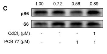
1). Activation of integrated stress response and disordered iron homeostasis upon combined exposure to cadmium and PCB77. JOURNAL OF HAZARDOUS MATERIALS, 2020 (PubMed: 31837937) [IF=13.6]

Application: WB    Species: Human    Sample: HEL cells

Fig. 5. Disordered iron homeostasis and inhibited mTORC1 activity upon exposure to CdCl2 and PCB77 at low dose. (A) The relative fluorescence intensity of CAeAM for measuring LIP to reflect intracellular iron availability (n = 3–4), and (B) Representative blots of FTH1 protein content to reflect iron storage. Analyses were performed after single or combined exposure to CdCl2 and PCB77 at 1 μM for 48 h. (C) Phosphorylated S6 and total S6 content to reflect mTORC1 activity as measured by Western blot. Ratio of FTH1 to eIF2αP and ratio of pS6 to S6 in the control group were defined as 1. Analyses were performed after single or combined exposure to CdCl2 and PCB77 at 1 μM for 48 h. a- significantly different from the control group. Data were presented in mean ± SE. P < 0.05 was considered statistically significant.

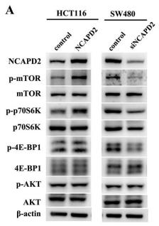
2). NCAPD2 inhibits autophagy by regulating Ca2+/CAMKK2/AMPK/mTORC1 pathway and PARP-1/SIRT1 axis to promote colorectal cancer. CANCER LETTERS, 2021 (PubMed: 34229059) [IF=9.7]

Application: WB    Species: Human    Sample: CRC cells

Fig. 2. NCAPD2 inhibited cell autophagy and disrupted autophagic flux via Ca2+/CAMKK2/AMPK/mTORC1 pathway. (A) Western blot analyses for phosphorylated mTOR (p-mTOR, S2448), phosphorylated p70S6K (p-p70S6K, T389/412), phosphorylated 4E-BP1 (p-4E-BP1, T70) and phosphorylated AKT (p-AKT, S473) in CRCC cells with different treatments as indicated. (B) Western blot of indicated proteins in cells treated with mTORC1 inhibitor Rapamycin (3 nM, 24h). (C) Immunofluorescence staining of LC3II (red) and P62 (red) in CRC cells with different treatments as indicated. Merged images represented overlays of LC3II or P62 and nuclear staining by DAPI (blue). (D) Intracellular Ca2+ levels were analyzed by flow cytometry after staining with the fluorescent probe Fluo-3, AM in CRC cells. (E) Representative Western blot gel documents of phosphorylated CAMKK2(S511), phosphorylated AMPK(T172), phosphorylated mTOR(S2448), Beclin, ATG5, P62, LC3II in CRC cells with different treatments. (F) Western blots of indicated proteins in cells treated with an inhibitor of microsomal Ca2+-ATPase Thapsigargin (1 μM, 6h) and Ca2+ chelator BAPTA-AM (10 μM, 12h) respectively. Results are shown as mean ± s.d, *P < 0.05, **P < 0.01, ***P < 0.001, based on Student’s t-test. . (For interpretation of the references to color in this figure legend, the reader is referred to the Web version of this article.)

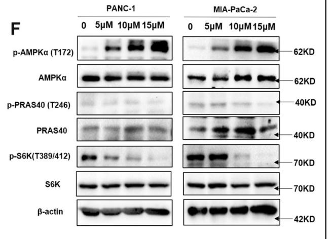
3). Inhibition of PTP1B blocks pancreatic cancer progression by targeting the PKM2/AMPK/mTOC1 pathway. Cell Death & Disease, 2019 (PubMed: 31745071) [IF=9.0]

Application: WB    Species: Human    Sample: pancreatic cancer tissue

Fig. 6 The relationship between PTP1B and AMPK. a PTP1B overexpression resulted in decreased p-AMPK (alpha). b, c The negative correlation between PTP1B and p-AMPKα was showed in pancreatic cancer patient tissue samples (p < 0.001, p value was obtained by a Pearson χ2 test; scale bar, 200 μm and 50 μm). d PTP1B inhibition either by shRNAs or by LXQ46 increased the phosphorylation of PKM2. e, f The inactivated PKM2 resulted in increased phosphorylation of AMPKα and decreased the phosphorylation of PRAS40, causing the inhibition of mTOC1 activity. g PTP1B inhibition caused AMPK activation and decreased p-p70S6K in vivo (scale bar, 200 and 50 μm).

4). PPM1H is down-regulated by ATF6 and dephosphorylates p-RPS6KB1 to inhibit progression of hepatocellular carcinoma. Molecular therapy. Nucleic acids, 2023 (PubMed: 37456776) [IF=8.8]

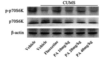
5). Patchouli alcohol protects against chronic unpredictable mild stress-induced depressant-like behavior through inhibiting excessive autophagy via activation of mTOR signaling pathway. BIOMEDICINE & PHARMACOTHERAPY, 2020 (PubMed: 32244196) [IF=7.5]

Application: WB    Species: rat    Sample: hippocampus

Fig. 5.| The effect of PA on the level of expression of p-p70S6K protein in the hippocampus. (A) The effect of PA on p-p70S6K and p70S6K protein levels in hippocampus were investigated by western blot analysis.

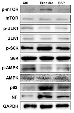
6). Exosomes derived from miR-26a-modified MSCs promote axonal regeneration via the PTEN/AKT/mTOR pathway following spinal cord injury. Stem Cell Research & Therapy, 2021 (PubMed: 33820561) [IF=7.5]

Application: WB    Species: rat    Sample: PC16 cells

FIGURE S2 | miR-26a-overexpressing exosomes inhibited autophagic activity and promoted axonal generation in PC12 cells. (a) The ability of Exos-26a to generate neurofilament (red fluorescent dye) in PC12 cells, which could be reversed by rapamycin. (b, c) Representative images of western blots used to determine the expression levels of NF, mTOR, p-mTOR, AMPK, p-AMPK, S6K, p-S6K, ULK1, p-ULK1, and p62 and semiquantification of the data. RAP indicates miR-26a exosome and rapamycin (100 nM) treatment for 48 h before lysis. *P < 0.05, **P < 0.01, ***P < 0.001 compared with the control group by t test or ANOVA. #P < 0.05 and ##P < 0.01 compared with the RAP group by t test. n = 3 for each group.

7). Timosaponin BII improved osteoporosis caused by hyperglycemia through promoting autophagy of osteoblasts via suppressing the mTOR/NFκB signaling pathway. Free Radical Biology and Medicine, 2021 (PubMed: 33992678) [IF=7.4]

Application: WB    Species: Rat    Sample: osteoblasts

Fig. 5. Timosaponin BII downregulated mTOR signaling in tibias of diabetic rats and in the high glucose-induced osteoblasts. A: Photomicrographs of the immunohistochemistry staining ( × 200, scale bar: 100 μm; × 400, scale bar: 50 μm) and the quantification of the p-mTOR expression in tibias of different groups of rats. Error bars indicated SEM (n = 6). **P < 0.01 compared with the Mod group. The arrows denoted positive staining. B: Representative western blot images and the quantification of p-mTOR, mTOR, p-S6K and S6K expression in the high glucose-induced osteoblasts with the treatment with TBII (0.1 μmol/L, 1 μmol/L, 10 μmol/L) and in those cells with NAC (25 μmol/L) incubation for 48 h **P < 0.01 compared with the high glucose group.

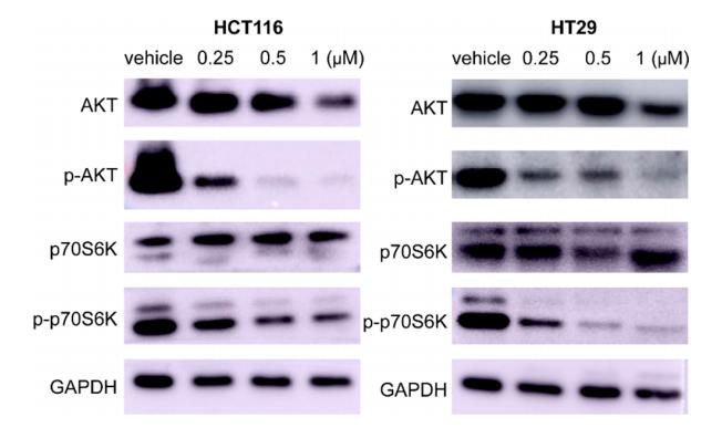
8). Design, Synthesis, and Biological Evaluation of Imidazo[1,2-a]pyridine Derivatives as Novel PI3K/mTOR Dual Inhibitors. JOURNAL OF MEDICINAL CHEMISTRY, 2020 (PubMed: 32069401) [IF=7.3]

Application: WB    Species: Human    Sample: HCT116 and HT-29 cells

Figure 5. Effects of 15a on pAKT, AKT, p-p70S6K, and p70S6K in HCT116 and HT-29 cells. HCT116 cells and HT-29 cells were treated at the indicated concentrations of 15a for 24 h. The expression levels of AKT, p70S6K, and their phosphorylated forms were analyzed by Western blotting. GAPDH was used as a loading control.

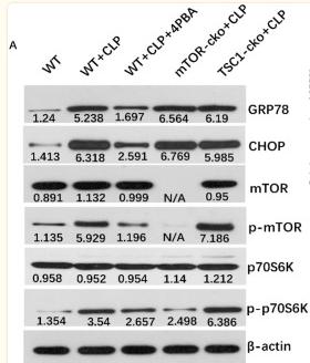
9). mTOR pathway mediates endoplasmic reticulum stress-induced CD4+ T cell apoptosis in septic mice. APOPTOSIS, 2022 (PubMed: 35759162) [IF=7.2]

Application: WB    Species: Mice    Sample: CD4+ T cells

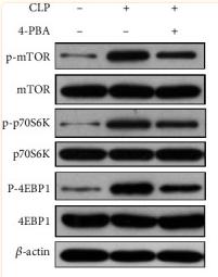
Fig. 4 Expression of ERS-UPR- and mTOR-related proteins in splenic CD4+ T cells of septic mice. Protein expression of GRP78, CHOP, mTOR, p-mTOR, p70S6k, p-p70S6k (A–E) in CD4+ T cells were quantified by western blotting and showed as the relative expression values of β-actin, which was used as a loading control to normalize the protein levels. In order to highlight the activation level of p-mTOR and p-p70S6K in this signaling pathway, the ratio of p-mTOR to mTOR (p/t mTOR) and p-p70S6K to p70S6K (p/t p70S6K) were used for statistics. Data are shown as Mean ± SD (n = 6). Statistically significant differences were determined by two-tailed Student’s t-test. **P < 0.01, ***P < 0.001, ****P < 0.0001

10). Fraxetin pretreatment alleviates cisplatin-induced kidney injury by antagonizing autophagy and apoptosis via mTORC1 activation. Phytotherapy research : PTR, 2024 (PubMed: 38558449) [IF=7.2]

もっと読みます

Restrictive clause


Affinity Biosciences tests all products strictly. Citations are provided as a resource for additional applications that have not been validated by Affinity Biosciences. Please choose the appropriate format for each application and consult Materials and Methods sections for additional details about the use of any product in these publications.

For Research Use Only.
Not for use in diagnostic or therapeutic procedures. Not for resale. Not for distribution without written consent. Affinity Biosciences will not be held responsible for patent infringement or other violations that may occur with the use of our products. Affinity Biosciences, Affinity Biosciences Logo and all other trademarks are the property of Affinity Biosciences LTD.