製品: AhR Antibody
カタログ: AF6278
タンパク質の説明: Rabbit polyclonal antibody to AhR
アプリケーション: WB IHC IF/ICC
Cited expt.: WB, IHC, IF/ICC
反応性: Human, Mouse, Rat
予測: Pig, Bovine, Sheep, Rabbit, Dog, Chicken, Xenopus
分子量: 96kDa; 96kD(Calculated).
ユニプロット: P35869
RRID: AB_2835131

類似製品を見る>>

   サイズ 価格 在庫状況
 100ul $280 在庫あり
 200ul $350 在庫あり

リードタイム: 当日配達

For pricing and ordering contact:
お問い合わせ先

製品説明

ソース:
Rabbit
アプリケーション:
WB 1:500-1:2000, IHC 1:50-1:200, IF/ICC 1:100-1:500
*The optimal dilutions should be determined by the end user.
*Tips:

WB: For western blot detection of denatured protein samples. IHC: For immunohistochemical detection of paraffin sections (IHC-p) or frozen sections (IHC-f) of tissue samples. IF/ICC: For immunofluorescence detection of cell samples. ELISA(peptide): For ELISA detection of antigenic peptide.

反応性:
Human,Mouse,Rat
予測:
Pig(100%), Bovine(100%), Sheep(100%), Rabbit(100%), Dog(100%), Chicken(100%), Xenopus(100%)
クローナリティ:
Polyclonal
特異性:
AhR Antibody detects endogenous levels of total AhR.
RRID:
AB_2835131
引用形式: Affinity Biosciences Cat# AF6278, RRID:AB_2835131.
コンジュゲート:
Unconjugated.
精製:
The antiserum was purified by peptide affinity chromatography using SulfoLink™ Coupling Resin (Thermo Fisher Scientific).
保存:
Rabbit IgG in phosphate buffered saline , pH 7.4, 150mM NaCl, 0.02% sodium azide and 50% glycerol. Store at -20 °C. Stable for 12 months from date of receipt.
別名:

折りたたみ/展開

Ah receptor; AhR; AHR_HUMAN; Aromatic hydrocarbon receptor; Aryl hydrocarbon receptor; Aryl hydrocarbon receptor precursor; bHLHe76; Class E basic helix loop helix protein 76; Class E basic helix-loop-helix protein 76; HGNC:348;

免疫原

免疫原:

A synthesized peptide derived from human AhR, corresponding to a region within N-terminal amino acids.

Uniprot:
遺伝子(ID):
発現特異性:
P35869 AHR_HUMAN:

Expressed in all tissues tested including blood, brain, heart, kidney, liver, lung, pancreas and skeletal muscle. Expressed in retinal photoreceptors (PubMed:29726989).

タンパク質の説明:
The Aryl Hydrocarbon Receptor (AHR), also known as the dioxin receptor, is a ligand-activated helix/loop/helix transcription factor found in a variety of vertebrate species. The known ligands for AHR are foreign planar aromatic compounds, such as polycyclic aromatic compounds and halogenated aromatic compounds such as 2,3,7,8-tetrachlorodibenzo-p-dioxin (TCDD).
タンパク質配列:
MNSSSANITYASRKRRKPVQKTVKPIPAEGIKSNPSKRHRDRLNTELDRLASLLPFPQDVINKLDKLSVLRLSVSYLRAKSFFDVALKSSPTERNGGQDNCRAANFREGLNLQEGEFLLQALNGFVLVVTTDALVFYASSTIQDYLGFQQSDVIHQSVYELIHTEDRAEFQRQLHWALNPSQCTESGQGIEEATGLPQTVVCYNPDQIPPENSPLMERCFICRLRCLLDNSSGFLAMNFQGKLKYLHGQKKKGKDGSILPPQLALFAIATPLQPPSILEIRTKNFIFRTKHKLDFTPIGCDAKGRIVLGYTEAELCTRGSGYQFIHAADMLYCAESHIRMIKTGESGMIVFRLLTKNNRWTWVQSNARLLYKNGRPDYIIVTQRPLTDEEGTEHLRKRNTKLPFMFTTGEAVLYEATNPFPAIMDPLPLRTKNGTSGKDSATTSTLSKDSLNPSSLLAAMMQQDESIYLYPASSTSSTAPFENNFFNESMNECRNWQDNTAPMGNDTILKHEQIDQPQDVNSFAGGHPGLFQDSKNSDLYSIMKNLGIDFEDIRHMQNEKFFRNDFSGEVDFRDIDLTDEILTYVQDSLSKSPFIPSDYQQQQSLALNSSCMVQEHLHLEQQQQHHQKQVVVEPQQQLCQKMKHMQVNGMFENWNSNQFVPFNCPQQDPQQYNVFTDLHGISQEFPYKSEMDSMPYTQNFISCNQPVLPQHSKCTELDYPMGSFEPSPYPTTSSLEDFVTCLQLPENQKHGLNPQSAIITPQTCYAGAVSMYQCQPEPQHTHVGQMQYNPVLPGQQAFLNKFQNGVLNETYPAELNNINNTQTTTHLQPLHHPSEARPFPDLTSSGFL

種類予測

種類予測:

Score>80(red) has high confidence and is suggested to be used for WB detection. *The prediction model is mainly based on the alignment of immunogen sequences, the results are for reference only, not as the basis of quality assurance.

Species
Results
Score
Pig
100
Bovine
100
Sheep
100
Dog
100
Xenopus
100
Chicken
100
Rabbit
100
Horse
0
Zebrafish
0
Model Confidence:
High(score>80) Medium(80>score>50) Low(score<50) No confidence

研究背景

機能:

Ligand-activated transcriptional activator. Binds to the XRE promoter region of genes it activates. Activates the expression of multiple phase I and II xenobiotic chemical metabolizing enzyme genes (such as the CYP1A1 gene). Mediates biochemical and toxic effects of halogenated aromatic hydrocarbons. Involved in cell-cycle regulation. Likely to play an important role in the development and maturation of many tissues. Regulates the circadian clock by inhibiting the basal and circadian expression of the core circadian component PER1. Inhibits PER1 by repressing the CLOCK-ARNTL/BMAL1 heterodimer mediated transcriptional activation of PER1. The heterodimer ARNT:AHR binds to core DNA sequence 5'-TGCGTG-3' within the dioxin response element (DRE) of target gene promoters and activates their transcription.

PTMs:

Mono-ADP-ribosylated, leading to inhibit transcription activator activity of AHR.

細胞の位置付け:

Cytoplasm. Nucleus.
Note: Initially cytoplasmic; upon binding with ligand and interaction with a HSP90, it translocates to the nucleus.

Extracellular region or secreted Cytosol Plasma membrane Cytoskeleton Lysosome Endosome Peroxisome ER Golgi apparatus Nucleus Mitochondrion Manual annotation Automatic computational assertionSubcellular location
組織特異性:

Expressed in all tissues tested including blood, brain, heart, kidney, liver, lung, pancreas and skeletal muscle. Expressed in retinal photoreceptors.

タンパク質ファミリー:

The PAS 1 domain is essential for dimerization and also required for AHR:ARNT heterodimerization.

研究領域

· Organismal Systems > Immune system > Th17 cell differentiation.   (View pathway)

参考文献

1). A postbiotic exopolysaccharide synergizes with Lactobacillus acidophilus to reduce intestinal inflammation in a mouse model of colitis. International journal of biological macromolecules, 2025 (PubMed: 39732236) [IF=7.7]

2). Disturbances of the gut microbiota-derived tryptophan metabolites as key actors in vagotomy-induced mastitis in mice. Cell reports, 2024 (PubMed: 39110590) [IF=7.5]

Application: WB    Species: Mouse    Sample:

Figure 6 The alleviation of Vag-induced mastitis in mice by 5-HIAA is dependent on AhR (A and B) Vag induced reduction of AhR protein expression and relative intensity in mammary glands (n = 3). (C and D) The antagonist CH223191 effectively suppressed AhR protein expression and relative intensity in the mammary gland (n = 3). (E) Representative H&E images among the different groups indicated that the antagonism of AhR abolished the protective function of 5-HIAA (n = 7; scale bar, 50 μm). (F) Histopathological score of the mammary gland (n = 6). (G–I) CH223191 treatment increased TNF-α (G), IL-1β (H), and MPO activity (I) in comparison to Vag+5-HIAA treatment (n = 7). (J–M) Pretreatment with CH223191 reduced the expression and relative intensity of TJ proteins ZO-1, Occludin, and Claudin-3 (n = 3). (N) The expression location and level of TJ protein were detected by immunohistochemistry (scale bar, 20 μm). (O and P) Pretreatment with CH223191 eliminated the decreasing effect of 5-HIAA on TNF-α and IL-1β mRNA levels in LPS-induced mouse mammary epithelial cells (n = 6). Data are presented as means ± SDs, and one-way ANOVA was performed for statistical analysis. ∗p < 0.05; ∗∗p < 0.01; ∗∗∗p < 0.001; and ∗∗∗∗p < 0.0001 indicate significant differences. See also Figure S8.

3). Emodin Inhibits the Proliferation of MCF-7 Human Breast Cancer Cells Through Activation of Aryl Hydrocarbon Receptor (AhR). Frontiers in Pharmacology, 2021 (PubMed: 33542691) [IF=5.6]

Application: WB    Species: Human    Sample: MCF-7 cells

FIGURE 7 Effects of emodin and AhR inhibitor CH223191 on the protein expression levels of AhR and CYP1A1 in MCF-7 cells (mean ± SD, n = 3) (A) In the cells treated with emodin, the expression of AhR protein using GAPDH as a loading control and the graphical representations of the AhR/GAPDH ratio; (B) In the cells treated with emodin, the expression of CYP1A1 protein using GAPDH as a loading control and the graphical representations of the CYP1A1/GAPDH ratio; (C) In the cells treated with CH223191, the expression of CYP1A1 protein using GAPDH as a loading control and the graphical representations of the CYP1A1/GAPDH ratio; (D) In the cells intervened with emodin and 10 μmol/L of CH223191, the expression of CYP1A1 protein using GAPDH as a loading control and the graphical representations of the CYP1A1/GAPDH ratio.

4). Fecal Microbiota Transplantation Ameliorates Experimentally Induced Colitis in Mice by Upregulating AhR. Frontiers in Microbiology, 2018 (PubMed: 30197631) [IF=5.2]

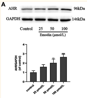
Application: WB    Species: mouse    Sample: colon

FIGURE 6 | Compared with that in the control group, decreased IL-10 and TGF-β expression in colon tissues of mice was induced by DSS and detected by immunohistochemistry.Representative photographs of immunohistochemical staining of IL-10 and TGF-β at the indicated time point in each group (A). Scale bars = 50 µm. (B) The mRNA level of IL-10 and TGF-β in colon tissues of different groups. (C) Protein expression of AhR,IL-10, and TGF-β in colon tissues of different groups.

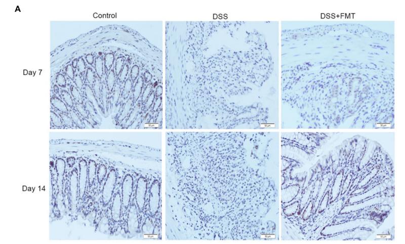
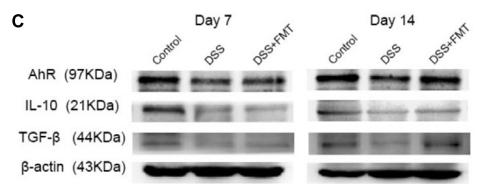
Application: IHC    Species: mouse    Sample: colon

FIGURE 5 | Compared with that in the control group, decreased AHR expression in colon tissues of mice was induced by DSS and detected by immunohistochemistry (A), Representative colon images at the indicated time point in each group. Scale bars = 50 µm. Representative example of an immunofluorescence double staining of Foxp3 (Red) and AhR (Green) in colon tissues (B).

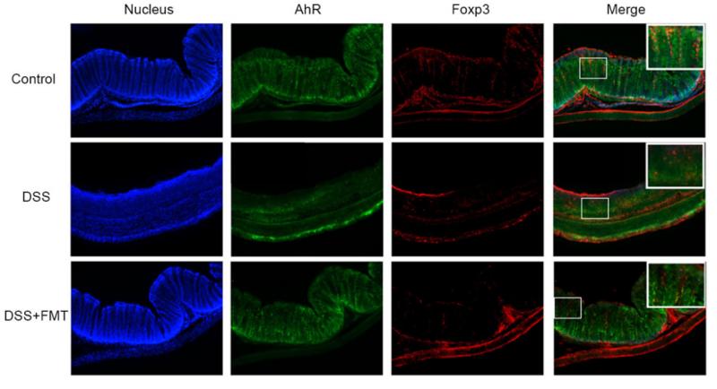
Application: IF/ICC    Species: mouse    Sample: colon

FIGURE 5 | Compared with that in the control group, decreased AHR expression in colon tissues of mice was induced by DSS and detected by immunohistochemistry (A), Representative colon images at the indicated time point in each group. Scale bars = 50 µm. Representative example of an immunofluorescence double staining of Foxp3 (Red) and AhR (Green) in colon tissues (B).

5). Hydrogen ameliorates lung injury in a rat model of subacute exposure to concentrated ambient PM2.5 via Aryl hydrocarbon receptor. International Immunopharmacology, 2019 (PubMed: 31718930) [IF=4.8]

Application: WB    Species: rat    Sample: lung

Fig. 7. |Hydrogen inhibited pulmonary AhR protein decline in the lung tissues of rats when exposed to PM2.5. Pulmonary AhR protein expression was determined by western blotting (A). AhR band relative intensities were quantified and normalized to GAPDH (B).

6). Psoralen and isopsoralen from Psoraleae Fructus aroused hepatotoxicity via induction of aryl hydrocarbon receptor-mediated CYP1A2 expression. Journal of Ethnopharmacology, 2022 (PubMed: 35872289) [IF=4.8]

7). NCOA4‐mediated ferritinophagy promoted inflammatory responses in periodontitis. JOURNAL OF PERIODONTAL RESEARCH, 2021 (PubMed: 33533512) [IF=3.4]

Application: WB    Species: Human    Sample: Periodontal ligament fibroblasts (PDLFs)

FIGURE 3 Porphyromonas gingivalis enhanced ferritinophagy in PDLFs. (A) PDLFs were treated with P. gingivalis stimulation (MOI =10, 50, 250) (n = 3) for 24 h, and gene transcription was assessed by real-time PCR. (B) Transcript levels of genes at 2 h, 12 h, and 24 h after bacteria treatment (MOI =250). (C) Protein expression of ferritin, SLC40A1, NCOA4, TFR1, LC3B, and P62 was analyzed by immunoblotting after P. gingivalis infection (MOI =250) (n = 3). (D) Labile iron pool in control or PDLFs treated P. gingivalis (MOI =250) was detected by flow cytometry using calcein-AM method (n = 3). (E) Labile iron pool was detected by the confocal 12 h after bacteria infection. Scale bar represents 200 µm. (F) Representative images of PDLFs showing colocalization (yellow) of NCOA4 (green) with LC3B (red). PDLFs were treated with P. gingivalis for 48 h. Scale bar represents 20 µm. Data from representative results were expressed as mean ±SD from a minimum of three replicates per experiment. *p < .05; **p < .01 vs control group

Restrictive clause


Affinity Biosciences tests all products strictly. Citations are provided as a resource for additional applications that have not been validated by Affinity Biosciences. Please choose the appropriate format for each application and consult Materials and Methods sections for additional details about the use of any product in these publications.

For Research Use Only.
Not for use in diagnostic or therapeutic procedures. Not for resale. Not for distribution without written consent. Affinity Biosciences will not be held responsible for patent infringement or other violations that may occur with the use of our products. Affinity Biosciences, Affinity Biosciences Logo and all other trademarks are the property of Affinity Biosciences LTD.