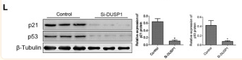
製品: p21 Cip1 Antibody
カタログ: AF6290
タンパク質の説明: Rabbit polyclonal antibody to p21 Cip1
アプリケーション: WB IF/ICC
Cited expt.: WB
反応性: Human, Mouse, Rat
予測: Bovine, Horse, Sheep, Rabbit, Dog
分子量: 21kDa; 18kD(Calculated).
ユニプロット: P38936
RRID: AB_2827699

類似製品を見る>>

   サイズ 価格 在庫状況
 100ul $280 在庫あり
 200ul $350 在庫あり

リードタイム: 当日配達

For pricing and ordering contact:
お問い合わせ先

製品説明

ソース:
Rabbit
アプリケーション:
WB 1:500-1:2000, IF/ICC 1:100-1:500
*The optimal dilutions should be determined by the end user. For optimal experimental results, antibody reuse is not recommended.
*Tips:

WB: For western blot detection of denatured protein samples. IHC: For immunohistochemical detection of paraffin sections (IHC-p) or frozen sections (IHC-f) of tissue samples. IF/ICC: For immunofluorescence detection of cell samples. ELISA(peptide): For ELISA detection of antigenic peptide.

反応性:
Human,Mouse,Rat
予測:
Bovine(100%), Horse(91%), Sheep(100%), Rabbit(83%), Dog(100%)
クローナリティ:
Polyclonal
特異性:
p21 Cip1 Antibody detects endogenous levels of total p21 Cip1.
RRID:
AB_2827699
引用形式: Affinity Biosciences Cat# AF6290, RRID:AB_2827699.
コンジュゲート:
Unconjugated.
精製:
The antiserum was purified by peptide affinity chromatography using SulfoLink™ Coupling Resin (Thermo Fisher Scientific).
保存:
Rabbit IgG in phosphate buffered saline , pH 7.4, 150mM NaCl, 0.02% sodium azide and 50% glycerol. Store at -20 °C. Stable for 12 months from date of receipt.
別名:

折りたたみ/展開

CAP20; CDK-interacting protein 1; CDKI; CDKN1; Cdkn1a; CDN1A_HUMAN; CIP1; Cyclin Dependent Kinase Inhibitor 1A; Cyclin-dependent kinase inhibitor 1; Cyclin-dependent kinase inhibitor 1A (P21); Cyclin-dependent kinase inhibitor 1A (p21, Cip1); DNA Synthesis Inhibitor; MDA-6; MDA6; Melanoma differentiation-associated protein 6; Melanoma differentiation-associated protein; p21; P21 protein; p21CIP1; p21Cip1/Waf1; p21WAF; PIC1; SDI1; SLC12A9; WAF1; Wild type p53 activated fragment 1 (WAF1); Wild type p53 activated fragment 1; Wildtype p53-activated fragment 1;

免疫原

免疫原:

A synthesized peptide derived from human p21 Cip1, corresponding to a region within C-terminal amino acids.

Uniprot:
遺伝子(ID):
発現特異性:
P38936 CDN1A_HUMAN:

Expressed in all adult tissues, with 5-fold lower levels observed in the brain.

タンパク質の説明:
This gene encodes a potent cyclin-dependent kinase inhibitor. The encoded protein binds to and inhibits the activity of cyclin-CDK2 or -CDK4 complexes, and thus functions as a regulator of cell cycle progression at G1. The expression of this gene is tightly controlled by the tumor suppressor protein p53, through which this protein mediates the p53-dependent cell cycle G1 phase arrest in response to a variety of stress stimuli.
タンパク質配列:
MSEPAGDVRQNPCGSKACRRLFGPVDSEQLSRDCDALMAGCIQEARERWNFDFVTETPLEGDFAWERVRGLGLPKLYLPTGPRRGRDELGGGRRPGTSPALLQGTAEEDHVDLSLSCTLVPRSGEQAEGSPGGPGDSQGRKRRQTSMTDFYHSKRRLIFSKRKP

種類予測

種類予測:

Score>80(red) has high confidence and is suggested to be used for WB detection. *The prediction model is mainly based on the alignment of immunogen sequences, the results are for reference only, not as the basis of quality assurance.

Species
Results
Score
Bovine
100
Sheep
100
Dog
100
Horse
91
Rabbit
83
Zebrafish
67
Xenopus
56
Pig
0
Chicken
0
Model Confidence:
High(score>80) Medium(80>score>50) Low(score<50) No confidence

研究背景

機能:

May be involved in p53/TP53 mediated inhibition of cellular proliferation in response to DNA damage. Binds to and inhibits cyclin-dependent kinase activity, preventing phosphorylation of critical cyclin-dependent kinase substrates and blocking cell cycle progression. Functions in the nuclear localization and assembly of cyclin D-CDK4 complex and promotes its kinase activity towards RB1. At higher stoichiometric ratios, inhibits the kinase activity of the cyclin D-CDK4 complex. Inhibits DNA synthesis by DNA polymerase delta by competing with POLD3 for PCNA binding. Plays an important role in controlling cell cycle progression and DNA damage-induced G2 arrest.

PTMs:

Phosphorylation of Thr-145 by Akt or of Ser-146 by PKC impairs binding to PCNA. Phosphorylation at Ser-114 by GSK3-beta enhances ubiquitination by the DCX(DTL) complex. Phosphorylation of Thr-145 by PIM2 enhances CDKN1A stability and inhibits cell proliferation. Phosphorylation of Thr-145 by PIM1 results in the relocation of CDKN1A to the cytoplasm and enhanced CDKN1A protein stability. UV radiation-induced phosphorylation at Thr-80 by LKB1 and at Ser-146 by NUAK1 leads to its degradation.

Ubiquitinated by MKRN1; leading to polyubiquitination and 26S proteasome-dependent degradation. Ubiquitinated by the DCX(DTL) complex, also named CRL4(CDT2) complex, leading to its degradation during S phase or following UV irradiation. Ubiquitination by the DCX(DTL) complex is essential to control replication licensing and is PCNA-dependent: interacts with PCNA via its PIP-box, while the presence of the containing the 'K+4' motif in the PIP box, recruit the DCX(DTL) complex, leading to its degradation. Ubiquitination at Ser-2 leads to degradation by the proteasome pathway. Ubiquitinated by RNF114; leading to proteasomal degradation.

Acetylation leads to protein stability. Acetylated in vitro on Lys-141, Lys-154, Lys-161 and Lys-163. Deacetylation by HDAC1 is prevented by competitive binding of C10orf90/FATS to HDAC1 (By similarity).

細胞の位置付け:

Cytoplasm. Nucleus.

Extracellular region or secreted Cytosol Plasma membrane Cytoskeleton Lysosome Endosome Peroxisome ER Golgi apparatus Nucleus Mitochondrion Manual annotation Automatic computational assertionSubcellular location
組織特異性:

Expressed in all adult tissues, with 5-fold lower levels observed in the brain.

タンパク質ファミリー:

The PIP-box K+4 motif mediates both the interaction with PCNA and the recruitment of the DCX(DTL) complex: while the PIP-box interacts with PCNA, the presence of the K+4 submotif, recruits the DCX(DTL) complex, leading to its ubiquitination.

The C-terminal is required for nuclear localization of the cyclin D-CDK4 complex.

Belongs to the CDI family.

研究領域

· Cellular Processes > Cell growth and death > Cell cycle.   (View pathway)

· Cellular Processes > Cell growth and death > p53 signaling pathway.   (View pathway)

· Cellular Processes > Cell growth and death > Cellular senescence.   (View pathway)

· Environmental Information Processing > Signal transduction > ErbB signaling pathway.   (View pathway)

· Environmental Information Processing > Signal transduction > HIF-1 signaling pathway.   (View pathway)

· Environmental Information Processing > Signal transduction > FoxO signaling pathway.   (View pathway)

· Environmental Information Processing > Signal transduction > PI3K-Akt signaling pathway.   (View pathway)

· Environmental Information Processing > Signal transduction > Jak-STAT signaling pathway.   (View pathway)

· Human Diseases > Drug resistance: Antineoplastic > Endocrine resistance.

· Human Diseases > Drug resistance: Antineoplastic > Platinum drug resistance.

· Human Diseases > Infectious diseases: Viral > Hepatitis C.

· Human Diseases > Infectious diseases: Viral > Hepatitis B.

· Human Diseases > Infectious diseases: Viral > Human papillomavirus infection.

· Human Diseases > Infectious diseases: Viral > HTLV-I infection.

· Human Diseases > Infectious diseases: Viral > Epstein-Barr virus infection.

· Human Diseases > Cancers: Overview > Pathways in cancer.   (View pathway)

· Human Diseases > Cancers: Overview > Transcriptional misregulation in cancer.

· Human Diseases > Cancers: Overview > Viral carcinogenesis.

· Human Diseases > Cancers: Overview > Proteoglycans in cancer.

· Human Diseases > Cancers: Overview > MicroRNAs in cancer.

· Human Diseases > Cancers: Specific types > Colorectal cancer.   (View pathway)

· Human Diseases > Cancers: Specific types > Renal cell carcinoma.   (View pathway)

· Human Diseases > Cancers: Specific types > Pancreatic cancer.   (View pathway)

· Human Diseases > Cancers: Specific types > Endometrial cancer.   (View pathway)

· Human Diseases > Cancers: Specific types > Glioma.   (View pathway)

· Human Diseases > Cancers: Specific types > Prostate cancer.   (View pathway)

· Human Diseases > Cancers: Specific types > Thyroid cancer.   (View pathway)

· Human Diseases > Cancers: Specific types > Basal cell carcinoma.   (View pathway)

· Human Diseases > Cancers: Specific types > Melanoma.   (View pathway)

· Human Diseases > Cancers: Specific types > Bladder cancer.   (View pathway)

· Human Diseases > Cancers: Specific types > Chronic myeloid leukemia.   (View pathway)

· Human Diseases > Cancers: Specific types > Small cell lung cancer.   (View pathway)

· Human Diseases > Cancers: Specific types > Non-small cell lung cancer.   (View pathway)

· Human Diseases > Cancers: Specific types > Breast cancer.   (View pathway)

· Human Diseases > Cancers: Specific types > Hepatocellular carcinoma.   (View pathway)

· Human Diseases > Cancers: Specific types > Gastric cancer.   (View pathway)

· Organismal Systems > Endocrine system > Oxytocin signaling pathway.

参考文献
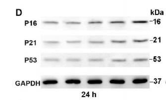
1). Sirt3-mediated mitophagy regulates AGEs-induced BMSCs senescence and senile osteoporosis. Redox Biology, 2021 (PubMed: 33662874) [IF=10.7]

Application: WB    Species: mice    Sample: bone marrow mesenchymal stem (BMSCs)

Fig. 1. Effects of AGEs in different concentrations on the senescence of BMSCs. The BMSCs were treated with AGEs (50–200 μg/mL) or BSA for 24–72 h. (A) SA-β-gal assay for detection of BMSCs senescence. Scale bar: 100 μm. (B) Detection of H3K9me3 by immunofluorescence in BMSCs. Scale bar: 100 μm. (C) Detection of γ-H2AX by immunofluorescence in BMSCs. Scale bar: 100 μm. (D–I) Representative Western blotting assay and quantitation of the level of P16, P21, P53. **p < 0.01 versus BSA.

2). Targeting FAP-positive chondrocytes in osteoarthritis: a novel lipid nanoparticle siRNA approach to mitigate cartilage degeneration. Journal of nanobiotechnology, 2024 (PubMed: 39456041) [IF=10.2]

3). BM-MSC-derived migrasomes reverse stroke-induced thymic atrophy and immunosuppression via Pin1 delivery to thymic epithelial cells. Journal of neuroinflammation, 2025 (PubMed: 41241718) [IF=9.3]

Application: WB    Species: Mouse    Sample:

Fig. 2 BM-MSC transplantation promotes the proliferation of medullary thymic epithelial cells (mTEC). Male wild-type C57/Bl6 mice underwent 60 minutes of tMCAO followed by reperfusion. BM-MSC (2 × 10⁶ cells/mouse) or vehicle (Veh) were administered intravenously as a single dose 2 hours postreperfusion. Thymic tissues were harvested at 7 days post-tMCAO. A-B Bulk RNA sequencing (RNA-seq) analysis of thymic tissues at 7 days post-tMCAO (n = 4 per group). A Volcano plot of differentially expressed genes (DEGs) between the BM-MSC and Veh groups. B Gene Ontology (GO) enrichment analysis of the top 20 enriched biological processes (BP) among the DEG. C Western blot analysis of proliferation-related (Ki67) and senescence-related (NAMPT, p21, and γH2AX) protein expression. The bar graphs show the relative protein levels (n = 4 per group). *p 

4). d-Borneol enhances cisplatin sensitivity via p21/p27-mediated S-phase arrest and cell apoptosis in non-small cell lung cancer cells and a murine xenograft model. Cellular & Molecular Biology Letters, 2022 (PubMed: 35883026) [IF=9.2]

5). The brain-protective mechanism of fecal microbiota transplantation from young donor mice in the natural aging process via exosome, gut microbiota, and metabolomics analyses. Pharmacological research, 2024 (PubMed: 39053865) [IF=9.1]

Application: WB    Species: Mouse    Sample:

Fig. 3. The cerebral protection effects of FMT in mice against age-associated proteins expression levels. (A, B) Quantified protein levels of the cell cycle arrest related proteins of p53, p21, p16/p14, and Rb in mice hippocampus tissues. Data are shown as the mean ± SD, n = 3 per group. Significance was determined by one-way ANOVA followed by the Dunnett T3 test for comparison of multiple groups, *P < 0.05, **P < 0.01, and ***P < 0.001. (C, D) Quantified protein levels of the DNA damage-related protein ATM, and the cognitive-related proteins synapsin I, synaptophysin and PSD95 in mice hippocampus tissues. Data are shown as the mean ± SD, n = 3 per group. Significance was determined by one-way ANOVA followed by the Dunnett T3 test for comparison of multiple groups, *P < 0.05, **P < 0.01, and ***P < 0.001. (E, F) Quantified protein levels of the cell senescence-related proteins CREB, p-CREB, ERK, p-ERK, AKT, p-AKT in mice hippocampus tissues. Data are shown as the mean ± SD, n = 3 per group. Significance was determined by one-way ANOVA followed by the Dunnett T3 test for comparison of multiple groups, *P < 0.05, **P < 0.01, and ***P < 0.001. (G, H) Quantified protein levels of the c-H2AX and TP53BP1 proteins in bone marrow mesenchymal stem cells. Data are shown as the mean ± SD, n = 3 per group. Significance was determined by one-way ANOVA followed by the Dunnett T3 test for comparison of multiple groups, *P < 0.05, **P < 0.01, and ***P < 0.001. (I, J) Quantified protein levels of the β-galactosidase, c-H2AX, and TP53BP1 proteins in in mice hippocampus tissues. Data are shown as the mean ± SD, n = 3 per group. Significance was determined by one-way ANOVA followed by the Dunnett T3 test for comparison of multiple groups, *P < 0.05, **P < 0.01, and ***P < 0.001.

6). Enhanced oral nanomedicine utilizing biomineralized oncolytic virus for synergistic gastrointestinal cancer therapy. Materials today. Bio, 2025 (PubMed: 41377584) [IF=8.7]

7). Curcumin-loaded milk-derived sEVs fused with platelet membrane attenuate endothelial senescence and promote spinal cord injury recovery in diabetic mice. Materials today. Bio, 2025 (PubMed: 40688662) [IF=8.7]

Application: WB    Species: human    Sample:
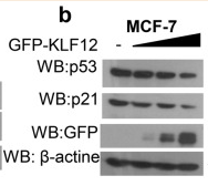
Fig. 3. Reduction in HG/IL-1β-induced senescence markers and TJ protein degradation in HUVECs by Cur treatment. (A) Typical images of HUVECs stained with SA-β-gal (blue stain indicates senescent cells). The number of SA-β-gal+ cells decreased in the Cur group compared to the HG/IL-1β + DMSO group, whereas it increased after brusatol treatment. Scale bar = 200 μm. (B) Quantification of SA-β-gal+ cells (n = 5). (C) Typical images of Claudin-5 staining (green, TJ protein). The intensity of Claudin-5 increased in the Cur group compared to the HG/IL-1β + DMSO group, which was decreased by brusatol treatment. Scale bar = 100 μm. (D) Quantification of the relative fluorescence intensity of Claudin-5 (n = 5). (E) Analysis of VE-Cadherin, ZO-1, Occludin and Claudin-5 expression in HUVECs by WB (TJ protein). The level of TJ protein was increased in the Cur group compared to the HG/IL-1β + DMSO group, which was decreased by brusatol treatment. (F–I) Quantification of the level of VE-Cadherin, ZO-1, Claudin-5, Occludin (n = 3). (J) Analysis of NRF2, HO-1, P21 and P16 expression in HUVECs by WB. Note: (1) The levels of NRF2 and HO-1 were upregulated in the Cur group compared to the HG/IL-1β + DMSO group, which was reduced by brusatol treatment. (2) The levels of P21 and P16 were decreased in the Cur group compared to the HG/IL-1β + DMSO group, which were increased by brusatol treatment. (K–N) Quantification of NRF2, HO-1, P21, and P16 levels (n = 3). ∗P < 0.05, ∗∗P < 0.01. (For interpretation of the references to colour in this figure legend, the reader is referred to the Web version of this article.)

8). Histone acetylation plays an important role in MC-LR-induced apoptosis and cycle disorder in SD rat testicular cells. Chemosphere, 2020 (PubMed: 31683423) [IF=8.1]

Application: WB    Species: Rat    Sample: testicular tissue

Fig4. The effect of TSA and MC-LR on cell cycle-related genes and proteins 434 levels in co-cultured Sertoli-germ cells and SD rat testicular tissues. (A) 435 Expressions of cell cycle-related genes in vitro and (B) expressions of cell 436 cycle-related genes in vivo were detected by RT-qPCR. (C) Expressions of cell cycle-related proteins in vitro and (D) expressions of cell cycle-related proteins in 438 vivo were detected by western blotting. (E) Quantitative analysis of proteins 439 expression levels in vitro. (F) Quantitative analysis of proteins expression levels in 440 vivo. (*P<0.05 vs. the control group; #P<0.05 vs. the 36 µM MC-LR group or the 40 441 µg kg-1 MC-LR group)

9). KLF12 promotes the proliferation of breast cancer cells by reducing the transcription of p21 in a p53-dependent and p53-independent manner. Cell Death & Disease, 2023 (PubMed: 37156774) [IF=8.1]

Application: WB    Species: Human    Sample: MCF7 cells
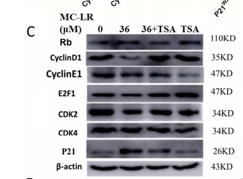
Fig. 4 KLF12 reduces the stability of p53. a Western blot assay showed changes of exogenous p53 in HEK293T cells transfected with increasing doses of KLF12. b Changes in endogenous p53 and p21 in MCF7 cells transfected with increasing doses of KLF12 by Western blot assay. c Changes in the endogenous level of p53 and p21 in ZR-75-30 cells transfected with NC, shKLF12#1, or shKLF12#2 by Western blot assay. d Changes in exogenous p53 in HEK293T cells transfected with GFP-KLF12 without and with 20 μg/ml MG132 treatment by Western blot assay. e, f The half-life of p53 in MCF-7 cells with overexpressed KLF12 treated with CHX (50 μg/ml) by Western blot assay. g CoIP assay showed changes in the level of ubiquitinated p53 in HEK293T cells transfected with Flag-KLF12 plus 4 h treatment with 20 μg/ml MG132. h CoIP assay detected changes in K48-linked polyubiquitylation of p53 and K63-linked polyubiquitylation of p53 in HEK293T cells. The cells were treated with MG132 for 4 h following transfection.

10). Downregulation of VEGFA accelerates AGEs-mediated nucleus pulposus degeneration through inhibiting protective mitophagy in high glucose environments. International journal of biological macromolecules, 2024 (PubMed: 38320636) [IF=7.7]

もっと読みます

Restrictive clause


Affinity Biosciences tests all products strictly. Citations are provided as a resource for additional applications that have not been validated by Affinity Biosciences. Please choose the appropriate format for each application and consult Materials and Methods sections for additional details about the use of any product in these publications.

For Research Use Only.
Not for use in diagnostic or therapeutic procedures. Not for resale. Not for distribution without written consent. Affinity Biosciences will not be held responsible for patent infringement or other violations that may occur with the use of our products. Affinity Biosciences, Affinity Biosciences Logo and all other trademarks are the property of Affinity Biosciences LTD.