製品: STAT1 Antibody
カタログ: AF6300
タンパク質の説明: Rabbit polyclonal antibody to STAT1
アプリケーション: WB IHC IF/ICC IP
反応性: Human, Mouse, Rat, Monkey
予測: Pig, Bovine, Horse, Rabbit, Dog, Chicken, Xenopus
分子量: 84kDa; 87kD(Calculated).
ユニプロット: P42224
RRID: AB_2835149

類似製品を見る>>

   サイズ 価格 在庫状況
 100ul $280 在庫あり
 200ul $350 在庫あり

リードタイム: 当日配達

For pricing and ordering contact:
お問い合わせ先

製品説明

ソース:
Rabbit
アプリケーション:
WB 1:500-1:2000, IHC 1:50-1:200, IP, IF/ICC 1:100-1:500
*The optimal dilutions should be determined by the end user.
*Tips:

WB: For western blot detection of denatured protein samples. IHC: For immunohistochemical detection of paraffin sections (IHC-p) or frozen sections (IHC-f) of tissue samples. IF/ICC: For immunofluorescence detection of cell samples. ELISA(peptide): For ELISA detection of antigenic peptide.

反応性:
Human,Mouse,Rat,Monkey
予測:
Pig(100%), Bovine(100%), Horse(100%), Rabbit(100%), Dog(100%), Chicken(80%), Xenopus(100%)
クローナリティ:
Polyclonal
特異性:
STAT1 Antibody detects endogenous levels of total STAT1.
RRID:
AB_2835149
引用形式: Affinity Biosciences Cat# AF6300, RRID:AB_2835149.
コンジュゲート:
Unconjugated.
精製:
The antiserum was purified by peptide affinity chromatography using SulfoLink™ Coupling Resin (Thermo Fisher Scientific).
保存:
Rabbit IgG in phosphate buffered saline , pH 7.4, 150mM NaCl, 0.02% sodium azide and 50% glycerol. Store at -20 °C. Stable for 12 months from date of receipt.
別名:

折りたたみ/展開

CANDF7; DKFZp686B04100; ISGF 3; ISGF3; OTTHUMP00000163552; OTTHUMP00000165046; OTTHUMP00000165047; OTTHUMP00000205845; Signal transducer and activator of transcription 1; Signal transducer and activator of transcription 1, 91kDa; Signal transducer and activator of transcription 1-alpha/beta; Stat1; STAT1_HUMAN; STAT91; Transcription factor ISGF-3 components p91/p84;

免疫原

免疫原:
Uniprot:
遺伝子(ID):
タンパク質の説明:
The protein encoded by this gene is a member of the STAT protein family. In response to cytokines and growth factors, STAT family members are phosphorylated by the receptor associated kinases, and then form homo- or heterodimers that translocate to the cell nucleus where they act as transcription activators.
タンパク質配列:
MSQWYELQQLDSKFLEQVHQLYDDSFPMEIRQYLAQWLEKQDWEHAANDVSFATIRFHDLLSQLDDQYSRFSLENNFLLQHNIRKSKRNLQDNFQEDPIQMSMIIYSCLKEERKILENAQRFNQAQSGNIQSTVMLDKQKELDSKVRNVKDKVMCIEHEIKSLEDLQDEYDFKCKTLQNREHETNGVAKSDQKQEQLLLKKMYLMLDNKRKEVVHKIIELLNVTELTQNALINDELVEWKRRQQSACIGGPPNACLDQLQNWFTIVAESLQQVRQQLKKLEELEQKYTYEHDPITKNKQVLWDRTFSLFQQLIQSSFVVERQPCMPTHPQRPLVLKTGVQFTVKLRLLVKLQELNYNLKVKVLFDKDVNERNTVKGFRKFNILGTHTKVMNMEESTNGSLAAEFRHLQLKEQKNAGTRTNEGPLIVTEELHSLSFETQLCQPGLVIDLETTSLPVVVISNVSQLPSGWASILWYNMLVAEPRNLSFFLTPPCARWAQLSEVLSWQFSSVTKRGLNVDQLNMLGEKLLGPNASPDGLIPWTRFCKENINDKNFPFWLWIESILELIKKHLLPLWNDGCIMGFISKERERALLKDQQPGTFLLRFSESSREGAITFTWVERSQNGGEPDFHAVEPYTKKELSAVTFPDIIRNYKVMAAENIPENPLKYLYPNIDKDHAFGKYYSRPKEAPEPMELDGPKGTGYIKTELISVSEVHPSRLQTTDNLLPMSPEEFDEVSRIVGSVEFDSMMNTV

種類予測

種類予測:

Score>80(red) has high confidence and is suggested to be used for WB detection. *The prediction model is mainly based on the alignment of immunogen sequences, the results are for reference only, not as the basis of quality assurance.

Species
Results
Score
Pig
100
Horse
100
Bovine
100
Dog
100
Xenopus
100
Rabbit
100
Chicken
80
Sheep
0
Zebrafish
0
Model Confidence:
High(score>80) Medium(80>score>50) Low(score<50) No confidence

PTMs - P42224 基板として

Site PTM Type Enzyme
S2 Acetylation
S2 Phosphorylation
R31 Methylation
K40 Ubiquitination
Y68 Phosphorylation
Y106 Phosphorylation
K114 Methylation
K114 Sumoylation
K114 Ubiquitination
K138 Ubiquitination
K140 Ubiquitination
K145 Ubiquitination
K152 Ubiquitination
K161 Ubiquitination
S162 Phosphorylation
Y170 Phosphorylation
K173 Acetylation
K173 Ubiquitination
K175 Methylation
K175 Ubiquitination
T184 Phosphorylation
K193 Sumoylation
K193 Ubiquitination
K200 Ubiquitination
K201 Acetylation
K201 Ubiquitination
K209 Ubiquitination
K240 Ubiquitination
K286 Ubiquitination
K296 Methylation
K296 Ubiquitination
K298 Ubiquitination
K350 Ubiquitination
K359 Ubiquitination
K361 Ubiquitination
K366 Methylation
K366 Ubiquitination
T373 Phosphorylation
K375 Ubiquitination
K379 Ubiquitination
K388 Ubiquitination
K410 Acetylation
K410 Ubiquitination
K413 Acetylation
T417 Phosphorylation
K511 Ubiquitination
K525 Methylation
K525 Ubiquitination
S532 Phosphorylation
K544 Ubiquitination
S583 Phosphorylation
K592 Ubiquitination
K636 Ubiquitination
K637 Methylation
K637 Ubiquitination
S640 Phosphorylation
K652 Sumoylation
K652 Ubiquitination
K665 Methylation
K665 Ubiquitination
Y668 Phosphorylation
K673 Ubiquitination
K679 Sumoylation
K679 Ubiquitination
K685 Ubiquitination
K697 Ubiquitination
T699 Phosphorylation
Y701 Phosphorylation P52333 (JAK3) , P22455 (FGFR4) , P23458 (JAK1) , P22607 (FGFR3) , P00533 (EGFR) , O60674 (JAK2) , P12931 (SRC)
K703 Sumoylation
K703 Ubiquitination
T704 Phosphorylation
S708 Phosphorylation
S710 Phosphorylation
S715 Phosphorylation
S727 Phosphorylation Q13554 (CAMK2B) , P49336 (CDK8) , Q05655 (PRKCD) , P68400 (CSNK2A1) , Q14164 (IKBKE) , Q15759 (MAPK11) , P19525 (EIF2AK2) , P27361 (MAPK3) , P28482 (MAPK1) , P45983 (MAPK8) , P45984 (MAPK9) , O75582 (RPS6KA5) , Q16539 (MAPK14)
S740 Phosphorylation
S745 Phosphorylation
T749 Phosphorylation

研究背景

機能:

Signal transducer and transcription activator that mediates cellular responses to interferons (IFNs), cytokine KITLG/SCF and other cytokines and other growth factors. Following type I IFN (IFN-alpha and IFN-beta) binding to cell surface receptors, signaling via protein kinases leads to activation of Jak kinases (TYK2 and JAK1) and to tyrosine phosphorylation of STAT1 and STAT2. The phosphorylated STATs dimerize and associate with ISGF3G/IRF-9 to form a complex termed ISGF3 transcription factor, that enters the nucleus. ISGF3 binds to the IFN stimulated response element (ISRE) to activate the transcription of IFN-stimulated genes (ISG), which drive the cell in an antiviral state. In response to type II IFN (IFN-gamma), STAT1 is tyrosine- and serine-phosphorylated. It then forms a homodimer termed IFN-gamma-activated factor (GAF), migrates into the nucleus and binds to the IFN gamma activated sequence (GAS) to drive the expression of the target genes, inducing a cellular antiviral state. Becomes activated in response to KITLG/SCF and KIT signaling. May mediate cellular responses to activated FGFR1, FGFR2, FGFR3 and FGFR4.

PTMs:

Phosphorylated on tyrosine and serine residues in response to a variety of cytokines/growth hormones including IFN-alpha, IFN-gamma, PDGF and EGF. Activated KIT promotes phosphorylation on tyrosine residues and subsequent translocation to the nucleus. Upon EGF stimulation, phosphorylation on Tyr-701 (lacking in beta form) by JAK1, JAK2 or TYK2 promotes dimerization and subsequent translocation to the nucleus. Growth hormone (GH) activates STAT1 signaling only via JAK2. Tyrosine phosphorylated in response to constitutively activated FGFR1, FGFR2, FGFR3 and FGFR4. Phosphorylation on Ser-727 by several kinases including MAPK14, ERK1/2 and CAMKII on IFN-gamma stimulation, regulates STAT1 transcriptional activity. Phosphorylation on Ser-727 promotes sumoylation though increasing interaction with PIAS. Phosphorylation on Ser-727 by PRKCD induces apoptosis in response to DNA-damaging agents. Phosphorylated on tyrosine residues when PTK2/FAK1 is activated; most likely this is catalyzed by a SRC family kinase. Dephosphorylation on tyrosine residues by PTPN2 negatively regulates interferon-mediated signaling. Upon viral infection or IFN induction, phosphorylation on Ser-708 occurs much later than phosphorylation on Tyr-701 and is required for the binding of ISGF3 on the ISREs of a subset of IFN-stimulated genes IKBKE-dependent. Phosphorylation at Tyr-701 and Ser-708 are mutually exclusive, phosphorylation at Ser-708 requires previous dephosphorylation of Tyr-701.

Sumoylated with SUMO1, SUMO2 and SUMO3. Sumoylation is enhanced by IFN-gamma-induced phosphorylation on Ser-727, and by interaction with PIAS proteins. Enhances the transactivation activity.

ISGylated.

Mono-ADP-ribosylated at Glu-657 and Glu-705 by PARP14; ADP-ribosylation prevents phosphorylation at Tyr-701. However, the role of ADP-ribosylation in the prevention of phosphorylation has been called into question and the lack of phosphorylation may be due to sumoylation of Lys-703.

Monomethylated at Lys-525 by SETD2; monomethylation is necessary for phosphorylation at Tyr-701, translocation into the nucleus and activation of the antiviral defense.

細胞の位置付け:

Cytoplasm. Nucleus.
Note: Translocated into the nucleus upon tyrosine phosphorylation and dimerization, in response to IFN-gamma and signaling by activated FGFR1, FGFR2, FGFR3 or FGFR4 (PubMed:15322115). Monomethylation at Lys-525 is required for phosphorylation at Tyr-701 and translocation into the nucleus (PubMed:28753426). Translocates into the nucleus in response to interferon-beta stimulation (PubMed:26479788).

Extracellular region or secreted Cytosol Plasma membrane Cytoskeleton Lysosome Endosome Peroxisome ER Golgi apparatus Nucleus Mitochondrion Manual annotation Automatic computational assertionSubcellular location
サブユニット構造:

Isoform alpha homodimerizes upon IFN-gamma induced phosphorylation. Heterodimer with STAT2 upon IFN-alpha/beta induced phosphorylation. The heterodimer STAT1:STAT2 forms the interferon-stimulated gene factor 3 complex (ISGF3) with IRF9 (By similarity). Interacts (phosphorylated at Ser-727) with PIAS1; the interaction results in release of STAT1 from its target gene. Interacts with IFNAR1; the interaction requires the phosphorylation of IFNAR1 at 'Tyr-466'. Interacts with IFNAR2. Found in a complex with NMI and CREBBP/CBP. Interacts with NMI which is required for CREBBP/CBP recruitment to the complex. Interacts with PTK2/FAK1. Interacts with SRC (By similarity). Interacts with ERBB4 (phosphorylated). Interacts with PARP9 and DTX3L independently of IFN-beta or IFN-gamma-mediated STAT1 'Tyr-701' phosphorylation. Interacts with histone acetyltransferase EP300/p300 in response to INF-gamma stimulation. Interacts with OTOP1 (By similarity).

(Microbial infection) Interacts with Sendai virus C', C, Y1 and Y2 proteins, preventing activation of ISRE and GAS promoter.

(Microbial infection) Interacts with Nipah virus P, V and W proteins preventing activation of ISRE and GAS promoter.

(Microbial infection) Interacts with Rabies virus phosphoprotein preventing activation of ISRE and GAS promoter.

(Microbial infection) Interacts with HCV core protein; the interaction results in STAT1 degradation.

(Microbial infection) Interacts with ebolavirus protein VP24.

タンパク質ファミリー:

Belongs to the transcription factor STAT family.

研究領域

· Cellular Processes > Cell growth and death > Necroptosis.   (View pathway)

· Environmental Information Processing > Signal transduction > Jak-STAT signaling pathway.   (View pathway)

· Human Diseases > Infectious diseases: Parasitic > Leishmaniasis.

· Human Diseases > Infectious diseases: Parasitic > Toxoplasmosis.

· Human Diseases > Infectious diseases: Bacterial > Tuberculosis.

· Human Diseases > Infectious diseases: Viral > Hepatitis C.

· Human Diseases > Infectious diseases: Viral > Hepatitis B.

· Human Diseases > Infectious diseases: Viral > Measles.

· Human Diseases > Infectious diseases: Viral > Influenza A.

· Human Diseases > Infectious diseases: Viral > Human papillomavirus infection.

· Human Diseases > Infectious diseases: Viral > Herpes simplex infection.

· Human Diseases > Cancers: Overview > Pathways in cancer.   (View pathway)

· Human Diseases > Cancers: Specific types > Pancreatic cancer.   (View pathway)

· Human Diseases > Immune diseases > Inflammatory bowel disease (IBD).

· Organismal Systems > Immune system > Chemokine signaling pathway.   (View pathway)

· Organismal Systems > Development > Osteoclast differentiation.   (View pathway)

· Organismal Systems > Immune system > Toll-like receptor signaling pathway.   (View pathway)

· Organismal Systems > Immune system > NOD-like receptor signaling pathway.   (View pathway)

· Organismal Systems > Immune system > Th1 and Th2 cell differentiation.   (View pathway)

· Organismal Systems > Immune system > Th17 cell differentiation.   (View pathway)

· Organismal Systems > Endocrine system > Prolactin signaling pathway.   (View pathway)

· Organismal Systems > Endocrine system > Thyroid hormone signaling pathway.   (View pathway)

参考文献

1). Calcium silicate-human serum albumin composite hydrogel decreases random pattern skin flap necrosis by attenuating vascular endothelial cell apoptosis and inflammation. Chemical Engineering Journal, 2021 [IF=15.1]

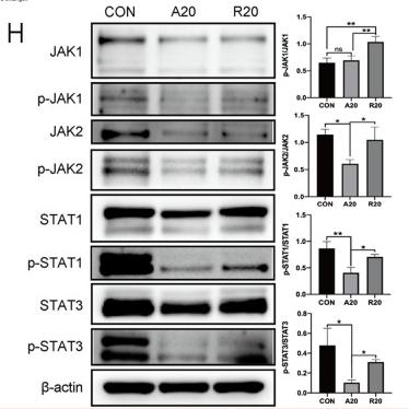
2). Aligned electrospun poly(l-lactide) nanofibers facilitate wound healing by inhibiting macrophage M1 polarization via the JAK-STAT and NF-κB pathways. JOURNAL OF NANOBIOTECHNOLOGY, 2022 (PubMed: 35883095) [IF=10.2]

Application: WB    Species: Mice    Sample:

Fig. 3 The underlying mechanism by which aligned fibers affected macrophage polarization. A Venn diagram showing differentially expressed genes. B KEGG pathway analysis between the A20 and R20 groups. C Heatmap of differentially expressed genes among the three groups. D Heatmap of macrophage polarization-related genes between the A20 and R20 groups. E Volcano diagram of differentially expressed genes. F Western blot analysis of the NF-κB signaling pathway. G Immunofluorescence staining showing the nuclear translocation of NF-κB p65. The nucleus is stained blue, and NF-κB p65 protein is stained red. H Western blot images and semiquantitative analysis of the JAK-STAT signaling pathway (*p < 0.05, **p < 0.01, n = 3)

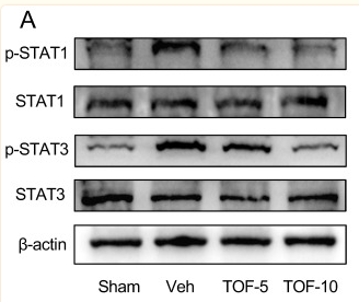
3). Tofacitinib Promotes Functional Recovery after Spinal Cord Injury by Regulating Microglial Polarization via JAK/STAT Signaling Pathway. International Journal of Biological Sciences, 2023 (PubMed: 37781508) [IF=9.2]

Application: WB    Species: Rat    Sample: spinal cord

Figure 8 TOF normalizes the activated STAT signaling in injured spinal cord and ex vivo microglia under inflammatory conditions. (A) Western blot analysis performed for p-STAT1, STAT1, p-STAT3 and STAT3 expression in spinal cord obtained on day 7 post-injury. (B) Western blot analysis of p-STAT1, STAT1, p-STAT3 and STAT3 expression in primary microglia. (C) Quantification of the ratio of p-STAT1/ STAT1 and p-STAT3/ STAT3 (The values are presented as mean ± SD; *p

4). Phillygenin inhibited M1 macrophage polarization and reduced hepatic stellate cell activation by inhibiting macrophage exosomal miR-125b-5p. BIOMEDICINE & PHARMACOTHERAPY, 2023 (PubMed: 36652738) [IF=7.5]

Application: WB    Species: Mouse    Sample: RAW264.7 cells

Fig. 2. PHI inhibited the JAK/STAT and Notch1 signaling pathways in RAW264.7 cells. (a) The 3D and 2D models of molecular docking of PHI and JAK1. (b) The 3D and 2D models of molecular docking of PHI and JAK2. (c) The 3D and 2D models of molecular docking of PHI and Notch1. (d-g) The expression of JAK1, JAK2, STAT1 and Notch1 mRNA in RAW264.7 cells after treatment with LPS/IFNγ and PHI for 12 h was detected by RT-qPCR (n = 3). (h) The expression of JAK1, JAK2, p-JAK1, p-JAK2, STAT1, p-STAT1 and Notch1 proteins in RAW264.7 cells after treatment with LPS/IFNγ and PHI for 12 h was detected by western blotting. (i-l) The relative quantification of p-JAK1/JAK1, p-JAK2/JAK2, p-STAT1/STAT1, and Notch1 protein expression in western blotting results was analyzed by ImageJ software (n = 3). Results are presented as mean ± SD. ###P 

5). Novel Insight Into Nutritional Regulation in Enhancement of Immune Status and Mediation of Inflammation Dynamics Integrated Study In Vivo and In Vitro of Teleost Grass Carp (Ctenopharyngodon idella): Administration of Threonine. Frontiers in Immunology, 2022 (PubMed: 35359991) [IF=7.3]

6). CTLA4Ig/VISTAIg combination therapy selectively induces CD4+ T cell-mediated immune tolerance by targeting the SOCS1 signaling pathway in porcine islet xenotransplantation. IMMUNOLOGY, 2022 (PubMed: 35263451) [IF=6.4]

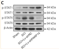
7). Poly(ADP-ribose) polymerase family member 14 promotes functional recovery after spinal cord injury through regulating microglia M1/M2 polarization via STAT1/6 pathway. Neural Regeneration Research, 2023 (PubMed: 36751810) [IF=6.1]

Application: WB    Species: Mouse    Sample:

Figure 5 PARP14 deficiency activates the STAT1 pathway but blocks the STAT6 pathway in mice 7 days post-SCI. (A) Representative images and quantitative analysis showing p-STAT1 (Try701)+/Iba1+ immunofluorescence staining. Lv-shPARP14 injection further promoted SCI-induced STAT1 pathway activation. White arrows indicate p-STAT1 (Try701)+ (green, FITC-labeled)/Iba1+ (red, Cy3-labeled, microglia marker) cells. (B) Representative images and quantitative analysis showing p-STAT6 (Tyr641)+/Iba1+ immunofluorescence staining. Lv-shPARP14 injection inhibited SCI-induced STAT6 pathway activation. White arrows indicated p-STAT6 (Tyr641)+ (green, FITC-labeled)/Iba1+ (red, Cy3-labeled) cells. Scale bars: 50 µm. (C) Relative protein levels of p-STAT1 (Try701), and p-STAT6 (Tyr641) in each group were detected by western blot analysis. p-STAT1 (Try701) expression was increased by Lv-shPARP14 injection, but p-STAT6 (Tyr641) expression was decreased by PARP14 silencing. Values are shown as mean ± SD (n = 6). **P < 0.01 (one-way analysis of variance followed by Tukey’s post hoc test). Images were taken from the gray matter ventral horn at the injury site. Spinal cord tissues from the injury site were used for western blot detection. DAPI: 4′,6-Diamidino-2-phenylindole; Iba1: ionized calcium-binding adaptor molecule 1; PARP14: poly(ADP-ribose)polymerase, member 14; SCI: spinal cord injury; STAT1: signal transducer and activator of transcription.

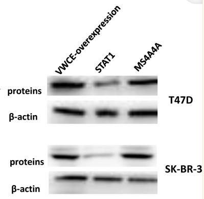
8). VWCE as a potential biomarker associated with immune infiltrates in breast cancer. Cancer Cell International, 2021 (PubMed: 34020650) [IF=5.8]

Application: WB    Species: Human    Sample: breast cancer tissues

Fig. 9 The expression of STAT1 and MS4A4A after VWCE-overexpression treatment. a The expression of STAT1 and MS4A4A after VWCE-overexpression was analyzed by RT-PCR. b The correlation between VWCE-overexpression and STAT1 and MS4A4A in T47D and SK-BR-3 breast cancer cell lines was analyzed by western blotting. C Correlation between VWCE expression and immune infiltration level of Th1 and M2 macrophages in breast cancer. a The expression of VWCE in normal tissues (++), b The expression of VWCE in breast cancer tissues (+), c The expression level of STAT1 was high in breast cancer tissues (++), d The expression level of MS4A4A was relatively low in breast cancer tissues (+). The expression density of VWCE, STAT1, and MS4A4A in breast cancer tissue was quantitated by scoring staining intensity, including negative (−) and weak (+) staining, moderate (++) and strong (+++) staining, respectively.

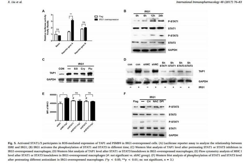
9). IRG1 increases MHC class I level in macrophages through STAT-TAP1 axis depending on NADPH oxidase mediated reactive oxygen species. International Immunopharmacology, 2017 (PubMed: 28477473) [IF=5.6]

Application: WB    Species: human    Sample:

Fig. 5. Activated STAT1/3 participates in ROS-mediated expression of TAP1 and PSMB9 in IRG1-overexpressed cells. (A) Luciferase reporter assay to analyze the relationship between ISRE and IRG1; (B) IRG1 activates the phosphorylation of STAT1 and STAT3 in different time; (C) Western blot analysis of TAP1 level after pretreating STAT1 or STAT3 inhibitors in IRG1-overexpressed macrophages; (D) Western blot analysis of TAP1 level after STAT1 or STAT3 knockdown in IRG1-overexpressed macrophages; (E) Flow cytometry analysis of MHC I level after STAT1 or STAT3 knockdown in IRG1-overexpressed macrophages (#: not significant vs. shNC group). (F) Western blot analysis of phosphorylation of STAT1 and STAT3 level after pretreating different antioxidant in IRG1-overexpressed macrophages. (*p < 0.05; **p < 0.01; ns: not significant, n = 3.)

10). Monotropein inhibits colitis associated cancer through VDR/JAK1/STAT1 regulation of macrophage polarization. International Immunopharmacology, 2023 (PubMed: 37633235) [IF=5.6]

もっと読みます

Restrictive clause


Affinity Biosciences tests all products strictly. Citations are provided as a resource for additional applications that have not been validated by Affinity Biosciences. Please choose the appropriate format for each application and consult Materials and Methods sections for additional details about the use of any product in these publications.

For Research Use Only.
Not for use in diagnostic or therapeutic procedures. Not for resale. Not for distribution without written consent. Affinity Biosciences will not be held responsible for patent infringement or other violations that may occur with the use of our products. Affinity Biosciences, Affinity Biosciences Logo and all other trademarks are the property of Affinity Biosciences LTD.