
 and mouse anti-beta tubulin Ab(T0023 1:200) for 1 hour at 37°C. An AlexaFluor594 conjugated goat anti-rabbit IgG(H+L) Ab(Red) and an AlexaFluor488 conjugated goat anti-mouse IgG(H+L) Ab(Green) were used as the secondary antibody.
The nuclear counter stain is DAPI(blue).")
| 製品: | PKD1/PKC mu Antibody |
| カタログ: | AF6443 |
| タンパク質の説明: | Rabbit polyclonal antibody to PKD1/PKC mu |
| アプリケーション: | WB IHC IF/ICC |
| Cited expt.: | IHC |
| 反応性: | Human, Mouse, Rat |
| 予測: | Pig, Zebrafish, Bovine, Horse, Sheep, Rabbit, Dog, Chicken, Xenopus |
| 分子量: | 115kDa(Observed); 102kD(Calculated). |
| ユニプロット: | Q15139 |
| RRID: | AB_2835267 |
Control Products

製品説明
*The optimal dilutions should be determined by the end user. For optimal experimental results, antibody reuse is not recommended.
*Tips:
WB: For western blot detection of denatured protein samples. IHC: For immunohistochemical detection of paraffin sections (IHC-p) or frozen sections (IHC-f) of tissue samples. IF/ICC: For immunofluorescence detection of cell samples. ELISA(peptide): For ELISA detection of antigenic peptide.
引用形式: Affinity Biosciences Cat# AF6443, RRID:AB_2835267.
折りたたみ/展開
KPCD1_HUMAN; nPKC D1; nPKC mu; nPKC-D1; nPKC-mu; nPKCD1; nPKCmu; PKC; PKC MU; PKCM; PKCmu; PKD 1; PKD; PKD1; PRKCM; PRKD 1; Prkd1; Protein kinase C mu; Protein kinase C mu type; Protein kinase D; Protein kinase D1; Serine/threonine protein kinase D1; Serine/threonine-protein kinase D1;
免疫原
A synthesized peptide derived from human PKD1/PKC mu, corresponding to a region within the internal amino acids.
- Q15139 KPCD1_HUMAN:
- Protein BLAST With
- NCBI/
- ExPASy/
- Uniprot
MSAPPVLRPPSPLLPVAAAAAAAAAALVPGSGPGPAPFLAPVAAPVGGISFHLQIGLSREPVLLLQDSSGDYSLAHVREMACSIVDQKFPECGFYGMYDKILLFRHDPTSENILQLVKAASDIQEGDLIEVVLSASATFEDFQIRPHALFVHSYRAPAFCDHCGEMLWGLVRQGLKCEGCGLNYHKRCAFKIPNNCSGVRRRRLSNVSLTGVSTIRTSSAELSTSAPDEPLLQKSPSESFIGREKRSNSQSYIGRPIHLDKILMSKVKVPHTFVIHSYTRPTVCQYCKKLLKGLFRQGLQCKDCRFNCHKRCAPKVPNNCLGEVTINGDLLSPGAESDVVMEEGSDDNDSERNSGLMDDMEEAMVQDAEMAMAECQNDSGEMQDPDPDHEDANRTISPSTSNNIPLMRVVQSVKHTKRKSSTVMKEGWMVHYTSKDTLRKRHYWRLDSKCITLFQNDTGSRYYKEIPLSEILSLEPVKTSALIPNGANPHCFEITTANVVYYVGENVVNPSSPSPNNSVLTSGVGADVARMWEIAIQHALMPVIPKGSSVGTGTNLHRDISVSISVSNCQIQENVDISTVYQIFPDEVLGSGQFGIVYGGKHRKTGRDVAIKIIDKLRFPTKQESQLRNEVAILQNLHHPGVVNLECMFETPERVFVVMEKLHGDMLEMILSSEKGRLPEHITKFLITQILVALRHLHFKNIVHCDLKPENVLLASADPFPQVKLCDFGFARIIGEKSFRRSVVGTPAYLAPEVLRNKGYNRSLDMWSVGVIIYVSLSGTFPFNEDEDIHDQIQNAAFMYPPNPWKEISHEAIDLINNLLQVKMRKRYSVDKTLSHPWLQDYQTWLDLRELECKIGERYITHESDDLRWEKYAGEQGLQYPTHLINPSASHSDTPETEETEMKALGERVSIL
種類予測
Score>80(red) has high confidence and is suggested to be used for WB detection. *The prediction model is mainly based on the alignment of immunogen sequences, the results are for reference only, not as the basis of quality assurance.
High(score>80) Medium(80>score>50) Low(score<50) No confidence
研究背景
Serine/threonine-protein kinase that converts transient diacylglycerol (DAG) signals into prolonged physiological effects downstream of PKC, and is involved in the regulation of MAPK8/JNK1 and Ras signaling, Golgi membrane integrity and trafficking, cell survival through NF-kappa-B activation, cell migration, cell differentiation by mediating HDAC7 nuclear export, cell proliferation via MAPK1/3 (ERK1/2) signaling, and plays a role in cardiac hypertrophy, VEGFA-induced angiogenesis, genotoxic-induced apoptosis and flagellin-stimulated inflammatory response. Phosphorylates the epidermal growth factor receptor (EGFR) on dual threonine residues, which leads to the suppression of epidermal growth factor (EGF)-induced MAPK8/JNK1 activation and subsequent JUN phosphorylation. Phosphorylates RIN1, inducing RIN1 binding to 14-3-3 proteins YWHAB, YWHAE and YWHAZ and increased competition with RAF1 for binding to GTP-bound form of Ras proteins (NRAS, HRAS and KRAS). Acts downstream of the heterotrimeric G-protein beta/gamma-subunit complex to maintain the structural integrity of the Golgi membranes, and is required for protein transport along the secretory pathway. In the trans-Golgi network (TGN), regulates the fission of transport vesicles that are on their way to the plasma membrane. May act by activating the lipid kinase phosphatidylinositol 4-kinase beta (PI4KB) at the TGN for the local synthesis of phosphorylated inositol lipids, which induces a sequential production of DAG, phosphatidic acid (PA) and lyso-PA (LPA) that are necessary for membrane fission and generation of specific transport carriers to the cell surface. Under oxidative stress, is phosphorylated at Tyr-463 via SRC-ABL1 and contributes to cell survival by activating IKK complex and subsequent nuclear translocation and activation of NFKB1. Involved in cell migration by regulating integrin alpha-5/beta-3 recycling and promoting its recruitment in newly forming focal adhesion. In osteoblast differentiation, mediates the bone morphogenetic protein 2 (BMP2)-induced nuclear export of HDAC7, which results in the inhibition of HDAC7 transcriptional repression of RUNX2. In neurons, plays an important role in neuronal polarity by regulating the biogenesis of TGN-derived dendritic vesicles, and is involved in the maintenance of dendritic arborization and Golgi structure in hippocampal cells. May potentiate mitogenesis induced by the neuropeptide bombesin or vasopressin by mediating an increase in the duration of MAPK1/3 (ERK1/2) signaling, which leads to accumulation of immediate-early gene products including FOS that stimulate cell cycle progression. Plays an important role in the proliferative response induced by low calcium in keratinocytes, through sustained activation of MAPK1/3 (ERK1/2) pathway. Downstream of novel PKC signaling, plays a role in cardiac hypertrophy by phosphorylating HDAC5, which in turn triggers XPO1/CRM1-dependent nuclear export of HDAC5, MEF2A transcriptional activation and induction of downstream target genes that promote myocyte hypertrophy and pathological cardiac remodeling. Mediates cardiac troponin I (TNNI3) phosphorylation at the PKA sites, which results in reduced myofilament calcium sensitivity, and accelerated crossbridge cycling kinetics. The PRKD1-HDAC5 pathway is also involved in angiogenesis by mediating VEGFA-induced specific subset of gene expression, cell migration, and tube formation. In response to VEGFA, is necessary and required for HDAC7 phosphorylation which induces HDAC7 nuclear export and endothelial cell proliferation and migration. During apoptosis induced by cytarabine and other genotoxic agents, PRKD1 is cleaved by caspase-3 at Asp-378, resulting in activation of its kinase function and increased sensitivity of cells to the cytotoxic effects of genotoxic agents. In epithelial cells, is required for transducing flagellin-stimulated inflammatory responses by binding and phosphorylating TLR5, which contributes to MAPK14/p38 activation and production of inflammatory cytokines. May play a role in inflammatory response by mediating activation of NF-kappa-B. May be involved in pain transmission by directly modulating TRPV1 receptor. Plays a role in activated KRAS-mediated stabilization of ZNF304 in colorectal cancer (CRC) cells. Regulates nuclear translocation of transcription factor TFEB in macrophages upon live S.enterica infection (By similarity).
Phosphorylated at Ser-397 and Ser-401 by MAPK13 during regulation of insulin secretion in pancreatic beta cells. Phosphorylated by DAPK1. Phosphorylated at Tyr-95 and by ABL at Tyr-463, which primes the kinase in response to oxidative stress, and promotes a second step activating phosphorylation at Ser-738/Ser-742 by PKRD. Phosphorylated on Ser-910 upon S.enterica infection in macrophages (By similarity).
Cytoplasm. Cell membrane. Golgi apparatus>trans-Golgi network.
Note: Translocation to the cell membrane is required for kinase activation.
Belongs to the protein kinase superfamily. CAMK Ser/Thr protein kinase family. PKD subfamily.
研究領域
· Environmental Information Processing > Signal transduction > Rap1 signaling pathway. (View pathway)
· Organismal Systems > Endocrine system > Aldosterone synthesis and secretion.
参考文献
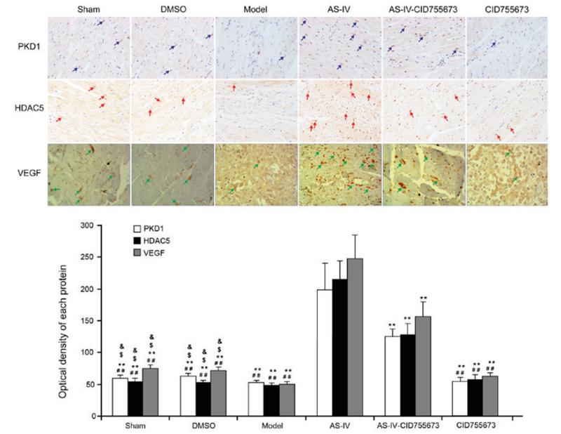
Application: IHC Species: rat Sample: myocardium
Restrictive clause
Affinity Biosciences tests all products strictly. Citations are provided as a resource for additional applications that have not been validated by Affinity Biosciences. Please choose the appropriate format for each application and consult Materials and Methods sections for additional details about the use of any product in these publications.
For Research Use Only.
Not for use in diagnostic or therapeutic procedures. Not for resale. Not for distribution without written consent. Affinity Biosciences will not be held responsible for patent infringement or other violations that may occur with the use of our products. Affinity Biosciences, Affinity Biosciences Logo and all other trademarks are the property of Affinity Biosciences LTD.