
, using Cox2 Antibody at 1/1000 dilution.
5ug/NC membrane strip.
Exposure for 10s with Affinity™ ECL Kit(#KF8003).
Bands result from membrane strip incubation.")
 The protein levels of E4BP4 and COX2 in 3T3-L1 cells transfected with pCDNA3.1, pCDNA3.1-E4BP4, pCDNA3.1-E4BP4 þ pCDNA3.1-COX2 or pCDNA3.1-COX2.")
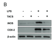
 and effects of compounds on protein expressions of iNOS in LPS-induced C6 cell
lines (B). BV2 cell lines were exposed to the indicated compounds at concentrations of 10, 20 and 40 mM for 24 h in the presence of LPS (1 mg/mL). C6 cell lines were exposed to the
indicated compounds at concentrations of 10, 20 and 40 mM for 24 h in the presence of LPS (10 mg/mL). The expression of iNOS and COX-2 was assayed and calculated by image J.
Each error bar represents the mean ± SD of three independent experiments. #, p < 0.05 versus the control group; ##, p < 0.01 versus the control group; ###, p < 0.001 versus the
control group. *, p < 0.05, **, p < 0.01 when compared to the LPS-treated cells. All data are the mean ± SD of at least three independent experiments.")
 Levels of exosomal proteins HIF-1α and COX-2 were determined by Western blot, with CD63 serving as the internal control. The data reported are the representative results of three independent experiments.")

 The production of nitric oxide (NO) in cells supernatant. NO was measured by Griess assay. The protein levels of (B) inducible-nitric oxide synthase (iNOS) and (C) cyclooxygenase (COX)-2. Protein levels were measured by western blotting. GAPDH was used as internal control. The data represent the mean ± SD (NO, n = 6; iNOS, COX-2, n = 3). * p < 0.05 vs. control group. # p < 0.05 vs. LPS group.")
 of COX-2 in
diferent groups (*P<0.05,
**P<0.01; one-way ANOVA)")
")
. ##p < 0.01 compared with control group; *p < 0.05, **p < 0.01 compared with LPS group")
. NO levels in the cell culture media of
Raw264.7 cells were determined using a Griess assay. B–E). Western blot analysis of iNOS, COX-2, NF-κB and p-NF-κB expression. These assays were performed in
three in dependent experiments. ANOVA followed by Bonferroni’s multiple comparison tests was used. (*) p < 0.05, (***) p < 0.001 and (****) p < 0.0001 versus
control group treated with vehicle; (##) p < 0.01, (###) p < 0.001 and (####) p < 0.0001 versus model group.")
 for the groups (n = 8/group). Statistical results (Kruskal–Wallis nonparametric test with Dunn's multiple post-hoc comparison) were shown in the bar graphs, with “∗∗” indicating significant intergroup difference. Data are expressed as the mean ± standard error of the mean (SEM) (n = 8/group). (a) Expression levels of p-P38 in AD + NS, AD + donepezil, and AD + BM25 groups; (b) expression levels of P38 in AD + NS, AD + donepezil, and AD + BM25 groups; (c) expression levels of p-IκBα in AD + NS, AD + donepezil, and AD + BM25 groups; (d) expression levels of Caspase1 in AD + NS, AD + donepezil, and AD + BM25 groups; (e) expression levels of COX2 in AD + NS, AD + donepezil, and AD + BM25 groups; (f) expression levels of iNOS in AD + NS, AD + donepezil, and AD + BM25 groups. BM25: Byu d Mar 25; AD: Alzheimer's disease; NS: normal saline.")

Representative immunofluorescence staining of Iba-1-positive microglia. Scale bar = 50 lm. (B) The morphology of resting (up) and activated
(down) microglia. Scale bar = 25 lm. (C) Quantification shows that teriflunomide significantly decreased the number of activated microglia
(*p < 0.05 vs. tMCAO + vehicle group, n = 8 per group). (D) Western blot analysis of IL-1b, COX-2 and 3-NT levels. b-Actin was used as loading
control. (E, G) Quantification of IL-1b, COX-2 and 3-NT expression (*p < 0.05 vs. Sham + vehicle group, #
p < 0.05 vs. tMCAO + vehicle group,
n = 10 per group). Data are presented as mean ± SD.")
 IL-6, (B) IL-1β and (C) IL-10 in H/R-A549 cells treated with different concentrations of MAF were detected by ELISA. (D) Western blot analysis of Cox-2 and iNOS protein expression in H/R-A549 cells treated with different concentrations of MAF. ***P<0.001 vs. Control; #P<0.05, ##P<0.01 and ###P<0.001 vs. H/R. MAF, mangiferin; H/R, hypoxia-reoxygenation; Cox-2, cyclooxygenase-2; iNOS, inducible nitric oxide synthase.")
 Protein expression of COX2 in the TAL. (B) PGE2 level in the TAL. **P<0.01 vs. control group; #P<0.05 and ##P<0.01 vs. diabetic group. COX2, cyclooxygenase-2; PGE2, prostaglandin E2; TAL, thick ascending limb; TNFR:Fc, TNF receptor fusion protein.")
 RAW264.7 cells were treated in previous mentioned ways, and the gene and protein expression of iNOS and COX-2 were examined.")
 Representative iNOS, COX-2, TNF-α, IL-1β, IL-6, and
β-actin bands. (B), (C), (D), (E), (F) The relative ratios of grayscale values of iNOS, COX-2, TNF-α, IL-1β, and IL-6 to that of corresponding β-actin, n = 4. ###P <
0.001, CUMS + saline vs. sham; *P < 0.05; **P < 0.01; ***P < 0.001, CUMS + saline vs. CUMS + AAV-siNCALD. All data are shown as the mean ± SEM.")
 Level of expression of cyclooxygenase-2 (COX-2) and inducible nitric oxide synthase (iNOS) in chondrocytes of the three groups were evaluated by Western blot. The data were expressed as mean ± SD. Significant differences between different groups are indicated as ##p < 0.01, vs. control group; ∗∗ p < 0.01, vs. IL-1β alone stimulation group, n = 3.")
. Protein levels of SOD1/GAPDH (B), SOD2/GAPDH (C), iNOS/GAPDH (D), nNOS/GAPDH (E), eNOS/GAPDH (F), COX2/GAPDH (G), p38/GAPDH (H) in mice gastric tissues. SOD1, Superoxide dismutase one or Cu/Zn-superoxide dismutase; SOD2/Mn SOD, superoxide dismutase 2. Control: the group administered zero ethanol; Model: the group administered ethanol intragastrically (13.25 ml/kg BW); MLG-L: low-dose (5 g/kg body weight) MLG-treated group; MLG-M: medium-dose (10 g/kg body weight) MLG-treated group; MLG-H: high-dose (20 g/kg body weight) MLG-treated group; CBP: the group receiving Colloid Bismuth Pectin (57 mg/kg body weight). Each group’s data was expressed as mean ± standard deviation (SD), n = 6. By one-way analysis of variance (ANOVA) test, *p < 0.05, **p < 0.01, ***p < 0.001, ****p < 0.0001 vs. each group. Whole page of western blot can be found in Supplementary Figures S1, S3, S7, S8, S10, S14, S15, respectively.")
 Morphological alteration of RAW264.7 cells. (B) The expressions of inflammation-related genes iNOS, COX-2, TNF-α, and IL-1β as determined using qRT-PCR. (C) The expressions of inflammation-related proteins iNOS, COX-2, TNF-α, and IL-1β as determined using Western blotting. Cells were treated with LPS (1 μg/mL) and GLP (RSGLP or BSGLP) at 0–2.5 mg/mL for 24 h. Data are presented as mean ± SE from three independent experiments. ***p < 0.001, compared with LPS treatment group. ###p < 0.001, compared with Con group.")
, C: LPS + wogonin (1.25 μg/μL); D: LPS + wogonin (2.5 μg/μL); E: LPS + wogonin (5 μg/μL). The data presented are means ± SD (n = 3). #P < 0.05 and ##P < 0.01 relative to the control group. Compared with the model group,")
 CCK-8 assay examining the proliferation-suppressing effect of ABP on CT26 cells. (B–C) EdU uptake assay examining the proliferation-suppressing effect of ABP on CT26 cells. Scale bar = 50 μm (×200 magnification). (D–E) Western blot examining the effect of ABP on the protein levels of β-catenin, Cyclin D1, c-Myc, PCNA, COX-2, and GAPDH in CT26 cells. Compared with the untreated group")
 Western blotting of STAT3, P-STAT3, COX-2, Cyclin D1, c-Myc and GAPDH in colon tissue treated with ABP for 3 weeks (A) and 11 weeks (B). (C-D) P-STAT3 protein expression relative to STAT3. COX-2, c-Myc and Cyclin D1 protein expressions relative to GAPDH in colon tissues treated with ABP for 3 weeks (C) and 11 weeks (D). Compared with the A/D group")
 Representative electrocardiogram shows the successful establishment of myocardial I/R injury model (n = 5 per group). The red arrows represent changes in the ST segment. (B) Representative images show TTC staining of the myocardium from rats (n = 5 per group) belonging to the sham, I/R, and I/R + 50 mg/kg aspirin groups. The infarct size quantification of the 3 groups of rats is also shown. (C) ELISA assay results show the serum levels of creatine kinase-MB (CK-MB) and cardiac troponin T (cTnT) in the sham, I/R, and I/R + aspirin group rats (n = 5 per group). (D) Laggendorff in vitro cardiac perfusion results show the left ventricular developed pressure (LVDP), maximum and minimum rates of pressure development (max dP/dt and min dP/dt), and heart rate (HR) in the sham, I/R, and I/R + aspirin group rats (n = 5 per group). (E) Representative H&E staining images (×200 and ×400) show the histological details of the myocardium from the sham, I/R, and I/R + aspirin group rats (n = 5 per group). (F,G) Representative immunoblots and quantification of the expression levels of COX-1 and COX-2 in the sham, I/R, and I/R + aspirin group rats (n = 5 per group). (H) ELISA results show the tumor necrosis factor-α (TNF-α) and interleukin-6 (IL-6) level in the myocardial tissues of the sham, I/R, and I/R + aspirin group rats (n = 5 per group). The tissues were harvested 2 h post-reperfusion. The data are represented as means ± SD; *p < 0.05 vs. the sham group; # p < 0.05 vs. the I/R group.")
 Representative images of the harvest stomach specimens from the sham, I/R, I/R + aspirin, I/R + 50 mg/kg aspirin + 200 mg/kg gastrodin group, and I/R + 200 mg/kg gastrodin group rats (n = 5 per group). (B) Byron gastric mucosa injury scores of the sham, I/R, I/R + aspirin, I/R + aspirin + gastrodin group, and I/R + gastrodin group rats (n = 5 per group). The scoring criteria are shown in Table 1. (C) Representative images (×100 and ×200) of the H&E-staining gastric tissue sections from the sham, I/R, I/R + aspirin, I/R + aspirin + gastrodin group, and I/R + gastrodin group rats (n = 5 per group). (D) Representative immunofluorescence staining images (×200) show the COX-1 and COX-2 expression levels in the gastric tissues of the sham, I/R, I/R + aspirin, I/R + aspirin + gastrodin group, and I/R + gastrodin group rats (n = 5 per group). The green denotes COX-1, red denotes COX-2, and blue denotes the nucleus. (E–G) ELISA assay results show the levels of cyclooxygenase-1 (COX-1), cyclooxygenase-2 (COX-2), tumor necrosis factor-α (TNF-α), interleukin-6 (IL-6), prostaglandin E2 (PGE2) and endothelin (ET) in the gastric tissues of the sham, I/R, I/R + aspirin, I/R + aspirin + gastrodin group,and I/R + gastrodin group rats (n = 5 per group). The data are represented as means ± SD; * p < 0.05 vs. the sham group; # p < 0.05 vs. the I/R group; ^ p < 0.05 vs. the I/R + aspirin group; & p < 0.05 vs. the I/R + aspirin + gastrodin group.")
 HMC3 cells were treated with NETs 500 (ng/mL) for 24 hours. Western blot assays of COX2, IL-6, and TNF-α expression in HMC3 cells were performed (n = 3 per group; unpaired t test). The protein samples were quantified using densitometry through ImageJ software and normalized. (B) HMC3 cells were treated with NETs 500 (ng/mL) for 24 hours. RT-qPCR of COX2, IL-6, and TNF-α expression in HMC3 cells was performed (n = 3 per group; unpaired t test). (C) HMC3 cells were treated with NETs 500 (ng/mL) for 24 hours in the absence or presence of DNase I. Western blot assays of COX2, IL-6, and TNF-α expression in HMC3 cells were performed (n = 3 per group; one-way ANOVA test). The protein samples were quantified using densitometry through ImageJ software and normalized. (D) HMC3 cells were treated with NETs 500 (ng/mL) for 24 hours in the absence or presence of DNase I. RT-qPCR of COX2, IL-6, and TNF-α expression in HMC3 cells was performed (n = 3 per group; one-way ANOVA test). (E) CD4+ T cells were treated with NETs 500 (ng/mL). Representative flow cytometry images of Th1 (IFN-γ+CD4+) and Th17 (IL-17A+CD4+) cells. (n = 3 per group; unpaired t-test). (F) CD4+ T cells were treated with NETs 500 (ng/mL). RT-qPCR of IL-17 and IFN-γ expression in CD4+ T cells was performed (n = 3 per group; unpaired t test). The data were expressed as mean ± SD.")
 Representative immunohistochemistry images of Iba-1 and COX-2 among Con, MPTP and MPTP + B. producta groups, Magnification: 200 ×, Scale bars: 100 μm. (B) Representative Western blot images of Iba-1 among three groups. (C) Quantitative analysis of Iba-1level; the ratio of Iba-1/β-actin in the Con group was used as a reference value. Error bars indicate the SEM, **P < 0.01 vs. Con group, #P < 0.05 vs. MPTP group.")
 control group (Con-Run, 3); (b) bee-pollen-supplemented group (BP-Run; 4). Concentration levels look similar in both photos, with almost equal distribution of pale, non-reactive cells, medium-toned cells that have shown standard levels of enzymes and the darkest cells with notable amounts of target-antibody complexes.")
 Fe2+ concentrations, (B and C) lipid ROS levels, and (D) mRNA levels of Tfrc, Slc11a2, Slc7a11, Gpx4 and Ptgs2, and (E) protein expression of GPX4 and PTGS2 were measured. ***P")

 and Atsttrin (500 ng/ml) for 8 h. mRNA expression of inflammatory molecules (IL-1β, IL-6, COX-2, and iNOS) was tested by real-time PCR (n = 3). e–g RAW264.7 cells treated with 10 ng/ml TNF-α without or with 200 ng/ml Atsttrin. The expression of COX-2 and iNOS was tested by Western blot assay. Quantitative analyses of the normalized protein levels (n = 3). h, i BMDMs were treated with M-CSF (10 ng/mL) for 6 days and then cultured with or without TNF-α (10 ng/mL) in the absence or presence of Atsttrin (500 ng/ml) for 48 h. The levels of IL-1β and IL-6 in the medium were detected by ELISA (n = 3). j–n RAW264.7 cells were treated with TNF-α (10 ng/mL) in the absence or presence of Atsttrin (500 ng/ml) for various durations. Protein levels of p-p38, t-p38, p-JNK, t-JNK, p-p65, t-p65, p-IκBα, and IκBα were detected by Western blot with corresponding antibodies (n = 3). The bands in the figure are not all derived from the same membrane. o–q RAW264.7 cells were treated with TNF-α (10 ng/mL) in the absence or presence of Atsttrin (500 ng/ml) for various time points. Nuclear translocation of NF-κB p65 in RAW264.7 cells as determined by Western blotting with an anti-p65 antibody (n = 3). Each experiment was performed 3 times independently. Significant differences are indicated as follows:")
 WT C57/BL6J mice were pretreated with PTS (10 mg/kg) and afterwards with DOX (20 mg/kg cumulative dose) for 6 days. Immunochemistry staining of the liver sections with anti-NLRP3 (left, scale bar = 50 μm), the quantification of NLRP3 positive area in each group (right, n = 6); (B) qPCR analyses of NLRP3, IL-1β and IL-18 mRNA levels in each group (n = 6); (C) Western blot analyses of NLRP3, ASC, Caspase-1 p20, IL-1β, and IL-18 proteins and the quantification of the blots in each group (n = 4 per group); (D) Western blot analyses of Cleaved Caspase-3, BAX, and BCL-2 proteins (left) and quantification of the blots in each group (right, n = 4 per group).")
 Changes in the concentration of MDA and Fe2+ in MDA-MB-23 and SK-BR-3 cells were detected after NGR1 treatment. (B, C) Changes in the expression of ACSL4, COX2, FIH1, GPX4, and NOX1 proteins were detected by Western blot in MDA-MB-231 and SK-BR-3 cells. (* P < 0.05, ** P < 0.01, *** P < 0.001, **** P < 0.0001).")
 Changes in the levels of Fe2+ and MDA in gastric tissues of luteolin-treated rats. (B) Changes in the mRNA expression of FIH1, COX2, ACSL4, GPX4, and NOX1 were detected using qPCR. (C) Western blot analysis of the protein expression levels of FIH1, COX2, ACSL4, GPX4, and NOX1.")
 Steroid-synthesis-related factors expression level after CIRBP overexpression, n = 4. (B) The mRNA expression level of cumulus-diffusion-related factors, n = 4. (C) The secretion of E2 and P4, n = 4. (D) E2 and P4 synthesis-related protein bands and analysis, n = 3. (E) Cumulus-diffusion-related protein bands and analysis, n = 3. (F) Immunofluorescence detection of cumulus-diffusion-related proteins. Values are mean ± SE, *: p < 0.05, **: p < 0.01, NS: no difference. Original Western blot images are provided in the Supplementary Materials.")
 Steroid-synthesis-related factors expression level after CIRBP overexpression, n = 4. (B) The mRNA expression level of cumulus-diffusion-related factors, n = 4. (C) The secretion of E2 and P4, n = 4. (D) E2 and P4 synthesis-related protein bands and analysis, n = 3. (E) Cumulus-diffusion-related protein bands and analysis, n = 3. (F) Immunofluorescence detection of cumulus-diffusion-related proteins. Values are mean ± SE, *: p < 0.05, **: p < 0.01, NS: no difference. Original Western blot images are provided in the Supplementary Materials.")

. *p")
 Representative images of the liver subjected to H&E staining. The magnification is 200× and 400×. (B) The levels of the liver function indexes ALT and AST were measured. (C) Survival curves for mice treated with different concentrations of CCL4. (D) The levels of MDA and GSH in the serum were measured. (E) Immunolocalization of Ptgs2, Gpx4 and hepcidin proteins after CCL4 exposure. The magnification is 100×. (F) The mean optical density (MOD) of Ptgs2, Gpx4 and hepcidin proteins was calculated from 3 stained pictures of three mice in each group. (G) The level of total iron content in the liver was measured. Data are expressed as the mean±SEM. ns: no significant difference. *P")
 The level of iron in the liver was measured. (B) Representative images of Prussian blue iron staining in the liver. The magnification is 100×. (C) Immunolocalization of hepcidin protein in the NC, CCL4+PBS, and CCL4+MSC groups. The magnification is 100×. The MOD of hepcidin protein was calculated from 3 stained pictures of three mice in each group. (D) Western blot analysis of the protein levels of hepcidin, BMP6 and FPN1 in each group (n=3). Data are expressed as the mean±SEM. ns: no significant difference. *P")
 (A), cell viability (CCK-8) (B), proliferation (EdU) (C), apoptosis (flow cytometry) (D,E), cytokine levels (IL-1β, TNF-α via ELISAs) (F,G), SOD activity (kit assay) (H), and MDA content (kit assay) (I). Similarly, THP-1 cells were grouped likewise, examining iNOS and COX-2 proteins (Western blot) (J) and M1 macrophage ratio (flow cytometry) (K). *, P")
 Experimental process of in vitro experiments. (B) Cell viability. (C) NO production. (D–F) Levels of IL1β, IL6, and TNF-α in the culture supernatant of RAW264.7 cells. (G–J) Representative immunoblot and relative quantification of IL1β, IL6, and TNF-α in RAW264.7 cells. (K,L) Representative immunoblot and relative quantification of COX2 and iNOS in RAW264.7 cells. (M,N) Representative immunofluorescence images of COX2-positive and iNOS-positive in RAW264.7 cells. Scale bars: 25 μm. Data are expressed as mean ± SEM (n = 3). ** p < 0.01 and **** p < 0.0001 vs. the Con group. # p < 0.05, ## p < 0.01, ### p < 0.001, and #### p < 0.0001 vs. the LPS group.")
 SOD enzyme activity in mammary tissue of the LC group and HC dairy goats. (B) CAT enzyme activity in mammary tissue of the LC group and HC dairy goats. (C) GSH level was measured in different groups using GSH Assay Kit. (D and E) Fe2+ (D) and total iron (E) content of mammary tissue in both groups. (F) Measurement of MDA in mammary tissue using an MDA assay kit. (G and H) Representative images and relative intensities of ferroptosis-associated proteins in mammary tissue of dairy goats in LC and HC groups (n = 3). (I–N) MDA level (I), GSH content (J), enzyme activity of CAT (K), enzyme activity of SOD (L), and Fe2+ (M) and total iron (N) content in mammary tissue of mice in groups Abx, Abx+RMTLC, and Abx+RMTHC. (O and P) Serum levels of Fe2+ and total iron in three groups of mice. (Q and R) Representative images and relative intensities of ferroptosis-associated proteins in mammary tissue from three groups of mice (n = 3). Data are presented as the means ± SD, and one-way analysis of variance (ANOVA) was performed for statistical analysis. ∗p < 0.05, ∗∗p < 0.01, ∗∗∗p < 0.001, and ∗∗∗∗p < 0.0001 indicate significant differences.")
 across groups. B Intergroup comparison of COX-2 fluorescence intensity (n = 6). ***P")
. ***P")
 tumor size, (B) volume, and (C) weight in mice. (D) Immunohistochemical staining was used for detecting P4HB, LGALS3BP, GPX4, and PTGS2 expression levels. Detection of (E) ROS and (F) GSH in tissues.")
 Results of immunohistochemical staining. (B–D) Quantitative expression of COX2, GRP78, and PERK in rat liver tissue by immunohistochemistry. ##P < 0.01 compared with the control group. *P < 0.05, **P < 0.01 compared with the model group.")
 Perl's iron staining in days 14 and 28, scale bar = 200 μm; (B) Iron-stained positive area presence significantly differed between the two groups, p")

製品説明
*The optimal dilutions should be determined by the end user. For optimal experimental results, antibody reuse is not recommended.
*Tips:
WB: For western blot detection of denatured protein samples. IHC: For immunohistochemical detection of paraffin sections (IHC-p) or frozen sections (IHC-f) of tissue samples. IF/ICC: For immunofluorescence detection of cell samples. ELISA(peptide): For ELISA detection of antigenic peptide.
引用形式: Affinity Biosciences Cat# AF7003, RRID:AB_2835311.
折りたたみ/展開
COX 2; COX-2; COX2; Cyclooxygenase 2; Cyclooxygenase 2b; Cyclooxygenase; Cyclooxygenase-2; Cyclooxygenase2; EC 1.14.99.1; fj02a10; Glucocorticoid-regulated inflammatory cyclooxygenase; Glucocorticoid-regulated inflammatory Prostaglandin G/H synthase; GRIPGHS; hCox 2; Macrophage activation-associated marker protein P71/73; OTTHUMP00000033524; PES-2; PGG/HS; PGH synthase 2; PGH2_HUMAN; PGHS 2; PGHS-2; PGHS2; PHS 2; PHS II; PHS2; Prostaglandin endoperoxide synthase 2 (prostaglandin G/H synthase and cyclooxygenase); Prostaglandin endoperoxide synthase 2; Prostaglandin G/H synthase 2; Prostaglandin G/H synthase 2 precursor; Prostaglandin G/H synthase and cyclooxygenase; Prostaglandin G/H synthase; Prostaglandin H2 synthase 2; prostaglandin-endoperoxide synthase 2 (prostaglandin G/H synthase and cyclooxygenase); Prostaglandin-endoperoxide synthase 2; PTGS2; ptgs2a; TIS10; TIS10 protein; unp1239; wu:fj02a10;
免疫原
A synthesized peptide derived from human Cox2, corresponding to a region within C-terminal amino acids.
- P35354 PGH2_HUMAN:
- Protein BLAST With
- NCBI/
- ExPASy/
- Uniprot
MLARALLLCAVLALSHTANPCCSHPCQNRGVCMSVGFDQYKCDCTRTGFYGENCSTPEFLTRIKLFLKPTPNTVHYILTHFKGFWNVVNNIPFLRNAIMSYVLTSRSHLIDSPPTYNADYGYKSWEAFSNLSYYTRALPPVPDDCPTPLGVKGKKQLPDSNEIVEKLLLRRKFIPDPQGSNMMFAFFAQHFTHQFFKTDHKRGPAFTNGLGHGVDLNHIYGETLARQRKLRLFKDGKMKYQIIDGEMYPPTVKDTQAEMIYPPQVPEHLRFAVGQEVFGLVPGLMMYATIWLREHNRVCDVLKQEHPEWGDEQLFQTSRLILIGETIKIVIEDYVQHLSGYHFKLKFDPELLFNKQFQYQNRIAAEFNTLYHWHPLLPDTFQIHDQKYNYQQFIYNNSILLEHGITQFVESFTRQIAGRVAGGRNVPPAVQKVSQASIDQSRQMKYQSFNEYRKRFMLKPYESFEELTGEKEMSAELEALYGDIDAVELYPALLVEKPRPDAIFGETMVEVGAPFSLKGLMGNVICSPAYWKPSTFGGEVGFQIINTASIQSLICNNVKGCPFTSFSVPDPELIKTVTINASSSRSGLDDINPTVLLKERSTEL
種類予測
Score>80(red) has high confidence and is suggested to be used for WB detection. *The prediction model is mainly based on the alignment of immunogen sequences, the results are for reference only, not as the basis of quality assurance.
High(score>80) Medium(80>score>50) Low(score<50) No confidence
研究背景
Converts arachidonate to prostaglandin H2 (PGH2), a committed step in prostanoid synthesis. Constitutively expressed in some tissues in physiological conditions, such as the endothelium, kidney and brain, and in pathological conditions, such as in cancer. PTGS2 is responsible for production of inflammatory prostaglandins. Up-regulation of PTGS2 is also associated with increased cell adhesion, phenotypic changes, resistance to apoptosis and tumor angiogenesis. In cancer cells, PTGS2 is a key step in the production of prostaglandin E2 (PGE2), which plays important roles in modulating motility, proliferation and resistance to apoptosis. During neuroinflammation, plays a role in neuronal secretion of specialized preresolving mediators (SPMs), especially 15-R-lipoxin A4, that regulates phagocytic microglia (By similarity).
S-nitrosylation by NOS2 (iNOS) activates enzyme activity. S-nitrosylation may take place on different Cys residues in addition to Cys-526.
Acetylated at Ser-565 by SPHK1. During neuroinflammation, acetylation by SPHK1 promotes neuronal secretion of specialized preresolving mediators (SPMs), especially 15-R-lipoxin A4, which results in an increase of phagocytic microglia.
Microsome membrane>Peripheral membrane protein. Endoplasmic reticulum membrane>Peripheral membrane protein.
Belongs to the prostaglandin G/H synthase family.
研究領域
· Environmental Information Processing > Signal transduction > NF-kappa B signaling pathway. (View pathway)
· Environmental Information Processing > Signal transduction > TNF signaling pathway. (View pathway)
· Human Diseases > Infectious diseases: Parasitic > Leishmaniasis.
· Human Diseases > Infectious diseases: Viral > Human papillomavirus infection.
· Human Diseases > Cancers: Overview > Pathways in cancer. (View pathway)
· Human Diseases > Cancers: Overview > Chemical carcinogenesis.
· Human Diseases > Cancers: Overview > MicroRNAs in cancer.
· Human Diseases > Cancers: Specific types > Small cell lung cancer. (View pathway)
· Metabolism > Lipid metabolism > Arachidonic acid metabolism.
· Metabolism > Global and overview maps > Metabolic pathways.
· Organismal Systems > Immune system > IL-17 signaling pathway. (View pathway)
· Organismal Systems > Nervous system > Retrograde endocannabinoid signaling. (View pathway)
· Organismal Systems > Nervous system > Serotonergic synapse.
· Organismal Systems > Endocrine system > Ovarian steroidogenesis.
· Organismal Systems > Endocrine system > Oxytocin signaling pathway.
· Organismal Systems > Endocrine system > Regulation of lipolysis in adipocytes.
参考文献
Application: WB Species: Mouse Sample: RAW264.7 cells

Application: WB Species: Mouse Sample:
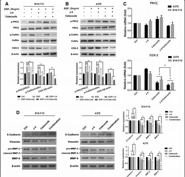
Application: WB Species: mouse Sample: B16F10

Application: WB Species: Mouse Sample: colon tissue

Application: WB Species: Rat Sample:

Application: WB Species: Rat Sample:
Restrictive clause
Affinity Biosciences tests all products strictly. Citations are provided as a resource for additional applications that have not been validated by Affinity Biosciences. Please choose the appropriate format for each application and consult Materials and Methods sections for additional details about the use of any product in these publications.
For Research Use Only.
Not for use in diagnostic or therapeutic procedures. Not for resale. Not for distribution without written consent. Affinity Biosciences will not be held responsible for patent infringement or other violations that may occur with the use of our products. Affinity Biosciences, Affinity Biosciences Logo and all other trademarks are the property of Affinity Biosciences LTD.