製品: Cyclin D1 Antibody
カタログ: AF0931
タンパク質の説明: Rabbit polyclonal antibody to Cyclin D1
アプリケーション: WB IHC IF/ICC
反応性: Human, Mouse, Rat
予測: Pig, Zebrafish, Bovine, Dog, Chicken, Xenopus
分子量: 33kDa; 34kD(Calculated).
ユニプロット: P24385
RRID: AB_2835325

類似製品を見る>>

   サイズ 価格 在庫状況
 100ul $280 在庫あり
 200ul $350 在庫あり

リードタイム: 当日配達

For pricing and ordering contact:
お問い合わせ先

製品説明

ソース:
Rabbit
アプリケーション:
WB 1:500-1:2000, IHC 1:50-1:200, IF/ICC 1:100-1:500
*The optimal dilutions should be determined by the end user.
*Tips:

WB: For western blot detection of denatured protein samples. IHC: For immunohistochemical detection of paraffin sections (IHC-p) or frozen sections (IHC-f) of tissue samples. IF/ICC: For immunofluorescence detection of cell samples. ELISA(peptide): For ELISA detection of antigenic peptide.

反応性:
Human,Mouse,Rat
予測:
Pig(100%), Zebrafish(88%), Bovine(100%), Dog(100%), Chicken(100%), Xenopus(100%)
クローナリティ:
Polyclonal
特異性:
Cyclin D1 Antibody detects endogenous levels of total Cyclin D1.
RRID:
AB_2835325
引用形式: Affinity Biosciences Cat# AF0931, RRID:AB_2835325.
コンジュゲート:
Unconjugated.
精製:
The antiserum was purified by peptide affinity chromatography using SulfoLink™ Coupling Resin (Thermo Fisher Scientific).
保存:
Rabbit IgG in phosphate buffered saline , pH 7.4, 150mM NaCl, 0.02% sodium azide and 50% glycerol. Store at -20 °C. Stable for 12 months from date of receipt.
別名:

折りたたみ/展開

AI327039;B cell CLL/lymphoma 1;B cell leukemia 1;B cell lymphoma 1 protein;B-cell lymphoma 1 protein;BCL 1;BCL-1;BCL-1 oncogene;BCL1;BCL1 oncogene;ccnd1;CCND1/FSTL3 fusion gene, included;CCND1/IGHG1 fusion gene, included;CCND1/IGLC1 fusion gene, included;CCND1/PTH fusion gene, included;CCND1_HUMAN;cD1;Cyl 1;D11S287E;G1/S specific cyclin D1;G1/S-specific cyclin-D1;Parathyroid adenomatosis 1;PRAD1;PRAD1 oncogene;U21B31;

免疫原

免疫原:
Uniprot:
遺伝子(ID):
タンパク質の説明:
The protein encoded by this gene belongs to the highly conserved cyclin family, whose members are characterized by a dramatic periodicity in protein abundance throughout the cell cycle. Cyclins function as regulators of CDK kinases. Different cyclins exhibit distinct expression and degradation patterns which contribute to the temporal coordination of each mitotic event.
タンパク質配列:
MEHQLLCCEVETIRRAYPDANLLNDRVLRAMLKAEETCAPSVSYFKCVQKEVLPSMRKIVATWMLEVCEEQKCEEEVFPLAMNYLDRFLSLEPVKKSRLQLLGATCMFVASKMKETIPLTAEKLCIYTDNSIRPEELLQMELLLVNKLKWNLAAMTPHDFIEHFLSKMPEAEENKQIIRKHAQTFVALCATDVKFISNPPSMVAAGSVVAAVQGLNLRSPNNFLSYYRLTRFLSRVIKCDPDCLRACQEQIEALLESSLRQAQQNMDPKAAEEEEEEEEEVDLACTPTDVRDVDI

種類予測

種類予測:

Score>80(red) has high confidence and is suggested to be used for WB detection. *The prediction model is mainly based on the alignment of immunogen sequences, the results are for reference only, not as the basis of quality assurance.

Species
Results
Score
Pig
100
Bovine
100
Dog
100
Xenopus
100
Chicken
100
Zebrafish
88
Horse
0
Sheep
0
Rabbit
0
Model Confidence:
High(score>80) Medium(80>score>50) Low(score<50) No confidence

PTMs - P24385 基板として

Site PTM Type Enzyme
K33 Ubiquitination
K46 Methylation
K46 Ubiquitination
K50 Ubiquitination
S90 Phosphorylation P17612 (PRKACA)
K95 Ubiquitination
K96 Ubiquitination
S111 Phosphorylation
K112 Ubiquitination
K114 Ubiquitination
S131 Phosphorylation
K167 Ubiquitination
K175 Ubiquitination
K180 Ubiquitination
S197 Phosphorylation P17612 (PRKACA)
S219 Phosphorylation
Y226 Phosphorylation
S234 Phosphorylation P17612 (PRKACA)
K238 Ubiquitination
K269 Ubiquitination
T286 Phosphorylation Q13627 (DYRK1A) , Q9Y463 (DYRK1B) , P49841 (GSK3B) , O15111 (CHUK)
T288 Phosphorylation P49841 (GSK3B) , Q9Y463 (DYRK1B)

研究背景

機能:

Regulatory component of the cyclin D1-CDK4 (DC) complex that phosphorylates and inhibits members of the retinoblastoma (RB) protein family including RB1 and regulates the cell-cycle during G(1)/S transition. Phosphorylation of RB1 allows dissociation of the transcription factor E2F from the RB/E2F complex and the subsequent transcription of E2F target genes which are responsible for the progression through the G(1) phase. Hypophosphorylates RB1 in early G(1) phase. Cyclin D-CDK4 complexes are major integrators of various mitogenenic and antimitogenic signals. Also substrate for SMAD3, phosphorylating SMAD3 in a cell-cycle-dependent manner and repressing its transcriptional activity. Component of the ternary complex, cyclin D1/CDK4/CDKN1B, required for nuclear translocation and activity of the cyclin D-CDK4 complex. Exhibits transcriptional corepressor activity with INSM1 on the NEUROD1 and INS promoters in a cell cycle-independent manner.

PTMs:

Phosphorylation at Thr-286 by MAP kinases is required for ubiquitination and degradation following DNA damage. It probably plays an essential role for recognition by the FBXO31 component of SCF (SKP1-cullin-F-box) protein ligase complex.

Ubiquitinated, primarily as 'Lys-48'-linked polyubiquitination. Ubiquitinated by a SCF (SKP1-CUL1-F-box protein) ubiquitin-protein ligase complex containing FBXO4 and CRYAB. Following DNA damage it is ubiquitinated by some SCF (SKP1-cullin-F-box) protein ligase complex containing FBXO31. SCF-type ubiquitination is dependent on Thr-286 phosphorylation (By similarity). Ubiquitinated also by UHRF2 apparently in a phosphorylation-independent manner. Ubiquitination leads to its degradation and G1 arrest. Deubiquitinated by USP2; leading to its stabilization.

細胞の位置付け:

Nucleus. Cytoplasm. Nucleus membrane.
Note: Cyclin D-CDK4 complexes accumulate at the nuclear membrane and are then translocated to the nucleus through interaction with KIP/CIP family members.

Extracellular region or secreted Cytosol Plasma membrane Cytoskeleton Lysosome Endosome Peroxisome ER Golgi apparatus Nucleus Mitochondrion Manual annotation Automatic computational assertionSubcellular location
サブユニット構造:

Interacts with FBXO4 (By similarity). Interacts with either CDK4 or CDK6 protein kinase to form a serine/threonine kinase holoenzyme complex. The cyclin subunit imparts substrate specificity to the complex. Component of the ternary complex CCND1/CDK4/CDKN1B required for nuclear translocation and modulation of CDK4-mediated kinase activity. Interacts directly with CDKN1B. Interacts with UHRF2; the interaction ubiquitinates CCND1 and appears to occur independently of phosphorylation. Can form similar complexes with either CDKN1A or CDKN2A. Interacts with USP2. Interacts (via cyclin N-terminal domain) with INSM1 (via N-terminal region); the interaction competes with the binding of CCND1 to CDK4 during cell cycle progression and inhibits CDK4 activity. Interacts with CDK4; the interaction is prevented with the binding of CCND1 to INSM1 during cell cycle progression.

タンパク質ファミリー:

Belongs to the cyclin family. Cyclin D subfamily.

研究領域

· Cellular Processes > Cell growth and death > Cell cycle.   (View pathway)

· Cellular Processes > Cell growth and death > p53 signaling pathway.   (View pathway)

· Cellular Processes > Cell growth and death > Cellular senescence.   (View pathway)

· Cellular Processes > Cellular community - eukaryotes > Focal adhesion.   (View pathway)

· Cellular Processes > Cellular community - eukaryotes > Tight junction.   (View pathway)

· Environmental Information Processing > Signal transduction > FoxO signaling pathway.   (View pathway)

· Environmental Information Processing > Signal transduction > PI3K-Akt signaling pathway.   (View pathway)

· Environmental Information Processing > Signal transduction > AMPK signaling pathway.   (View pathway)

· Environmental Information Processing > Signal transduction > Wnt signaling pathway.   (View pathway)

· Environmental Information Processing > Signal transduction > Hedgehog signaling pathway.   (View pathway)

· Environmental Information Processing > Signal transduction > Apelin signaling pathway.   (View pathway)

· Environmental Information Processing > Signal transduction > Hippo signaling pathway.   (View pathway)

· Environmental Information Processing > Signal transduction > Jak-STAT signaling pathway.   (View pathway)

· Human Diseases > Drug resistance: Antineoplastic > Endocrine resistance.

· Human Diseases > Infectious diseases: Viral > Hepatitis B.

· Human Diseases > Infectious diseases: Viral > Measles.

· Human Diseases > Infectious diseases: Viral > Human papillomavirus infection.

· Human Diseases > Infectious diseases: Viral > HTLV-I infection.

· Human Diseases > Cancers: Overview > Pathways in cancer.   (View pathway)

· Human Diseases > Cancers: Overview > Viral carcinogenesis.

· Human Diseases > Cancers: Overview > Proteoglycans in cancer.

· Human Diseases > Cancers: Overview > MicroRNAs in cancer.

· Human Diseases > Cancers: Specific types > Colorectal cancer.   (View pathway)

· Human Diseases > Cancers: Specific types > Pancreatic cancer.   (View pathway)

· Human Diseases > Cancers: Specific types > Endometrial cancer.   (View pathway)

· Human Diseases > Cancers: Specific types > Glioma.   (View pathway)

· Human Diseases > Cancers: Specific types > Prostate cancer.   (View pathway)

· Human Diseases > Cancers: Specific types > Thyroid cancer.   (View pathway)

· Human Diseases > Cancers: Specific types > Melanoma.   (View pathway)

· Human Diseases > Cancers: Specific types > Bladder cancer.   (View pathway)

· Human Diseases > Cancers: Specific types > Chronic myeloid leukemia.   (View pathway)

· Human Diseases > Cancers: Specific types > Acute myeloid leukemia.   (View pathway)

· Human Diseases > Cancers: Specific types > Small cell lung cancer.   (View pathway)

· Human Diseases > Cancers: Specific types > Non-small cell lung cancer.   (View pathway)

· Human Diseases > Cancers: Specific types > Breast cancer.   (View pathway)

· Human Diseases > Cancers: Specific types > Hepatocellular carcinoma.   (View pathway)

· Human Diseases > Cancers: Specific types > Gastric cancer.   (View pathway)

· Human Diseases > Cardiovascular diseases > Viral myocarditis.

· Organismal Systems > Endocrine system > Prolactin signaling pathway.   (View pathway)

· Organismal Systems > Endocrine system > Thyroid hormone signaling pathway.   (View pathway)

· Organismal Systems > Endocrine system > Oxytocin signaling pathway.

参考文献

1). Blockage of EGFR/AKT and mevalonate pathways synergize the antitumor effect of temozolomide by reprogramming energy metabolism in glioblastoma. Cancer communications (London, England), 2023 (PubMed: 37920878) [IF=16.2]

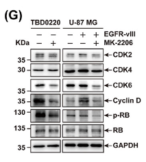
Application: WB    Species: Human    Sample: TBD0220, U-87 MG, and U-87 MG-EGFR-vIII cells

FIGURE 3 EGFR/AKT pathway regulates mitochondrial respiration and proliferation in GBM cells. (A-B) TBD0220 (A), and U-87 MG-EGFR-vIII cells (B) were treated with DMSO or 5 μmol/L MK-2206 for 24 h. The mitochondrial functions were monitored by Seahorse XF Cell Mito Stress test. The OCR, basal respiration, proton leak, and ATP production rates were measured as illustrated. ∗P < 0.05, ∗∗P < 0.01, ∗∗∗P < 0.001 (independent-sample Student's t-test for TBD0220; one-way ANOVA for U-87 MG). (C) ATP levels in TBD0220, U-87 MG, and U-87 MG-EGFR-vIII cells were analyzed after 24 h of treatments with DMSO or 5 μmol/L of MK-2206. ∗∗P < 0.01, ∗∗∗P < 0.001 (independent-sample Student's t-test for TBD0220; one-way ANOVA for U-87 MG). (D) The cell growth assay for TBD0220, U-87 MG, and U-87 MG-EGFR-vIII lines treated with DMSO, 5 μmol/L MK-2206, or 5 μmol/L MK-2206 plus 50 μmol/L ATP were performed. ∗∗∗P < 0.001 (two-way ANOVA). (E) The colony formation assay of TBD0220, U-87 MG, and U-87 MG-EGFR-vIII lines treated with DMSO or 1 μmol/L MK-2206. (F) Cell cycle distributions were analyzed by flow cytometry in TBD0220, U-87 MG, and U-87 MG-EGFR-vIII cells treated with DMSO or 5 μmol/L MK-2206. (G) Western blotting to show changes in expressions of CDK2, CDK4, CDK6, Cyclin D, RB, p-RB, and GAPDH in TBD0220, U-87 MG, and U-87 MG-EGFR-vIII cells treated with DMSO or MK-2206. Abbreviations: EGFR, epidermal growth factor receptor; AKT, AKT serine/threonine kinase 1; GBM, glioblastoma; ANOVA, analysis of variance; DMSO, dimethyl sulfoxide; CDK2, cyclin-dependent kinase 2; CDK4, cyclin-dependent kinase 4; CDK6, cyclin-dependent kinase 6; OCR, oxygen consumption rate.

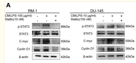
2). Gut dysbiosis promotes prostate cancer progression and docetaxel resistance via activating NF-κB-IL6-STAT3 axis. Microbiome, 2022 (PubMed: 35710492) [IF=13.8]

Application: WB    Species: Mouse    Sample: tumor tissue

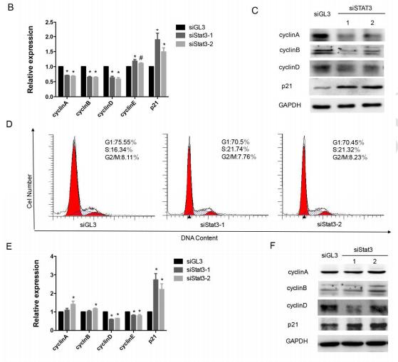
Fig. 4 The IL6-STAT3 pathway promoted prostate cancer proliferation. A Western blot of relative proteins in RM-1 and DU-145 cultured with CM or CM with Stattic for 24 h. B, C Edu (scale bar, 100 μm) and clone formation assay were conducted on RM-1 and DU-145 under condition as described. D Flowchart of the NC, Abx, and Abx+Stattic groups for in vivo study. Relevant tumor images and comparison of volume and weight for tumors in three groups (n = 5). E Immunohistochemistry of tumor tissues for p-STAT3-, c-myc-, and cyclin D1-positive cell in three groups (scale bar, 50 μm). Statistical significance was assessed by LSD in one-way ANOVA. *p < 0.05, **p < 0.01, and ***p < 0.001: compared to the NC group; #p < 0.05, ##p < 0.01, and ###p < 0.001: compared to the CM or Abx group

Application: IHC    Species: Mouse    Sample: tumor tissue

Fig. 4 The IL6-STAT3 pathway promoted prostate cancer proliferation. A Western blot of relative proteins in RM-1 and DU-145 cultured with CM or CM with Stattic for 24 h. B, C Edu (scale bar, 100 μm) and clone formation assay were conducted on RM-1 and DU-145 under condition as described. D Flowchart of the NC, Abx, and Abx+Stattic groups for in vivo study. Relevant tumor images and comparison of volume and weight for tumors in three groups (n = 5). E Immunohistochemistry of tumor tissues for p-STAT3-, c-myc-, and cyclin D1-positive cell in three groups (scale bar, 50 μm). Statistical significance was assessed by LSD in one-way ANOVA. *p < 0.05, **p < 0.01, and ***p < 0.001: compared to the NC group; #p < 0.05, ##p < 0.01, and ###p < 0.001: compared to the CM or Abx group

3). TCA-phospholipid-glycolysis targeted triple therapy effectively suppresses ATP production and tumor growth in glioblastoma. Theranostics, 2023 (PubMed: 36276638) [IF=12.4]

Application: WB    Species: Human    Sample: TBD0220 and U87MG cells

Figure 3 Reduction in ATP production hinders cell proliferation and contributes to G1/S arrest in GBM. (A-B) The relative viability of TBD0220 (A) and U87MG (B) cells was measured using a CCK-8 kit (n = 3). (C-D) Colony formation assay to detect GBM cell growth (D), and the quantification of colony numbers (C) (n = 3). (E) Cell cycle analysis using flow cytometry after incubation with different treatments like EPIC (20 µM), AA (25 µM), or EPIC (20 µM) + AA (25 µM) for 48 h, and the results are plotted as a histogram (n = 3) (F) Representative western blotting showing the expression of p21 and Rb and their downstream targets. The results were normalized to Tubulin with the control group as 1. Protein expression was quantified by ImageJ. (G) Representative confocal images of CDK6 after the treatment with AA (25 µM), EPIC (20 µM), or EPIC (20 µM) + AA (25 µM) for 48 h (n = 6). (H) The quantitative analysis of fluorescence images of CDK6 (n = 6). All data are shown as the mean values ± SD, and p values are based on one-way or two-way ANOVA. ****p < 0.0001, ***p < 0.001, **p < 0.01, *p < 0.05. Scale bar = 20 μm.

4). Precise editing of FGFR3-TACC3 fusion genes with CRISPR-Cas13a in glioblastoma. Molecular Therapy, 2021 (PubMed: 34274537) [IF=12.1]

5). Targeted p53 activation by saRNA suppresses human bladder cancer cells growth and metastasis. Journal of Experimental & Clinical Cancer Research, 2016 (PubMed: 27012825) [IF=11.3]

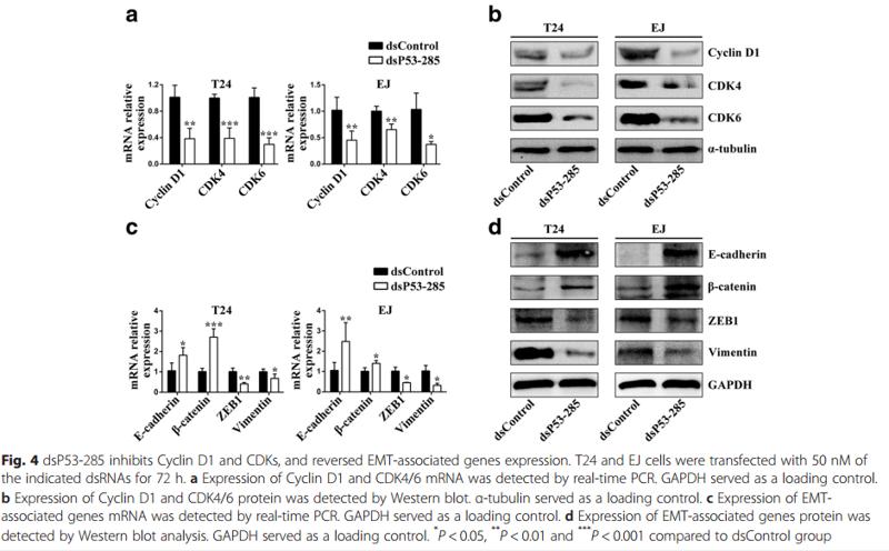
Application: WB    Species: human    Sample: T24 and EJ cells

Fig. 4 dsP53-285 inhibits Cyclin D1 and CDKs, and reversed EMT-associated genes expression. T24 and EJ cells were transfected with 50 nM of the indicated dsRNAs for 72 h. a Expression of Cyclin D1 and CDK4/6 mRNA was detected by real-time PCR. GAPDH served as a loading control. b Expression of Cyclin D1 and CDK4/6 protein was detected by Western blot. α-tubulin served as a loading control. c Expression of EMTassociated genes mRNA was detected by real-time PCR. GAPDH served as a loading control. d Expression of EMT-associated genes protein was detected by Western blot analysis. GAPDH served as a loading control. * P < 0.05, **P < 0.01 and ***P < 0.001 compared to dsControl group

6). Combination LSD1 and HOTAIR-EZH2 inhibition disrupts cell cycle processes and induces apoptosis in glioblastoma cells. PHARMACOLOGICAL RESEARCH, 2021 (PubMed: 34246782) [IF=9.1]

7). Immobilizing c(RGDfc) on the surface of metal-phenolic networks by thiol-click reaction for accelerating osteointegration of implant. Materials Today Bio, 2024 [IF=8.7]

8). Interfering with hyaluronic acid metabolism suppresses glioma cell proliferation by regulating autophagy. Cell Death & Disease, 2021 (PubMed: 33986244) [IF=8.1]

Application: WB    Species: human    Sample: U251 glioma cells

Fig. 5 |Treatments interfering with HAS3 and CD44 combined with autophagy inhibitors have a synergistic effect on the glioma cell cycle.C Relative levels of the CCNB1 and CCND1 proteins in U251 glioma cells transfected with the HAS3 siRNA or cultured with a CD44 antibody, followed by treatment with CQ (30 μmol/L) for 48 h. The data are presented as the mean ± SD; *P < 0.05, **P < 0.01,and ***P < 0.001, ****P < 0.0001.

9). ASIC1a promotes hepatic stellate cell activation through the exosomal miR-301a-3p/BTG1 pathway. International Journal of Biological Macromolecules, 2022 (PubMed: 35561854) [IF=7.7]

10). Long non-coding RNA RP11-197K6.1 as ceRNA promotes colorectal cancer progression via miR-135a-5p/DLX5 axis. Journal of translational medicine, 2024 (PubMed: 38760791) [IF=7.4]

Application: WB    Species: Human    Sample: HCT116 and SW480 cells

Fig. 2 Functional effects of lncRNA RP11-197K6.1 knockdown in CRC cells. A: Confirmation of lncRNA RP11-197K6.1 knockdown efficiency in HCT116 cells by RT-qPCR. B: Reduction in the proliferation of HCT116 and SW480 cells post-knockdown, as determined by proliferation assays. C: Increase in the apoptosis of HCT116 and SW480 cells following lncRNA RP11-197K6.1 knockdown, as measured by flow cytometry. D & E: Decrease in the migration and invasion of HCT116 and SW480 cells post-knockdown, as assessed by wound healing (scale = 200 μm) and Transwell assays (scale = 50 μm), respectively. 2 F: Changes in the levels of proteins related to the proliferation, apoptosis, and migration of HCT116 and SW480 cells after lncRNA RP11-197K6.1 knockdown, as analyzed by western blotting assay (**P 

もっと読みます

Restrictive clause


Affinity Biosciences tests all products strictly. Citations are provided as a resource for additional applications that have not been validated by Affinity Biosciences. Please choose the appropriate format for each application and consult Materials and Methods sections for additional details about the use of any product in these publications.

For Research Use Only.
Not for use in diagnostic or therapeutic procedures. Not for resale. Not for distribution without written consent. Affinity Biosciences will not be held responsible for patent infringement or other violations that may occur with the use of our products. Affinity Biosciences, Affinity Biosciences Logo and all other trademarks are the property of Affinity Biosciences LTD.