製品: NCOA4 Antibody
カタログ: DF4255
タンパク質の説明: Rabbit polyclonal antibody to NCOA4
アプリケーション: WB IHC IF/ICC
反応性: Human, Mouse, Rat
分子量: 70~90KD; 70kD(Calculated).
ユニプロット: Q13772
RRID: AB_2836606

類似製品を見る>>

   サイズ 価格 在庫状況
 100ul $280 在庫あり
 200ul $350 在庫あり

リードタイム: 当日配達

For pricing and ordering contact:
お問い合わせ先

製品説明

ソース:
Rabbit
アプリケーション:
WB 1:500-1:1000, IHC 1:50-1:200, IF/ICC 1:100-1:500
*The optimal dilutions should be determined by the end user.
*Tips:

WB: For western blot detection of denatured protein samples. IHC: For immunohistochemical detection of paraffin sections (IHC-p) or frozen sections (IHC-f) of tissue samples. IF/ICC: For immunofluorescence detection of cell samples. ELISA(peptide): For ELISA detection of antigenic peptide.

反応性:
Human,Mouse,Rat
クローナリティ:
Polyclonal
特異性:
NCOA4 Antibody detects endogenous levels of total NCOA4.
RRID:
AB_2836606
引用形式: Affinity Biosciences Cat# DF4255, RRID:AB_2836606.
コンジュゲート:
Unconjugated.
精製:
The antiserum was purified by peptide affinity chromatography using SulfoLink™ Coupling Resin (Thermo Fisher Scientific).
保存:
Rabbit IgG in phosphate buffered saline , pH 7.4, 150mM NaCl, 0.02% sodium azide and 50% glycerol. Store at -20 °C. Stable for 12 months from date of receipt.
別名:

折りたたみ/展開

70 kDa androgen receptor activator; 70 kDa androgen receptor coactivator; 70 kDa AR activator; 70 kDa AR-activator; Androgen receptor coactivator 70 kD; Androgen receptor coactivator 70 kDa protein; Androgen receptor-associated protein of 70 kDa; ARA 70; ARA70; DKFZp762E1112; ELE 1; ELE1; ELE1/ret TK; NCOA 4; NCoA-4; NCOA4; NCOA4_HUMAN; Nuclear receptor coactivator 4; Papillary thyroid carcinoma 3; PTC 3; PTC3; RET activating gene ELE1; Ret activating protein ELE1; Ret-activating protein ELE1; RFG;

免疫原

免疫原:
Uniprot:
遺伝子(ID):
発現特異性:
Q13772 NCOA4_HUMAN:

Widely expressed. Also detected in adipose tissues and in different cell lines. Isoform Beta is only expressed in testis.

タンパク質配列:
MNTFQDQSGSSSNREPLLRCSDARRDLELAIGGVLRAEQQIKDNLREVKAQIHSCISRHLECLRSREVWLYEQVDLIYQLKEETLQQQAQQLYSLLGQFNCLTHQLECTQNKDLANQVSVCLERLGSLTLKPEDSTVLLFEADTITLRQTITTFGSLKTIQIPEHLMAHASSANIGPFLEKRGCISMPEQKSASGIVAVPFSEWLLGSKPASGYQAPYIPSTDPQDWLTQKQTLENSQTSSRACNFFNNVGGNLKGLENWLLKSEKSSYQKCNSHSTTSSFSIEMEKVGDQELPDQDEMDLSDWLVTPQESHKLRKPENGSRETSEKFKLLFQSYNVNDWLVKTDSCTNCQGNQPKGVEIENLGNLKCLNDHLEAKKPLSTPSMVTEDWLVQNHQDPCKVEEVCRANEPCTSFAECVCDENCEKEALYKWLLKKEGKDKNGMPVEPKPEPEKHKDSLNMWLCPRKEVIEQTKAPKAMTPSRIADSFQVIKNSPLSEWLIRPPYKEGSPKEVPGTEDRAGKQKFKSPMNTSWCSFNTADWVLPGKKMGNLSQLSSGEDKWLLRKKAQEVLLNSPLQEEHNFPPDHYGLPAVCDLFACMQLKVDKEKWLYRTPLQM

PTMs - Q13772 基板として

Site PTM Type Enzyme
K42 Ubiquitination
K49 Ubiquitination
S57 Phosphorylation
S156 Phosphorylation
S171 Phosphorylation
S172 Phosphorylation
K181 Ubiquitination
S186 Phosphorylation
S194 Phosphorylation
K209 Ubiquitination
S212 Phosphorylation
K231 Ubiquitination
S237 Phosphorylation
S240 Phosphorylation
S241 Phosphorylation
K255 Ubiquitination
K263 Ubiquitination
K266 Ubiquitination
K271 Ubiquitination
T307 Phosphorylation
K313 Ubiquitination
K316 Ubiquitination
S334 Phosphorylation
K356 Ubiquitination
K367 Ubiquitination
K377 Ubiquitination
S412 Phosphorylation
K424 Ubiquitination
K429 Ubiquitination
K439 Ubiquitination
K447 Ubiquitination
K454 Ubiquitination
S456 Phosphorylation
K465 Ubiquitination
K472 Ubiquitination
K475 Ubiquitination
K490 Ubiquitination
S492 Phosphorylation
K504 Ubiquitination
S507 Phosphorylation
K509 Ubiquitination
K544 Ubiquitination
K545 Ubiquitination
S553 Phosphorylation
S554 Phosphorylation
K558 Ubiquitination

研究背景

機能:

Enhances the androgen receptor transcriptional activity in prostate cancer cells. Ligand-independent coactivator of the peroxisome proliferator-activated receptor (PPAR) gamma.

組織特異性:

Widely expressed. Also detected in adipose tissues and in different cell lines. Isoform Beta is only expressed in testis.

サブユニット構造:

Interacts with the androgen receptor and the retinoid X receptor (RXR) in a ligand-dependent manner.

研究領域

· Cellular Processes > Cell growth and death > Ferroptosis.   (View pathway)

· Human Diseases > Cancers: Overview > Pathways in cancer.   (View pathway)

· Human Diseases > Cancers: Specific types > Thyroid cancer.   (View pathway)

参考文献

1). Arsenic induces pancreatic dysfunction and ferroptosis via mitochondrial ROS-autophagy-lysosomal pathway. JOURNAL OF HAZARDOUS MATERIALS, 2020 (PubMed: 31735470) [IF=12.2]

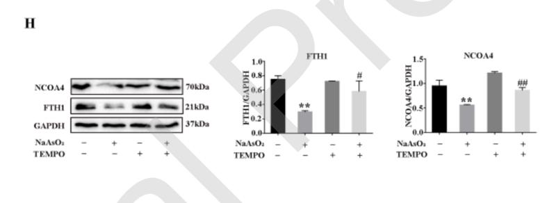
Application: WB    Species: Rat    Sample: MIN6 cells

Fig. 6 Ferroptosis was triggered by MtROS-dependent autophagy. (A) Effects of NaAsO2 (4  and TEMPO on LC3 and p62 in MIN6 cells. (B) Autophagic flux analysis. (C) The efficiency of CQ, and its effect on GPX4 and COX-2 in NaAsO2- Journal Pre-proof treated MIN6 cells. (D) Effects of NaAsO2 and CQ on relative GSH, T-SOD, and MDA content in MIN6 cells. (E) Effects of CQ on the NaAsO2-induced accumulation of lipid ROS by BODIPY 581/591 C11 staining (scale bar = 50 m). (F) Effects of CQ on the NaAsO2-induced insulin released using an ELISA kit. All results are expressed as the mean ± SD, n = 3. *P < 0.05, **P < 0.001 vs. control group, #P < 0.05, ##P < 0.001 vs. NaAsO2 group.

2). Induction of autophagy via the ROS-dependent AMPK-mTOR pathway protects copper-induced spermatogenesis disorder. Redox Biology, 2022 (PubMed: 34979450) [IF=10.7]

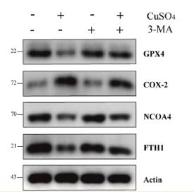
Application: WB    Species: Mouse    Sample: GC-1 spg cells

Fig. 8 Autophagy triggered ferroptosis in CuSO4-treated GC-1 spg cells. (a) Cells were treated with CuSO4 (0, 0.4, 0.8 and 1.6 mM) for 12h and 24h. The Western blot results of GPX4 and COX-2 protein expression. (b) The quantification of GPX4 and COX-2. (c) Cells were treated with CuSO4 (1.6 mM, 24h) in the presence/absence of 3-MA (1 mM, 1h), the changes of GPX4, COX-2, NCOA4 and FTH1. (d) Cells transfected with control shRNA and ATG5 shRNA exposed with CuSO4 (1.6 mM) for 24 h, changes of GPX4, COX-2, NCOA4 and FTH1. Data are presented with the means ± standard deviation. *p < 0.05 **p < 0.01.

3). ZnO NPs induce miR-342-5p mediated ferroptosis of spermatocytes through the NF-κB pathway in mice. JOURNAL OF NANOBIOTECHNOLOGY, 2024 [IF=10.2]

Application: WB    Species: Mouse    Sample: GC-2 cells

Fig. 6 ZnO NPs induce the ferroptosis of GC-2 cells. (A) Intracellular chelatable iron in GC-2 cells treated with or without ZnO NPs stained with PGSK (green). Statistical analysis of MFI of PGSK was shown. (B) Representative FACS data for lipid peroxidation level in GC-2 cells following ZnO NPs treatment using C11 BODIPY. Statistical analysis of MFI of the ratio of green/red was shown. (C) qRT-PCR analysis of ferroptosis-related gene expression in GC-2 cells after ZnO NPs treatment. (D) Western blot of ferroptosis-related protein levels in GC-2 cells treated with ZnO NPs. Statistical analysis of mean grey values ratios of the corresponding proteins/β-actin was shown, the same as below. (E) The cell viability of GC-2 cells following ZnO NPs treatment with or without Fer-1 (3.5 µM). (F) Intracellular chelatable iron in GC-2 cells following ZnO NPs treatment with or without Fer-1 stained with PGSK. Statistical analysis of MFI of PGSK was shown. (G) Representative FACS data of lipid peroxidation level in GC-2 cells following ZnO NPs treatment with or without Fer-1 stained with C11 BODIPY. Statistical analysis of MFI of the ratio of green/red was shown. (H and I) The levels of GSH and MDA in GC-2 cells following ZnO NPs treatment with or without Fer-1. (J) qRT-PCR analysis of ferroptosis-related gene expression in GC-2 cells following ZnO NPs treatment with or without Fer-1. (K) Western blot of ferroptosis-related protein levels in GC-2 cells following ZnO NPs treatment with or without Fer-1

4). Defective ferritinophagy and imbalanced iron metabolism in PBDE-47-triggered neuronal ferroptosis and salvage by Canolol. The Science of the total environment, 2024 (PubMed: 38750757) [IF=8.2]

5). Melatonin inhibits ferroptosis and delays age-related cataract by regulating SIRT6/p-Nrf2/GPX4 and SIRT6/NCOA4/FTH1 pathways. Biomedicine & Pharmacotherapy, 2023 (PubMed: 36463827) [IF=6.9]

6). Tiliroside disrupted iron homeostasis and induced ferroptosis via directly targeting calpain-2 in pancreatic cancer cells. Phytomedicine : international journal of phytotherapy and phytopharmacology, 2024 (PubMed: 38412575) [IF=6.7]

7). d-Borneol enhances cisplatin sensitivity via autophagy dependent EMT signaling and NCOA4-mediated ferritinophagy. PHYTOMEDICINE, 2022 (PubMed: 36030746) [IF=6.7]

8). Patulin Induces Acute Kidney Injury in Mice through Autophagy–Ferroptosis Pathway. JOURNAL OF AGRICULTURAL AND FOOD CHEMISTRY, 2022 (PubMed: 35543324) [IF=5.7]

9). YAP1 alleviates sepsis-induced acute lung injury via inhibiting ferritinophagy-mediated ferroptosis. Frontiers in Immunology, 2022 (PubMed: 35979359) [IF=5.7]

Application: WB    Species: Mice    Sample: MLE-12 cells.

Figure 3 YAP1 restrained the degradation of ferritin in lysosomes to suppress ferritinophagy. (A) Confocal images of ferritin (red) and LAMP2 (green) were shown in LPS-treated MLE-12 cells, while lysosomes were stained with LAMP2 (green). (B) Quantification measurement of lysosome fluorescence intensity. (C) Quantification measurement of ferritin fluorescence intensity. (D–G) Data showed the expression contents of LC3II/LC3I (E), NCOA4 (F), and FTH1 (G). Data are expressed as mean ± SD. n = 3; *p < 0.05, **p < 0.01, ***p < 0.001. NS: no significance, P>0.05.

10). Inhibition of Ferroptosis Ameliorates Photoreceptor Degeneration in Experimental Diabetic Mice. International journal of molecular sciences, 2023 (PubMed: 38069270) [IF=5.6]

Application: WB    Species: Mouse    Sample: 661W cells

Figure 2 HG causes changes in the expression of ferroptosis-related proteins in 661W cells. (A) Western blot analysis of ferroptosis-related protein expression levels in HG-induced 661W cells. GAPDH was used as a control. (B) Expression of GPX4 and SLC7A11 proteins was significantly downregulated in HG-stimulated 661W cells after 12, 18, and 24 h. HG induced obvious upregulation in the expression of ACSL4, FTH1, and NCOA4 in 661W cells compared with the Ctrl group. (C) Immunofluorescence staining of localization of ferroptosis-related proteins (red) and nuclear (blue) in HG-induced 661W cells after 18 h. Data are shown as mean ± SEM, n = 3 per group for Western blotting. p = not significant [ns], * p < 0.05, ** p < 0.01, *** p < 0.001 versus Ctrl group. Scale bar: 50 μm.

Application: IF/ICC    Species: Mouse    Sample: 661W cells

Figure 2 HG causes changes in the expression of ferroptosis-related proteins in 661W cells. (A) Western blot analysis of ferroptosis-related protein expression levels in HG-induced 661W cells. GAPDH was used as a control. (B) Expression of GPX4 and SLC7A11 proteins was significantly downregulated in HG-stimulated 661W cells after 12, 18, and 24 h. HG induced obvious upregulation in the expression of ACSL4, FTH1, and NCOA4 in 661W cells compared with the Ctrl group. (C) Immunofluorescence staining of localization of ferroptosis-related proteins (red) and nuclear (blue) in HG-induced 661W cells after 18 h. Data are shown as mean ± SEM, n = 3 per group for Western blotting. p = not significant [ns], * p < 0.05, ** p < 0.01, *** p < 0.001 versus Ctrl group. Scale bar: 50 μm.

もっと読みます

Restrictive clause


Affinity Biosciences tests all products strictly. Citations are provided as a resource for additional applications that have not been validated by Affinity Biosciences. Please choose the appropriate format for each application and consult Materials and Methods sections for additional details about the use of any product in these publications.

For Research Use Only.
Not for use in diagnostic or therapeutic procedures. Not for resale. Not for distribution without written consent. Affinity Biosciences will not be held responsible for patent infringement or other violations that may occur with the use of our products. Affinity Biosciences, Affinity Biosciences Logo and all other trademarks are the property of Affinity Biosciences LTD.