製品: NEK7 Antibody
カタログ: DF4467
タンパク質の説明: Rabbit polyclonal antibody to NEK7
アプリケーション: WB IHC IF/ICC
反応性: Human, Mouse, Rat
予測: Pig, Bovine, Horse, Sheep, Rabbit, Dog, Chicken, Xenopus
分子量: 35 KD; 35kD(Calculated).
ユニプロット: Q8TDX7
RRID: AB_2836822

類似製品を見る>>

   サイズ 価格 在庫状況
 100ul $280 在庫あり
 200ul $350 在庫あり

リードタイム: 当日配達

For pricing and ordering contact:
お問い合わせ先

製品説明

ソース:
Rabbit
アプリケーション:
WB 1:500-1:1000, IHC 1:50-1:200, IF/ICC 1:100-1:500
*The optimal dilutions should be determined by the end user.
*Tips:

WB: For western blot detection of denatured protein samples. IHC: For immunohistochemical detection of paraffin sections (IHC-p) or frozen sections (IHC-f) of tissue samples. IF/ICC: For immunofluorescence detection of cell samples. ELISA(peptide): For ELISA detection of antigenic peptide.

反応性:
Human,Mouse,Rat
予測:
Pig(100%), Bovine(100%), Horse(100%), Sheep(100%), Rabbit(100%), Dog(100%), Chicken(100%), Xenopus(100%)
クローナリティ:
Polyclonal
特異性:
NEK7 Antibody detects endogenous levels of total NEK7.
RRID:
AB_2836822
引用形式: Affinity Biosciences Cat# DF4467, RRID:AB_2836822.
コンジュゲート:
Unconjugated.
精製:
The antiserum was purified by peptide affinity chromatography using SulfoLink™ Coupling Resin (Thermo Fisher Scientific).
保存:
Rabbit IgG in phosphate buffered saline , pH 7.4, 150mM NaCl, 0.02% sodium azide and 50% glycerol. Store at -20 °C. Stable for 12 months from date of receipt.
別名:

折りたたみ/展開

EC 2.7.11.1; NEK7; NEK7_HUMAN; Never in mitosis A-related kinase 7; Never in mitosis gene a-related kinase 7; NIMA (never in mitosis gene a) related kinase 7; NIMA (never in mitosis gene a)-related expressed kinase 7; NimA related protein kinase 7; NIMA-related kinase 7; NimA-related protein kinase 7; Serine/threonine-protein kinase Nek7;

免疫原

免疫原:
Uniprot:
遺伝子(ID):
発現特異性:
Q8TDX7 NEK7_HUMAN:

Highly expressed in lung, muscle, testis, brain, heart, liver, leukocyte and spleen. Lower expression in ovary, prostate and kidney. No expression seen in small intestine.

タンパク質配列:
MDEQSQGMQGPPVPQFQPQKALRPDMGYNTLANFRIEKKIGRGQFSEVYRAACLLDGVPVALKKVQIFDLMDAKARADCIKEIDLLKQLNHPNVIKYYASFIEDNELNIVLELADAGDLSRMIKHFKKQKRLIPERTVWKYFVQLCSALEHMHSRRVMHRDIKPANVFITATGVVKLGDLGLGRFFSSKTTAAHSLVGTPYYMSPERIHENGYNFKSDIWSLGCLLYEMAALQSPFYGDKMNLYSLCKKIEQCDYPPLPSDHYSEELRQLVNMCINPDPEKRPDVTYVYDVAKRMHACTASS

種類予測

種類予測:

Score>80(red) has high confidence and is suggested to be used for WB detection. *The prediction model is mainly based on the alignment of immunogen sequences, the results are for reference only, not as the basis of quality assurance.

Species
Results
Score
Pig
100
Horse
100
Bovine
100
Sheep
100
Dog
100
Xenopus
100
Chicken
100
Rabbit
100
Zebrafish
78
Model Confidence:
High(score>80) Medium(80>score>50) Low(score<50) No confidence

PTMs - Q8TDX7 基板として

Site PTM Type Enzyme
M1 Acetylation
S5 Phosphorylation
K20 Ubiquitination
T30 Phosphorylation
K63 Ubiquitination
K64 Ubiquitination
K74 Ubiquitination
K81 Ubiquitination
K87 Ubiquitination
Y97 Phosphorylation
S147 Phosphorylation
K163 Ubiquitination
T170 Phosphorylation
T172 Phosphorylation
K176 Ubiquitination
S187 Phosphorylation
S188 Phosphorylation
K189 Ubiquitination
T190 Phosphorylation
T191 Phosphorylation
S195 Phosphorylation Q8TD19 (NEK9)
T199 Phosphorylation
Y201 Phosphorylation
Y202 Phosphorylation
S204 Phosphorylation
Y244 Phosphorylation
S245 Phosphorylation
K248 Ubiquitination
K249 Ubiquitination
K281 Ubiquitination
T286 Phosphorylation
Y287 Phosphorylation
Y289 Phosphorylation
K293 Ubiquitination

PTMs - Q8TDX7 酵素として

Substrate Site Source
P23443 (RPS6KB1) T412 Uniprot
P54274 (TERF1) S114 Uniprot

研究背景

機能:

Protein kinase which plays an important role in mitotic cell cycle progression. Required for microtubule nucleation activity of the centrosome, robust mitotic spindle formation and cytokinesis. Phosphorylates RPS6KB1.

PTMs:

Phosphorylation at Ser-195 required for its activation.

細胞の位置付け:

Nucleus. Cytoplasm. Cytoplasm>Cytoskeleton>Spindle pole. Cytoplasm>Cytoskeleton>Microtubule organizing center>Centrosome.
Note: Present at centrosome throughout the cell cycle (PubMed:17586473). Also detected at spindle midzone of the anaphase cells and eventually concentrates at the midbody (PubMed:17586473). Interaction with ANKS3 prevents its translocation to the nucleus (By similarity).

Extracellular region or secreted Cytosol Plasma membrane Cytoskeleton Lysosome Endosome Peroxisome ER Golgi apparatus Nucleus Mitochondrion Manual annotation Automatic computational assertionSubcellular location
組織特異性:

Highly expressed in lung, muscle, testis, brain, heart, liver, leukocyte and spleen. Lower expression in ovary, prostate and kidney. No expression seen in small intestine.

サブユニット構造:

Monomer. Interacts with NEK9. Interacts with ANKS3; this interaction alters the subcellular distribution of NEK7 by preventing its nuclear translocation.

タンパク質ファミリー:

Displays an autoinhibited conformation: Tyr-97 side chain points into the active site, interacts with the activation loop, and blocks the alphaC helix. The autoinhibitory conformation is released upon binding with NEK9.

The NTE (N-terminal extension) motif is a structural component of the catalytic domain and thus contributes to activity.

Belongs to the protein kinase superfamily. NEK Ser/Thr protein kinase family. NIMA subfamily.

研究領域

· Organismal Systems > Immune system > NOD-like receptor signaling pathway.   (View pathway)

参考文献

1). Quercetin-induced pyroptosis in colon cancer through NEK7-mediated NLRP3 inflammasome-GSDMD signaling pathway activation. American journal of cancer research, 2024 (PubMed: 38590424) [IF=5.3]

Application: WB    Species: Human    Sample: colon cancer tissues

Figure 7 Downregulated expression of NEK7 in human colon cancer tissues and correlated with NLRP3 expression. A. Gene correlation between NLRP3 and NEK7 in colon cancer investigated using the TIMER2.0 database based on Spearman’s correlation analysis. Rho represents the correlation index. B. Influence of NEK7 knockdown on NLRP3 protein expression determined using western blotting (n=3, where “n” represents the number of independent repeated experiments). Values are presented as mean ± SD. *P

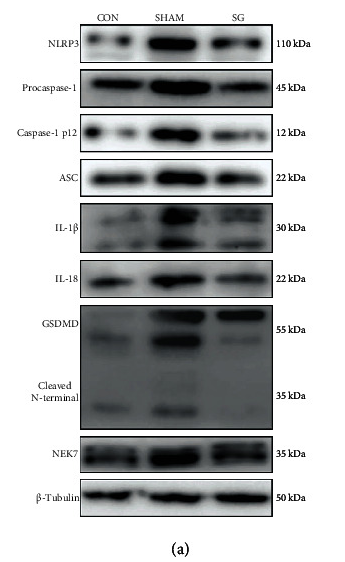
2). Attenuation of ROS/Chloride Efflux-Mediated NLRP3 Inflammasome Activation Contributes to Alleviation of Diabetic Cardiomyopathy in Rats after Sleeve Gastrectomy. Oxidative Medicine and Cellular Longevity, 2022 (PubMed: 35498125)

Application: WB    Species: Rat    Sample: diabetic myocardium

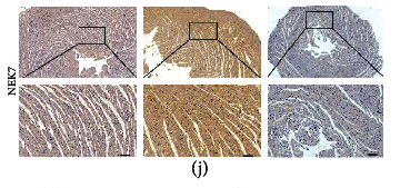
"Figure 4 SG attenuates the activation of NLRP3 in diabetic myocardium. (a) Protein levels of NLRP3-related molecules shown by western blot. β-Tubulin was an internal reference control. n = 3 in each group. (b) Relative mRNA levels of Nlrp3-relaterd genes shown by real-time RT-PCR. Tubb3 served as a reference gene. n = 3 in each group. (c) Ratio of caspase-1 p12/procaspase-1. n = 3 in each group. (d) Caspase-1 activity in heart tissues. n = 6 in each group. Immunohistochemical staining of (e) NLRP3, (f) ASC, (g) GSDMD, (h) IL-1β, (i) IL-18, and (j) NEK7. Scale bar, 50 μm. (k) Double-labeling immunofluorescence of NLRP3 and CD68. Scale bar, 50 μm. Data are presented as mean ± SD. ∗∗P < 0.01 CON vs. SHAM; ##P < 0.01 SG vs. SHAM. n = 3 in each group."

Application: IHC    Species: Rat    Sample: diabetic myocardium

"Figure 4 SG attenuates the activation of NLRP3 in diabetic myocardium. (a) Protein levels of NLRP3-related molecules shown by western blot. β-Tubulin was an internal reference control. n = 3 in each group. (b) Relative mRNA levels of Nlrp3-relaterd genes shown by real-time RT-PCR. Tubb3 served as a reference gene. n = 3 in each group. (c) Ratio of caspase-1 p12/procaspase-1. n = 3 in each group. (d) Caspase-1 activity in heart tissues. n = 6 in each group. Immunohistochemical staining of (e) NLRP3, (f) ASC, (g) GSDMD, (h) IL-1β, (i) IL-18, and (j) NEK7. Scale bar, 50 μm. (k) Double-labeling immunofluorescence of NLRP3 and CD68. Scale bar, 50 μm. Data are presented as mean ± SD. ∗∗P < 0.01 CON vs. SHAM; ##P < 0.01 SG vs. SHAM. n = 3 in each group."

Restrictive clause


Affinity Biosciences tests all products strictly. Citations are provided as a resource for additional applications that have not been validated by Affinity Biosciences. Please choose the appropriate format for each application and consult Materials and Methods sections for additional details about the use of any product in these publications.

For Research Use Only.
Not for use in diagnostic or therapeutic procedures. Not for resale. Not for distribution without written consent. Affinity Biosciences will not be held responsible for patent infringement or other violations that may occur with the use of our products. Affinity Biosciences, Affinity Biosciences Logo and all other trademarks are the property of Affinity Biosciences LTD.