製品: OPRM1 Antibody
カタログ: DF5045
タンパク質の説明: Rabbit polyclonal antibody to OPRM1
アプリケーション: WB IHC IF/ICC
反応性: Human, Mouse, Rat
予測: Pig, Bovine, Sheep, Rabbit, Dog, Chicken, Xenopus
分子量: 48 KD; 45kD(Calculated).
ユニプロット: P35372
RRID: AB_2837404

類似製品を見る>>

   サイズ 価格 在庫状況
 100ul $280 在庫あり
 200ul $350 在庫あり

リードタイム: 当日配達

For pricing and ordering contact:
お問い合わせ先

製品説明

ソース:
Rabbit
アプリケーション:
WB 1:500-1:1000, IHC 1:50-1:200, IF/ICC 1:100-500
*The optimal dilutions should be determined by the end user.
*Tips:

WB: For western blot detection of denatured protein samples. IHC: For immunohistochemical detection of paraffin sections (IHC-p) or frozen sections (IHC-f) of tissue samples. IF/ICC: For immunofluorescence detection of cell samples. ELISA(peptide): For ELISA detection of antigenic peptide.

反応性:
Human,Mouse,Rat
予測:
Pig(90%), Bovine(90%), Sheep(90%), Rabbit(90%), Dog(90%), Chicken(80%), Xenopus(80%)
クローナリティ:
Polyclonal
特異性:
OPRM1 Antibody detects endogenous levels of total OPRM1.
RRID:
AB_2837404
引用形式: Affinity Biosciences Cat# DF5045, RRID:AB_2837404.
コンジュゲート:
Unconjugated.
精製:
The antiserum was purified by peptide affinity chromatography using SulfoLink™ Coupling Resin (Thermo Fisher Scientific).
保存:
Rabbit IgG in phosphate buffered saline , pH 7.4, 150mM NaCl, 0.02% sodium azide and 50% glycerol. Store at -20 °C. Stable for 12 months from date of receipt.
別名:

折りたたみ/展開

hMOP; LMOR; M-OR-1; MOP; MOR; MOR-1; MOR1; Mu opiate receptor; Mu opioid receptor; Mu-type opioid receptor; Opioid receptor mu 1; OPRM; OPRM_HUMAN; OPRM1;

免疫原

免疫原:
Uniprot:
遺伝子(ID):
発現特異性:
P35372 OPRM_HUMAN:

Expressed in brain. Isoform 16 and isoform 17 are detected in brain.

タンパク質配列:
MDSSAAPTNASNCTDALAYSSCSPAPSPGSWVNLSHLDGNLSDPCGPNRTDLGGRDSLCPPTGSPSMITAITIMALYSIVCVVGLFGNFLVMYVIVRYTKMKTATNIYIFNLALADALATSTLPFQSVNYLMGTWPFGTILCKIVISIDYYNMFTSIFTLCTMSVDRYIAVCHPVKALDFRTPRNAKIINVCNWILSSAIGLPVMFMATTKYRQGSIDCTLTFSHPTWYWENLLKICVFIFAFIMPVLIITVCYGLMILRLKSVRMLSGSKEKDRNLRRITRMVLVVVAVFIVCWTPIHIYVIIKALVTIPETTFQTVSWHFCIALGYTNSCLNPVLYAFLDENFKRCFREFCIPTSSNIEQQNSTRIRQNTRDHPSTANTVDRTNHQLENLEAETAPLP

種類予測

種類予測:

Score>80(red) has high confidence and is suggested to be used for WB detection. *The prediction model is mainly based on the alignment of immunogen sequences, the results are for reference only, not as the basis of quality assurance.

Species
Results
Score
Pig
90
Bovine
90
Sheep
90
Dog
90
Rabbit
90
Xenopus
80
Chicken
80
Horse
0
Zebrafish
0
Model Confidence:
High(score>80) Medium(80>score>50) Low(score<50) No confidence

PTMs - P35372 基板として

Site PTM Type Enzyme
Y168 Phosphorylation
T182 Phosphorylation P35626 (GRK3)
S263 Phosphorylation
S268 Phosphorylation Q9UQM7 (CAMK2A)
S270 Phosphorylation Q9UQM7 (CAMK2A)
Y338 Phosphorylation
T356 Phosphorylation P25098 (GRK2)
S357 Phosphorylation P25098 (GRK2)
S358 Phosphorylation P25098 (GRK2)
S365 Phosphorylation
T372 Phosphorylation Q9UQM7 (CAMK2A)
S377 Phosphorylation P25098 (GRK2)
T378 Phosphorylation
T396 Phosphorylation

研究背景

機能:

Receptor for endogenous opioids such as beta-endorphin and endomorphin. Receptor for natural and synthetic opioids including morphine, heroin, DAMGO, fentanyl, etorphine, buprenorphin and methadone. Agonist binding to the receptor induces coupling to an inactive GDP-bound heterotrimeric G-protein complex and subsequent exchange of GDP for GTP in the G-protein alpha subunit leading to dissociation of the G-protein complex with the free GTP-bound G-protein alpha and the G-protein beta-gamma dimer activating downstream cellular effectors. The agonist- and cell type-specific activity is predominantly coupled to pertussis toxin-sensitive G(i) and G(o) G alpha proteins, GNAI1, GNAI2, GNAI3 and GNAO1 isoforms Alpha-1 and Alpha-2, and to a lesser extent to pertussis toxin-insensitive G alpha proteins GNAZ and GNA15. They mediate an array of downstream cellular responses, including inhibition of adenylate cyclase activity and both N-type and L-type calcium channels, activation of inward rectifying potassium channels, mitogen-activated protein kinase (MAPK), phospholipase C (PLC), phosphoinositide/protein kinase (PKC), phosphoinositide 3-kinase (PI3K) and regulation of NF-kappa-B. Also couples to adenylate cyclase stimulatory G alpha proteins. The selective temporal coupling to G-proteins and subsequent signaling can be regulated by RGSZ proteins, such as RGS9, RGS17 and RGS4. Phosphorylation by members of the GPRK subfamily of Ser/Thr protein kinases and association with beta-arrestins is involved in short-term receptor desensitization. Beta-arrestins associate with the GPRK-phosphorylated receptor and uncouple it from the G-protein thus terminating signal transduction. The phosphorylated receptor is internalized through endocytosis via clathrin-coated pits which involves beta-arrestins. The activation of the ERK pathway occurs either in a G-protein-dependent or a beta-arrestin-dependent manner and is regulated by agonist-specific receptor phosphorylation. Acts as a class A G-protein coupled receptor (GPCR) which dissociates from beta-arrestin at or near the plasma membrane and undergoes rapid recycling. Receptor down-regulation pathways are varying with the agonist and occur dependent or independent of G-protein coupling. Endogenous ligands induce rapid desensitization, endocytosis and recycling whereas morphine induces only low desensitization and endocytosis. Heterooligomerization with other GPCRs can modulate agonist binding, signaling and trafficking properties. Involved in neurogenesis. Isoform 12 couples to GNAS and is proposed to be involved in excitatory effects. Isoform 16 and isoform 17 do not bind agonists but may act through oligomerization with binding-competent OPRM1 isoforms and reduce their ligand binding activity.

PTMs:

Phosphorylated. Differentially phosphorylated in basal and agonist-induced conditions. Agonist-mediated phosphorylation modulates receptor internalization. Phosphorylated by GRK2 in a agonist-dependent manner. Phosphorylation at Tyr-168 requires receptor activation, is dependent on non-receptor protein tyrosine kinase Src and results in a decrease in agonist efficacy by reducing G-protein coupling efficiency. Phosphorylated on tyrosine residues; the phosphorylation is involved in agonist-induced G-protein-independent receptor down-regulation. Phosphorylation at Ser-377 is involved in G-protein-dependent but not beta-arrestin-dependent activation of the ERK pathway (By similarity).

Ubiquitinated. A basal ubiquitination seems not to be related to degradation. Ubiquitination is increased upon formation of OPRM1:OPRD1 oligomers leading to proteasomal degradation; the ubiquitination is diminished by RTP4.

細胞の位置付け:

Cell membrane>Multi-pass membrane protein. Cell projection>Axon. Perikaryon. Cell projection>Dendrite. Endosome.
Note: Is rapidly internalized after agonist binding.

Cytoplasm.

Extracellular region or secreted Cytosol Plasma membrane Cytoskeleton Lysosome Endosome Peroxisome ER Golgi apparatus Nucleus Mitochondrion Manual annotation Automatic computational assertionSubcellular location
組織特異性:

Expressed in brain. Isoform 16 and isoform 17 are detected in brain.

サブユニット構造:

Forms homooligomers and heterooligomers with other GPCRs, such as OPRD1, OPRK1, OPRL1, NPFFR2, ADRA2A, SSTR2, CNR1 and CCR5 (probably in dimeric forms). Interacts with heterotrimeric G proteins; interaction with a heterotrimeric complex containing GNAI1, GNB1 and GNG2 stabilizes the active conformation of the receptor and increases its affinity for endomorphin-2, the synthetic opioid peptide DAMGO and for morphinan agonists (By similarity). Interacts with PPL; the interaction disrupts agonist-mediated G-protein activation. Interacts (via C-terminus) with DNAJB4 (via C-terminus). Interacts with calmodulin; the interaction inhibits the constitutive activity of OPRM1; it abolishes basal and attenuates agonist-stimulated G-protein coupling. Interacts with FLNA, PLD2, RANBP9 and WLS and GPM6A. Interacts with RTP4 (By similarity). Interacts with SYP and GNAS (By similarity). Interacts with RGS9, RGS17, RGS20, RGS4, PPP1R9B and HINT1 (By similarity).

タンパク質ファミリー:

Belongs to the G-protein coupled receptor 1 family.

研究領域

· Environmental Information Processing > Signaling molecules and interaction > Neuroactive ligand-receptor interaction.

· Human Diseases > Substance dependence > Morphine addiction.

· Organismal Systems > Endocrine system > Estrogen signaling pathway.   (View pathway)

参考文献

1). CDKN2AIP is critical for spermiogenesis and germ cell development. Cell and Bioscience, 2022 (PubMed: 35989335) [IF=7.5]

Application: WB    Species: Mice    Sample:

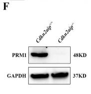
Fig. 5Disruption of the affect protamine replacement in Cdkn2aip-/- mice. A Immunofluorescence staining of histone H3 in seminiferous tubules of Cdkn2aip+/+ and Cdkn2aip−/− testes. histone H3(green) and DAPI (blue). Scale bar, 50 μm. B Immunofluorescence staining of TNP1 in seminiferous tubules of Cdkn2aip+/+ and Cdkn2aip−/− testes. TNP1 (green) and DAPI (blue). Scale bar, 50 μm. C and D Immunofluorescence staining of PRM1 and PRM2 in seminiferous tubules of Cdkn2aip+/+ and Cdkn2aip−/− testes. PRMs(green) and DAPI (blue). Scale bar, 50 μm. E qPCR analyses showing significantly reduced levels of Prm1 and Prm2 in 8-week-old Cdkn2aip+/+ and Cdkn2aip−/− testis. Data are presented as Data are presented as mean ± S.D. Student’s t test; *P < 0.05, ***P < 0.001. F Quantification of relative protein level of PRM1 and PRM2 by Western blotting in Cdkn2aip+/+ and Cdkn2aip−/− testis. GAPDH is serves as a loading control

Application: IF/ICC    Species: Mice    Sample:

Fig. 5Disruption of the affect protamine replacement in Cdkn2aip-/- mice. A Immunofluorescence staining of histone H3 in seminiferous tubules of Cdkn2aip+/+ and Cdkn2aip−/− testes. histone H3(green) and DAPI (blue). Scale bar, 50 μm. B Immunofluorescence staining of TNP1 in seminiferous tubules of Cdkn2aip+/+ and Cdkn2aip−/− testes. TNP1 (green) and DAPI (blue). Scale bar, 50 μm. C and D Immunofluorescence staining of PRM1 and PRM2 in seminiferous tubules of Cdkn2aip+/+ and Cdkn2aip−/− testes. PRMs(green) and DAPI (blue). Scale bar, 50 μm. E qPCR analyses showing significantly reduced levels of Prm1 and Prm2 in 8-week-old Cdkn2aip+/+ and Cdkn2aip−/− testis. Data are presented as Data are presented as mean ± S.D. Student’s t test; *P < 0.05, ***P < 0.001. F Quantification of relative protein level of PRM1 and PRM2 by Western blotting in Cdkn2aip+/+ and Cdkn2aip−/− testis. GAPDH is serves as a loading control

2). Agonists Specific for κ-Opioid Receptor Induces Apoptosis of HCC Cells Through Enhanced Endoplasmic Reticulum Stress. Frontiers in Oncology, 2022 (PubMed: 35433440) [IF=3.5]

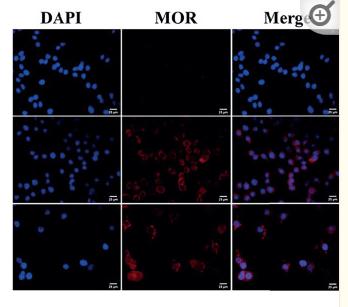
Application: IF/ICC    Species: Human    Sample: HCC tissues

Figure 1 The expression of KOR and MOR in HCC cell lines and human HCC tissues. (A) The expression of KOR and MOR were detected in LO2, Hep3B and Huh7 cells by immunofluorescent staining, scale bar, 25um. The expression of KOR and MOR was evaluated in 4 types of HCC cell lines (HepG2, Bel-7402, Hep3B and Huh-7) by RT-qPCR (B) and western blotting (C). (D) In human HCC tissues, KOR and MOR were detected by western blotting, GADPH was used as an internal control. N, Non-tumor; T, Tumor. Values are presented as the mean ± standard deviation (n = 3). KOR, kappa opioid receptor; MOR, mu opioid receptor; GADPH, glyceraldehyde-3-phosphate dehydrogenase.

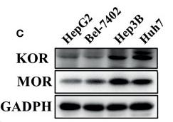
Application: WB    Species: Human    Sample: HCC tissues

Figure 1 The expression of KOR and MOR in HCC cell lines and human HCC tissues. (A) The expression of KOR and MOR were detected in LO2, Hep3B and Huh7 cells by immunofluorescent staining, scale bar, 25um. The expression of KOR and MOR was evaluated in 4 types of HCC cell lines (HepG2, Bel-7402, Hep3B and Huh-7) by RT-qPCR (B) and western blotting (C). (D) In human HCC tissues, KOR and MOR were detected by western blotting, GADPH was used as an internal control. N, Non-tumor; T, Tumor. Values are presented as the mean ± standard deviation (n = 3). KOR, kappa opioid receptor; MOR, mu opioid receptor; GADPH, glyceraldehyde-3-phosphate dehydrogenase.

3). Co-Administration of nalbuphine to improve morphine tolerance in mice with bone cancer pain. Molecular Pain, 2023 (PubMed: 37226458) [IF=3.3]

Application: WB    Species: Mouse    Sample:

Figure 5. Changes of spinal MOR and KOR protein expression over time in tumor-implanted mice and sham-implanted mice. Western blot for β-actin, MOR, and KOR resulted in products of 42, 45, and 43 kDa, as expected (markers show predicted band sizes) (a and b). Densitometric quantification of β-actin, MOR, and KOR immunoreactivity on Western blots (c and d). Data were presented as fold change of control (Naive) mean ± SD.*p < 0.05 vs. sham-implanted mice. (n = 4 per group). (Interaction: F-statistics = 15.92/17.71; Row Factor: F-statistics = 43.83/25.90; Column Factor: F-statistics = 115.70/113.90, MOR protein/KOR protein respectively).

Restrictive clause


Affinity Biosciences tests all products strictly. Citations are provided as a resource for additional applications that have not been validated by Affinity Biosciences. Please choose the appropriate format for each application and consult Materials and Methods sections for additional details about the use of any product in these publications.

For Research Use Only.
Not for use in diagnostic or therapeutic procedures. Not for resale. Not for distribution without written consent. Affinity Biosciences will not be held responsible for patent infringement or other violations that may occur with the use of our products. Affinity Biosciences, Affinity Biosciences Logo and all other trademarks are the property of Affinity Biosciences LTD.