製品: Bcl-2 Antibody
カタログ: BF9103
タンパク質の説明: Mouse monoclonal antibody to Bcl-2
アプリケーション: WB IHC IF/ICC ELISA
反応性: Human, Mouse, Rat
分子量: 26 kd; 26kD(Calculated).
ユニプロット: P10415
RRID: AB_2837570

類似製品を見る>>

   サイズ 価格 在庫状況
 100ul $280 在庫あり
 200ul $350 在庫あり

リードタイム: 当日配達

For pricing and ordering contact:
お問い合わせ先

製品説明

ソース:
Mouse
アプリケーション:
WB 1:1000, IHC 1:50-1:200, IF/ICC 1:100-1:500
*The optimal dilutions should be determined by the end user.
*Tips:

WB: For western blot detection of denatured protein samples. IHC: For immunohistochemical detection of paraffin sections (IHC-p) or frozen sections (IHC-f) of tissue samples. IF/ICC: For immunofluorescence detection of cell samples. ELISA(peptide): For ELISA detection of antigenic peptide.

反応性:
Human,Mouse,Rat
クローナリティ:
Monoclonal [AFfirm063]
特異性:
The Bcl-2 mouse monoclonal antibody can detect endogenous Bcl-2 proteins.
RRID:
AB_2837570
引用形式: Affinity Biosciences Cat# BF9103, RRID:AB_2837570.
コンジュゲート:
Unconjugated.
精製:
affinity purification.
保存:
Store at -20°C. Stable for one year from the date of shipment.1mg/ml in PBS, pH 7.4, containing 0.02% sodium azide and 50% glycerol. Store at -20 °C. Stable for 12 months from date of receipt.
別名:

折りたたみ/展開

Apoptosis regulator Bcl 2; Apoptosis regulator Bcl-2; Apoptosis regulator Bcl2; AW986256; B cell CLL/lymphoma 2; B cell leukemia/lymphoma 2; Bcl-2; Bcl2; BCL2_HUMAN; C430015F12Rik; D630044D05Rik; D830018M01Rik; Leukemia/lymphoma, B-cell, 2; Oncogene B-cell leukemia 2; PPP1R50; Protein phosphatase 1, regulatory subunit 50;

免疫原

免疫原:

Mouse monoclonal antibody is prepared by immunizing synthetic peptide coupled to KLH.

Uniprot:
遺伝子(ID):
発現特異性:
P10415 BCL2_HUMAN:

Expressed in a variety of tissues.

タンパク質の説明:
BCL2 is an integral outer mitochondrial membrane protein that blocks the apoptotic death of some cells such as lymphocytes. Constitutive expression of BCL2, such as in the case of translocation of BCL2 to Ig heavy chain locus, is thought to be the cause of follicular lymphoma. BCL2 suppresses apoptosis in a variety of cell systems including factor-dependent lymphohematopoietic and neural cells. It regulates cell death by controlling the mitochondrial membrane permeability.
タンパク質配列:
MAHAGRTGYDNREIVMKYIHYKLSQRGYEWDAGDVGAAPPGAAPAPGIFSSQPGHTPHPAASRDPVARTSPLQTPAAPGAAAGPALSPVPPVVHLTLRQAGDDFSRRYRRDFAEMSSQLHLTPFTARGRFATVVEELFRDGVNWGRIVAFFEFGGVMCVESVNREMSPLVDNIALWMTEYLNRHLHTWIQDNGGWDAFVELYGPSMRPLFDFSWLSLKTLLSLALVGACITLGAYLGHK

PTMs - P10415 基板として

Site PTM Type Enzyme
Y9 Phosphorylation
K22 Ubiquitination
S24 Phosphorylation
T56 Phosphorylation Q16539 (MAPK14) , P06493 (CDK1) , P53779 (MAPK10) , P28482 (MAPK1) , P27361 (MAPK3)
T69 Phosphorylation P45983 (MAPK8)
S70 Phosphorylation P27361 (MAPK3) , P06493 (CDK1) , P53779 (MAPK10) , P17252 (PRKCA) , Q00534 (CDK6) , P28482 (MAPK1) , P45983 (MAPK8)
T74 Phosphorylation P28482 (MAPK1) , P53779 (MAPK10) , P27361 (MAPK3)
S87 Phosphorylation Q16539 (MAPK14) , P45983 (MAPK8) , Q00534 (CDK6) , P27361 (MAPK3) , P28482 (MAPK1) , P53779 (MAPK10)
C158 S-Nitrosylation
C229 S-Nitrosylation
Y235 Phosphorylation

研究背景

機能:

Suppresses apoptosis in a variety of cell systems including factor-dependent lymphohematopoietic and neural cells. Regulates cell death by controlling the mitochondrial membrane permeability. Appears to function in a feedback loop system with caspases. Inhibits caspase activity either by preventing the release of cytochrome c from the mitochondria and/or by binding to the apoptosis-activating factor (APAF-1). May attenuate inflammation by impairing NLRP1-inflammasome activation, hence CASP1 activation and IL1B release.

PTMs:

Phosphorylation/dephosphorylation on Ser-70 regulates anti-apoptotic activity. Growth factor-stimulated phosphorylation on Ser-70 by PKC is required for the anti-apoptosis activity and occurs during the G2/M phase of the cell cycle. In the absence of growth factors, BCL2 appears to be phosphorylated by other protein kinases such as ERKs and stress-activated kinases. Phosphorylated by MAPK8/JNK1 at Thr-69, Ser-70 and Ser-87, wich stimulates starvation-induced autophagy. Dephosphorylated by protein phosphatase 2A (PP2A) (By similarity).

Proteolytically cleaved by caspases during apoptosis. The cleaved protein, lacking the BH4 motif, has pro-apoptotic activity, causes the release of cytochrome c into the cytosol promoting further caspase activity.

Monoubiquitinated by PRKN, leading to increase its stability. Ubiquitinated by SCF(FBXO10), leading to its degradation by the proteasome.

細胞の位置付け:

Mitochondrion outer membrane>Single-pass membrane protein. Nucleus membrane>Single-pass membrane protein. Endoplasmic reticulum membrane>Single-pass membrane protein.

Extracellular region or secreted Cytosol Plasma membrane Cytoskeleton Lysosome Endosome Peroxisome ER Golgi apparatus Nucleus Mitochondrion Manual annotation Automatic computational assertionSubcellular location
組織特異性:

Expressed in a variety of tissues.

サブユニット構造:

Forms homodimers, and heterodimers with BAX, BAD, BAK and Bcl-X(L). Heterodimerization with BAX requires intact BH1 and BH2 motifs, and is necessary for anti-apoptotic activity. Interacts with EI24 (By similarity). Also interacts with APAF1, BBC3, BCL2L1, BNIPL, MRPL41 and TP53BP2. Binding to FKBP8 seems to target BCL2 to the mitochondria and probably interferes with the binding of BCL2 to its targets. Interacts with BAG1 in an ATP-dependent manner. Interacts with RAF1 (the 'Ser-338' and 'Ser-339' phosphorylated form). Interacts (via the BH4 domain) with EGLN3; the interaction prevents the formation of the BAX-BCL2 complex and inhibits the anti-apoptotic activity of BCL2. Interacts with G0S2; this interaction also prevents the formation of the anti-apoptotic BAX-BCL2 complex. Interacts with RTL10/BOP. Interacts with the SCF(FBXO10) complex. Interacts (via the loop between motifs BH4 and BH3) with NLRP1 (via LRR repeats), but not with NLRP2, NLRP3, NLRP4, PYCARD, nor MEFV. Interacts with GIMAP3/IAN4, GIMAP4/IAN1 and GIMAP5/IAN5 (By similarity).

タンパク質ファミリー:

BH1 and BH2 domains are required for the interaction with BAX and for anti-apoptotic activity.

The BH4 motif is required for anti-apoptotic activity and for interaction with RAF1 and EGLN3.

The loop between motifs BH4 and BH3 is required for the interaction with NLRP1.

Belongs to the Bcl-2 family.

研究領域

· Cellular Processes > Transport and catabolism > Autophagy - animal.   (View pathway)

· Cellular Processes > Cell growth and death > Apoptosis.   (View pathway)

· Cellular Processes > Cell growth and death > Apoptosis - multiple species.   (View pathway)

· Cellular Processes > Cell growth and death > Necroptosis.   (View pathway)

· Cellular Processes > Cellular community - eukaryotes > Focal adhesion.   (View pathway)

· Environmental Information Processing > Signal transduction > NF-kappa B signaling pathway.   (View pathway)

· Environmental Information Processing > Signal transduction > HIF-1 signaling pathway.   (View pathway)

· Environmental Information Processing > Signal transduction > Sphingolipid signaling pathway.   (View pathway)

· Environmental Information Processing > Signal transduction > PI3K-Akt signaling pathway.   (View pathway)

· Environmental Information Processing > Signal transduction > Hedgehog signaling pathway.   (View pathway)

· Environmental Information Processing > Signal transduction > Jak-STAT signaling pathway.   (View pathway)

· Genetic Information Processing > Folding, sorting and degradation > Protein processing in endoplasmic reticulum.   (View pathway)

· Human Diseases > Drug resistance: Antineoplastic > EGFR tyrosine kinase inhibitor resistance.

· Human Diseases > Drug resistance: Antineoplastic > Endocrine resistance.

· Human Diseases > Drug resistance: Antineoplastic > Platinum drug resistance.

· Human Diseases > Neurodegenerative diseases > Amyotrophic lateral sclerosis (ALS).

· Human Diseases > Infectious diseases: Parasitic > Toxoplasmosis.

· Human Diseases > Infectious diseases: Bacterial > Tuberculosis.

· Human Diseases > Infectious diseases: Viral > Hepatitis B.

· Human Diseases > Infectious diseases: Viral > Epstein-Barr virus infection.

· Human Diseases > Cancers: Overview > Pathways in cancer.   (View pathway)

· Human Diseases > Cancers: Overview > MicroRNAs in cancer.

· Human Diseases > Cancers: Specific types > Colorectal cancer.   (View pathway)

· Human Diseases > Cancers: Specific types > Prostate cancer.   (View pathway)

· Human Diseases > Cancers: Specific types > Small cell lung cancer.   (View pathway)

· Human Diseases > Cancers: Specific types > Gastric cancer.   (View pathway)

· Organismal Systems > Circulatory system > Adrenergic signaling in cardiomyocytes.   (View pathway)

· Organismal Systems > Immune system > NOD-like receptor signaling pathway.   (View pathway)

· Organismal Systems > Nervous system > Neurotrophin signaling pathway.   (View pathway)

· Organismal Systems > Nervous system > Cholinergic synapse.

· Organismal Systems > Endocrine system > Estrogen signaling pathway.   (View pathway)

参考文献

1). Passively-targeted mitochondrial tungsten-based nanodots for efficient acute kidney injury treatment. Bioactive materials, 2023 (PubMed: 36185743) [IF=18.9]

Application: WB    Species: Mouse    Sample:

Fig. 4 TWNDs reduced mitochondrial-dependent apoptosis. (A–B) TEM image of mitochondria of tubular cells in normal mouse (A) and AKI mouse (B). Scale bar: 1 μm. (C) Schematic illustration of TWNDs passing through the glomerulus to the tubular site. (D) TEM image of mitochondria of tubule cells in AKI mouse after injection of TWNDs and the magnified image. Scale bar: 1 μm. (E) Schematic illustration of ROS induced mitochondrial-dependent cell apoptosis. (F) WB analysis of BAX, BCL-2, Cyt c, KIM-1 and NGAL proteins in renal tissue homogenate from each group. (G) Quantification of the protein immunoblots of BAX, BCL-2, Cyt c, KIM-1 and NGAL. (H) MitoSOX staining (red fluorescence), DAPI (blue fluorescence) staining, and their merge images of HK-2 cells from each group. Scale bar: 10 μm. (I) Quantification of MitoSOX-positive cells in (H). (J) TUNEL staining (green fluorescence), DAPI (blue fluorescence) staining, and their merge images of kidney tissues from each group. Scale bar: 100 μm. (K) Quantification of TUNEL-positive cells in (J). (L) Immunohistochemical staining of Cyt c from each group. Scale bar: 20 μm. (M) Quantification of Cyt-c-positive rate in (L). Data represent means ± S.D. from at least three independent replicates. (*P < 0.05, **P < 0.01, ***P < 0.001 vs AKI or H2O2 group; ##P < 0.01, ###P < 0.001vs sham or control group).

2). Oral Metal-Free Melanin Nanozymes for Natural and Durable Targeted Treatment of Inflammatory Bowel Disease (IBD). Small (Weinheim an der Bergstrasse, Germany), 2023 (PubMed: 36760016) [IF=13.0]

3). Aberrant activation of p53-TRIB3 axis contributes to diabetic myocardial insulin resistance and sulforaphane protection. Journal of advanced research, 2024 (PubMed: 39069209) [IF=11.4]

4). Protective mechanisms of 10-gingerol against myocardial ischemia may involve activation of JAK2/STAT3 pathway and regulation of Ca2+ homeostasis. BIOMEDICINE & PHARMACOTHERAPY, 2022 (PubMed: 35569350) [IF=6.9]

Application: WB    Species: Rat    Sample: myocardial tissues

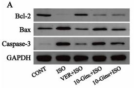
Fig. 6. Effects of 10-Gin on the Bax, Bcl-2, Caspase-3 protein expressions in myocardial tissues (A). The band gray values of Bcl-2 (B), Bax (C), and Caspase-3 (D) were analyzed by normalization to β-actin. Data are represented as mean ± SEM. * P < 0.05, ** P < 0.01 vs. CONT; # P < 0.05, ## P < 0.01 vs. ISO group, n = 5.

5). A snake cathelicidin enhances transcription factor EB-mediated autophagy and alleviates ROS-induced pyroptosis after ischaemia-reperfusion injury of island skin flaps. British journal of pharmacology, 2024 (PubMed: 37850255) [IF=6.8]

6). Celastrol upregulated ATG7 triggers autophagy via targeting Nur77 in colorectal cancer. Phytomedicine, 2022 (PubMed: 35752079) [IF=6.7]

7). Ginkgolide A targets forkhead box O1 to protect against lipopolysaccharide‐induced septic cardiomyopathy. Phytotherapy Research, 2023 (PubMed: 36932920) [IF=6.1]

8). Epigallocatechin-3-gallate Mo nanoparticles (EGM NPs) efficiently treat liver injury by strongly reducing oxidative stress, inflammation and endoplasmic reticulum stress. Frontiers in pharmacology, 2022 (PubMed: 36278211) [IF=5.6]

Application: WB    Species: Human    Sample: L02 cells

FIGURE 2 Antioxidant properties and cellular protection of EGM NPs. (A) Detection of ROS generation by DCFH-DA in L02 cells after different treatments. Scale bar: 20 μm. (B) H2O2 concentrations generated by L02 cells after different treatments. (C) Cell viability of L02 cells after different treatments. Data represent means ± S.D. from six independent replicates. (D) Fluorescence images of MitoSOX staining in L02 cells after different treatments. Scale bar: 20 μm. (E) Western blot analysis of p-JNK, JNK, BAX and BCL-2 proteins expression in L02 cells after different treatments. (F–H) Quantification of the protein immunoblots of p-JNK/JNK (F), BAX (G) and BCL-2 (H). Data represent means ± S.D. from three independent replicates. (** p < 0.01vs. APAP group; ## p < 0.01 vs. Control group).

9). Chang qing formula ameliorates colitis-associated colorectal cancer via suppressing IL-17/NF-κB/STAT3 pathway in mice as revealed by network pharmacology study. Frontiers in Pharmacology, 2022 (PubMed: 35991881) [IF=5.6]

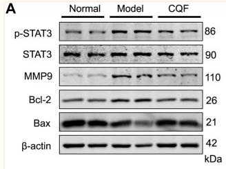
Application: WB    Species: Mice    Sample: colon tissue

FIGURE 7 CQF suppressed the phosphorylation of STAT3 and altered the expression of its downstream proteins. (A,B) Expression of MMP9, p-STAT3, STAT3, Bcl-2, and Bax proteins in colon tissue (n = 4 per group). (C) Immunofluorescence staining of colon tissue, green: p-STAT3, red: MMP9, blue: DAPI. Values are expressed as mean ± SEM. # p < 0.05, ## p < 0.01, compared with normal mice; * p < 0.05, ** p < 0.01, compared with AOM/DSS-treated mice.

10). Activation of STING Pathway Contributed to Cisplatin-Induced Cardiac Dysfunction via Promoting the Activation of TNF-α-AP-1 Signal Pathway. Frontiers in Pharmacology, 2021 (PubMed: 34483919) [IF=5.6]

Application: WB    Species: Mice    Sample: HL-1 cells

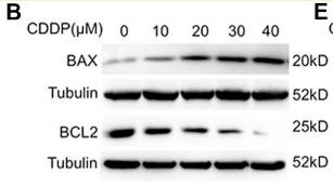
FIGURE 1 Activation of the cGAS-STING pathway was detected in cisplatin-induced HL-1 cells (A) HL-1 cells were incubated with CDDP (10, 20, 30, 40 μM) for 24 h and then the cell viability of HL-1 cells was examined by CCK8 analysis (n = 5 independent experiments; ### p < 0.001, vs. the 0 group). (B) HL-1 cells were incubated with CDDP (10, 20, 30, 40 μM) for 24 h, and then total protein was collected. The protein level of BAX and BCL2 in HL-1 cells was detected by Western blot. (C–D) HL-1 cells were incubated with CDDP (10, 20, 30, 40 μM) for 24 h, and then cell apoptosis induced by CDDP was detected by TUNEL staining. (C) Representative images of TUNEL staining in HL-1 cells (Green: TUNEL positive cell, DAPI: nucleus; Scale Bar: 25 μm, ×400 magnification). (D) Quantification of TUNEL-positive cells (E) HL-1 cells were incubated with CDDP (10, 20, 30, 40 μM) for 1 h, and total protein was collected. The protein level of p-STING, STING, p-TBK1, TBK1 in HL-1 cells was detected by Western blot (n = 3 independent experiments; # p < 0.05, ## p < 0.01, ### p < 0.001, vs. Vehicle group).

もっと読みます

Restrictive clause


Affinity Biosciences tests all products strictly. Citations are provided as a resource for additional applications that have not been validated by Affinity Biosciences. Please choose the appropriate format for each application and consult Materials and Methods sections for additional details about the use of any product in these publications.

For Research Use Only.
Not for use in diagnostic or therapeutic procedures. Not for resale. Not for distribution without written consent. Affinity Biosciences will not be held responsible for patent infringement or other violations that may occur with the use of our products. Affinity Biosciences, Affinity Biosciences Logo and all other trademarks are the property of Affinity Biosciences LTD.