製品: CD8 Antibody
カタログ: AF5126
タンパク質の説明: Rabbit polyclonal antibody to CD8
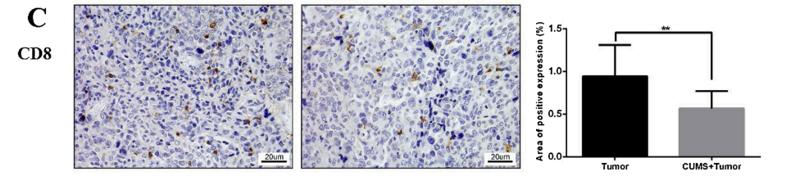
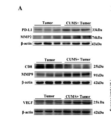
アプリケーション: WB IHC
Cited expt.: WB, IHC
反応性: Human, Mouse, Rat
分子量: 25 kDa; 26kD(Calculated).
ユニプロット: P01732
RRID: AB_2837612

類似製品を見る>>

   サイズ 価格 在庫状況
 100ul $280 在庫あり
 200ul $350 在庫あり

リードタイム: 当日配達

For pricing and ordering contact:
お問い合わせ先

製品説明

ソース:
Rabbit
アプリケーション:
WB 1:500-1:2000, IHC 1:50-1:200
*The optimal dilutions should be determined by the end user. For optimal experimental results, antibody reuse is not recommended.
*Tips:

WB: For western blot detection of denatured protein samples. IHC: For immunohistochemical detection of paraffin sections (IHC-p) or frozen sections (IHC-f) of tissue samples. IF/ICC: For immunofluorescence detection of cell samples. ELISA(peptide): For ELISA detection of antigenic peptide.

反応性:
Human,Mouse,Rat
クローナリティ:
Polyclonal
特異性:
CD8 Antibody detects endogenous levels of total CD8.
RRID:
AB_2837612
引用形式: Affinity Biosciences Cat# AF5126, RRID:AB_2837612.
コンジュゲート:
Unconjugated.
精製:
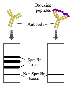
The antiserum was purified by peptide affinity chromatography using SulfoLink™ Coupling Resin (Thermo Fisher Scientific).
保存:
Rabbit IgG in phosphate buffered saline , pH 7.4, 150mM NaCl, 0.02% sodium azide and 50% glycerol. Store at -20 °C. Stable for 12 months from date of receipt.
別名:

折りたたみ/展開

alpha polypeptide (p32); CD8; CD8 antigen alpha polypeptide; CD8 antigen alpha polypeptide (p32); CD8a; CD8A antigen; CD8A molecule; CD8A_HUMAN; Leu2; Leu2 T lymphocyte antigen; Ly3; LYT3; MAL; OKT8 T cell antigen; OTTHUMP00000160760; OTTHUMP00000160764; OTTHUMP00000203528; OTTHUMP00000203721; p32; T cell antigen Leu2; T cell co receptor; T-cell surface glycoprotein CD8 alpha chain; T-lymphocyte differentiation antigen T8/Leu-2; T8 T cell antigen; T8/Leu-2 T-lymphocyte differentiation antigen;

免疫原

免疫原:

A synthesized peptide derived from human CD8, corresponding to a region within the internal amino acids.

Uniprot:
遺伝子(ID):
発現特異性:
P01732 CD8A_HUMAN:

CD8 on thymus-derived T-cells usually consists of a disulfide-linked alpha/CD8A and a beta/CD8B chain. Less frequently, CD8 can be expressed as a CD8A homodimer. A subset of natural killer cells, memory T-cells, intraepithelial lymphocytes, monocytes and dendritic cells expresses CD8A homodimers. Expressed at the cell surface of plasmacytoid dendritic cells upon herpes simplex virus-1 stimulation.

タンパク質の説明:
Identifies cytotoxic/suppressor T-cells that interact with MHC class I bearing targets. CD8 is thought to play a role in the process of T-cell mediated killing. CD8 alpha chains binds to class I MHC molecules alpha-3 domains.
タンパク質配列:
MALPVTALLLPLALLLHAARPSQFRVSPLDRTWNLGETVELKCQVLLSNPTSGCSWLFQPRGAAASPTFLLYLSQNKPKAAEGLDTQRFSGKRLGDTFVLTLSDFRRENEGYYFCSALSNSIMYFSHFVPVFLPAKPTTTPAPRPPTPAPTIASQPLSLRPEACRPAAGGAVHTRGLDFACDIYIWAPLAGTCGVLLLSLVITLYCNHRNRRRVCKCPRPVVKSGDKPSLSARYV

研究背景

機能:

Integral membrane glycoprotein that plays an essential role in the immune response and serves multiple functions in responses against both external and internal offenses. In T-cells, functions primarily as a coreceptor for MHC class I molecule:peptide complex. The antigens presented by class I peptides are derived from cytosolic proteins while class II derived from extracellular proteins. Interacts simultaneously with the T-cell receptor (TCR) and the MHC class I proteins presented by antigen presenting cells (APCs). In turn, recruits the Src kinase LCK to the vicinity of the TCR-CD3 complex. LCK then initiates different intracellular signaling pathways by phosphorylating various substrates ultimately leading to lymphokine production, motility, adhesion and activation of cytotoxic T-lymphocytes (CTLs). This mechanism enables CTLs to recognize and eliminate infected cells and tumor cells. In NK-cells, the presence of CD8A homodimers at the cell surface provides a survival mechanism allowing conjugation and lysis of multiple target cells. CD8A homodimer molecules also promote the survival and differentiation of activated lymphocytes into memory CD8 T-cells.

PTMs:

Palmitoylated, but association with CD8B seems to be more important for the enrichment of CD8A in lipid rafts.

O-glycosylated.

Phosphorylated in cytotoxic T-lymphocytes (CTLs) following activation.

細胞の位置付け:

Cell membrane>Single-pass type I membrane protein.
Note: CD8A localizes to lipid rafts only when associated with its partner CD8B.

Secreted.

Extracellular region or secreted Cytosol Plasma membrane Cytoskeleton Lysosome Endosome Peroxisome ER Golgi apparatus Nucleus Mitochondrion Manual annotation Automatic computational assertionSubcellular location
組織特異性:

CD8 on thymus-derived T-cells usually consists of a disulfide-linked alpha/CD8A and a beta/CD8B chain. Less frequently, CD8 can be expressed as a CD8A homodimer. A subset of natural killer cells, memory T-cells, intraepithelial lymphocytes, monocytes and dendritic cells expresses CD8A homodimers. Expressed at the cell surface of plasmacytoid dendritic cells upon herpes simplex virus-1 stimulation.

研究領域

· Environmental Information Processing > Signaling molecules and interaction > Cell adhesion molecules (CAMs).   (View pathway)

· Human Diseases > Immune diseases > Primary immunodeficiency.

· Organismal Systems > Immune system > Antigen processing and presentation.   (View pathway)

· Organismal Systems > Immune system > Hematopoietic cell lineage.   (View pathway)

· Organismal Systems > Immune system > T cell receptor signaling pathway.   (View pathway)

参考文献

1). Tumor cells impair immunological synapse formation via central nervous system-enriched metabolite. Cancer cell, 2024 (PubMed: 38821061) [IF=48.8]

2). Mitochondria-Targeting Polymer Micelle of Dichloroacetate Induced Pyroptosis to Enhance Osteosarcoma Immunotherapy. ACS nano, 2022 (PubMed: 35737477) [IF=15.8]

3). USP35 Acts as a Deubiquitinating Enzyme for ID3 to Promote Immune Escape in Colorectal Cancer. Advanced science (Weinheim, Baden-Wurttemberg, Germany), 2026 (PubMed: 41486422) [IF=15.1]

4). Accurate Transcription Factor Activity Inference to Decipher Cell Identity from Single-Cell Transcriptomic Data with MetaTF. Advanced science (Weinheim, Baden-Wurttemberg, Germany), 2025 (PubMed: 40397381) [IF=15.1]

Application: IF/ICC    Species: human    Sample:

Figure 5 metaTF reveals TF activity heterogeneity in human breast cancer T cells. a) UMAP plots visualizing cluster assignments of human breast cancer T cells based on TF activity profile (left) and highly variable gene expression profile (right). Cells are color-coded to represent different human breast cancer T-cell populations, including TEM (effector/memory T cells), TEX (experienced T cells), TN (naive T cells), TREG (regulatory T cells), natural killer (NK) cells), and Proliferating (proliferating T cells). b) UMAP plot of CD8+ TEM C1 and C2 marker genes expression. c) Heatmap plot showing the expression of the top 50 differentially expressed genes between CD8+ TEM C1 and C2 cell populations. d) GO biological process enrichment results for the differentially expressed genes between CD8+ TEM C1 (upper panel) and C2 (lower panel) cell populations respectively. e) Dot plot showing the significant expression of β2-adrenergic receptor (ADRB2) and adrenoceptor alpha 2B (ADRA2B) primarily in the CD8+ TEM C1 cell population. f) Dot plot indicating that BCL6 was specifically activated in the CD8+ TEM C1 cell population. g) Spatial co-mapping of CD8 and ADRB2 gene expression by spatial transcriptomics of a sample from a breast cancer patient recorded in a public dataset (GSE195665, patient35, right panel). The left and middle panels depict expression levels in fresh frozen sections. h) Expression of CD8 and ADRB2 in breast cancer tissue, and the colocalization (Pearson's coefficient) between CD8 and ADRB2 is shown, n = 12. i) Upregulated targets of BCL6 in the epinephrine treatment group compared to the control group from in vitro cytological experiments. j) In vitro cytological experiments confirmed that candidate TFs and their targets are highly expressed in the epinephrine-treated group through GSEA prerank analysis.

5). Multifunctional 3D-printed scaffolds eradiate orthotopic osteosarcoma and promote osteogenesis via microwave thermo-chemotherapy combined with immunotherapy. Biomaterials, 2023 (PubMed: 37506512) [IF=12.8]

6). Single-cell transcriptomics of peripheral blood reveals anti-tumor systemic immunity induced by oncolytic virotherapy. Theranostics, 2022 (PubMed: 36438484) [IF=12.4]

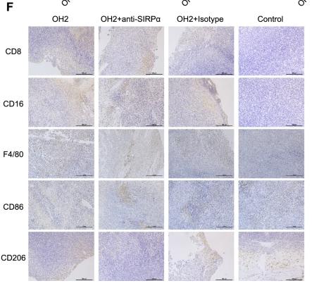
Application: IHC    Species: Mouse    Sample:

Figure 7. Effects of OH2 on distant tumor lesions. (A) Timeline of tumor injection and treatment. At each time point, only the right-side tumor was treated. i.t., intratumoral injection. (B) The Left and right panels show the growth curves for the left and right flank tumors from the two groups (PBS and OH2), respectively (n = 6). (C-E) A heatmap showing the infiltration level of 22 types of immune cells (C), the activation degree of 17 key immune pathways (D), and the scaled expression of key immunomodulators (E) in left flank tumors of the control group and the OH2 group. (F) Representative flow cytometric analysis results of Ccl5+CD8+ T cells. (G) The percentages of Ccl5+CD8+ T cells were determined 14 days after grouping. (H) Histological appearance of representative untreated flank tumor samples from each treatment group. Middle, original magnification, × 20. Top and bottom, original magnification, × 100.

7). Dietary palmitoleic acid reprograms gut microbiota and improves biological therapy against colitis. Gut microbes, 2023 (PubMed: 37203220) [IF=12.2]

8). Ion interference induced by Ca-Mn nanoplatform enhances ferroptosis and promotes immune response for osteosarcoma treatment. Journal of advanced research, 2025 (PubMed: 40518113) [IF=11.4]

9). Alantolactone-loaded chitosan/hyaluronic acid nanoparticles suppress psoriasis by deactivating STAT3 pathway and restricting immune cell recruitment. Asian Journal of Pharmaceutical Sciences, 2022 (PubMed: 35582636) [IF=10.7]

Application: IF/ICC    Species: mouse    Sample: skin

Fig. 8| CHALT attenuates splenomegaly and immune cell recruitment in IMQ-induced skin. On Day 7, (A) the spleens were collected and photographed, and (B) the weight ratio of spleen/body was calculated. Immunfluorescence staining of (C) CD 4 and (E) CD 8 in the skin, and the relative amount of (D) CD 4 and (F) CD 8 was quantified (the normal group as the control).

10). Harnessing the power of traceable system C-GAP: homologous-targeting to fire up T-cell immune responses with low-dose irradiation. Journal of nanobiotechnology, 2025 (PubMed: 40075499) [IF=10.6]

もっと読みます

Restrictive clause


Affinity Biosciences tests all products strictly. Citations are provided as a resource for additional applications that have not been validated by Affinity Biosciences. Please choose the appropriate format for each application and consult Materials and Methods sections for additional details about the use of any product in these publications.

For Research Use Only.
Not for use in diagnostic or therapeutic procedures. Not for resale. Not for distribution without written consent. Affinity Biosciences will not be held responsible for patent infringement or other violations that may occur with the use of our products. Affinity Biosciences, Affinity Biosciences Logo and all other trademarks are the property of Affinity Biosciences LTD.