製品: P Glycoprotein 1(MDR1) Antibody
カタログ: AF5185
タンパク質の説明: Rabbit polyclonal antibody to P Glycoprotein 1(MDR1)
アプリケーション: WB IHC
反応性: Human, Mouse, Rat
予測: Pig, Horse, Rabbit, Dog, Chicken, Xenopus
分子量: 141 kDa; 141kD(Calculated).
ユニプロット: P08183
RRID: AB_2837671

類似製品を見る>>

   サイズ 価格 在庫状況
 100ul $280 在庫あり
 200ul $350 在庫あり

リードタイム: 当日配達

For pricing and ordering contact:
お問い合わせ先

製品説明

ソース:
Rabbit
アプリケーション:
WB 1:500-1:1000, IHC 1:50-1:200
*The optimal dilutions should be determined by the end user.
*Tips:

WB: For western blot detection of denatured protein samples. IHC: For immunohistochemical detection of paraffin sections (IHC-p) or frozen sections (IHC-f) of tissue samples. IF/ICC: For immunofluorescence detection of cell samples. ELISA(peptide): For ELISA detection of antigenic peptide.

反応性:
Human,Mouse,Rat
予測:
Pig(92%), Horse(83%), Rabbit(100%), Dog(100%), Chicken(83%), Xenopus(83%)
クローナリティ:
Polyclonal
特異性:
P Glycoprotein 1(MDR1) Antibody detects endogenous levels of total P Glycoprotein 1(MDR1).
RRID:
AB_2837671
引用形式: Affinity Biosciences Cat# AF5185, RRID:AB_2837671.
コンジュゲート:
Unconjugated.
精製:
The antiserum was purified by peptide affinity chromatography using SulfoLink™ Coupling Resin (Thermo Fisher Scientific).
保存:
Rabbit IgG in phosphate buffered saline , pH 7.4, 150mM NaCl, 0.02% sodium azide and 50% glycerol. Store at -20 °C. Stable for 12 months from date of receipt.
別名:

折りたたみ/展開

ABC20; ABCB1; ATP binding cassette, sub family B (MDR/TAP), member 1; ATP-binding cassette sub-family B member 1; CD243; CLCS; Colchicin sensitivity; Doxorubicin resistance; GP170; MDR1; MDR1_HUMAN; Multidrug resistance 1; Multidrug resistance protein 1; P glycoprotein 1; P gp; P-glycoprotein 1; PGY1;

免疫原

免疫原:
Uniprot:
遺伝子(ID):
発現特異性:
P08183 MDR1_HUMAN:

Expressed in liver, kidney, small intestine and brain.

タンパク質の説明:
A chronic, relapsing inflammation of the gastrointestinal tract with a complex etiology. It is subdivided into Crohn disease and ulcerative colitis phenotypes. Crohn disease may affect any part of the gastrointestinal tract from the mouth to the anus, but most frequently it involves the terminal ileum and colon.
タンパク質配列:
MDLEGDRNGGAKKKNFFKLNNKSEKDKKEKKPTVSVFSMFRYSNWLDKLYMVVGTLAAIIHGAGLPLMMLVFGEMTDIFANAGNLEDLMSNITNRSDINDTGFFMNLEEDMTRYAYYYSGIGAGVLVAAYIQVSFWCLAAGRQIHKIRKQFFHAIMRQEIGWFDVHDVGELNTRLTDDVSKINEGIGDKIGMFFQSMATFFTGFIVGFTRGWKLTLVILAISPVLGLSAAVWAKILSSFTDKELLAYAKAGAVAEEVLAAIRTVIAFGGQKKELERYNKNLEEAKRIGIKKAITANISIGAAFLLIYASYALAFWYGTTLVLSGEYSIGQVLTVFFSVLIGAFSVGQASPSIEAFANARGAAYEIFKIIDNKPSIDSYSKSGHKPDNIKGNLEFRNVHFSYPSRKEVKILKGLNLKVQSGQTVALVGNSGCGKSTTVQLMQRLYDPTEGMVSVDGQDIRTINVRFLREIIGVVSQEPVLFATTIAENIRYGRENVTMDEIEKAVKEANAYDFIMKLPHKFDTLVGERGAQLSGGQKQRIAIARALVRNPKILLLDEATSALDTESEAVVQVALDKARKGRTTIVIAHRLSTVRNADVIAGFDDGVIVEKGNHDELMKEKGIYFKLVTMQTAGNEVELENAADESKSEIDALEMSSNDSRSSLIRKRSTRRSVRGSQAQDRKLSTKEALDESIPPVSFWRIMKLNLTEWPYFVVGVFCAIINGGLQPAFAIIFSKIIGVFTRIDDPETKRQNSNLFSLLFLALGIISFITFFLQGFTFGKAGEILTKRLRYMVFRSMLRQDVSWFDDPKNTTGALTTRLANDAAQVKGAIGSRLAVITQNIANLGTGIIISFIYGWQLTLLLLAIVPIIAIAGVVEMKMLSGQALKDKKELEGSGKIATEAIENFRTVVSLTQEQKFEHMYAQSLQVPYRNSLRKAHIFGITFSFTQAMMYFSYAGCFRFGAYLVAHKLMSFEDVLLVFSAVVFGAMAVGQVSSFAPDYAKAKISAAHIIMIIEKTPLIDSYSTEGLMPNTLEGNVTFGEVVFNYPTRPDIPVLQGLSLEVKKGQTLALVGSSGCGKSTVVQLLERFYDPLAGKVLLDGKEIKRLNVQWLRAHLGIVSQEPILFDCSIAENIAYGDNSRVVSQEEIVRAAKEANIHAFIESLPNKYSTKVGDKGTQLSGGQKQRIAIARALVRQPHILLLDEATSALDTESEKVVQEALDKAREGRTCIVIAHRLSTIQNADLIVVFQNGRVKEHGTHQQLLAQKGIYFSMVSVQAGTKRQ

種類予測

種類予測:

Score>80(red) has high confidence and is suggested to be used for WB detection. *The prediction model is mainly based on the alignment of immunogen sequences, the results are for reference only, not as the basis of quality assurance.

Species
Results
Score
Rabbit
100
Dog
100
Pig
92
Horse
83
Xenopus
83
Chicken
83
Bovine
0
Sheep
0
Zebrafish
0
Model Confidence:
High(score>80) Medium(80>score>50) Low(score<50) No confidence

PTMs - P08183 基板として

Site PTM Type Enzyme
S23 Phosphorylation
T263 Phosphorylation
K271 Ubiquitination
K279 Ubiquitination
K367 Ubiquitination
K372 Ubiquitination
K380 Ubiquitination
K384 Acetylation
K389 Ubiquitination
K405 Sumoylation
K408 Sumoylation
K411 Ubiquitination
T422 Phosphorylation
T496 Phosphorylation
K515 Ubiquitination
K519 Ubiquitination
T522 Phosphorylation
K536 Ubiquitination
K617 Ubiquitination
K619 Ubiquitination
S644 Phosphorylation
S658 Phosphorylation
S660 Phosphorylation
S661 Phosphorylation P17252 (PRKCA)
S667 Phosphorylation P17612 (PRKACA) , P17252 (PRKCA)
S671 Phosphorylation P17252 (PRKCA) , P17612 (PRKACA)
S675 Phosphorylation
S683 Phosphorylation P17612 (PRKACA)
T684 Phosphorylation
S696 Phosphorylation
K826 Ubiquitination
K915 Ubiquitination
T1015 Phosphorylation
T1023 Phosphorylation
T1030 Phosphorylation
K1062 Ubiquitination
Y1087 Phosphorylation
K1093 Ubiquitination
K1099 Ubiquitination
K1102 Methylation
K1172 Ubiquitination
K1181 Ubiquitination
K1212 Ubiquitination
Y1267 Phosphorylation
S1269 Phosphorylation

研究背景

機能:

Translocates drugs and phospholipids across the membrane. Catalyzes the flop of phospholipids from the cytoplasmic to the exoplasmic leaflet of the apical membrane. Participates mainly to the flop of phosphatidylcholine, phosphatidylethanolamine, beta-D-glucosylceramides and sphingomyelins. Energy-dependent efflux pump responsible for decreased drug accumulation in multidrug-resistant cells.

細胞の位置付け:

Cell membrane>Multi-pass membrane protein. Apical cell membrane.

Extracellular region or secreted Cytosol Plasma membrane Cytoskeleton Lysosome Endosome Peroxisome ER Golgi apparatus Nucleus Mitochondrion Manual annotation Automatic computational assertionSubcellular location
組織特異性:

Expressed in liver, kidney, small intestine and brain.

サブユニット構造:

Interacts with PSMB5. Finds in a complex with ABCB1, TFPI2 and PPP2R3C; leading to the dephosphorylation of ABCB1.

タンパク質ファミリー:

Belongs to the ABC transporter superfamily. ABCB family. Multidrug resistance exporter (TC 3.A.1.201) subfamily.

研究領域

· Environmental Information Processing > Membrane transport > ABC transporters.

· Human Diseases > Cancers: Overview > MicroRNAs in cancer.

· Human Diseases > Cancers: Specific types > Gastric cancer.   (View pathway)

参考文献

1). Expression of multidrug resistance marker MDR1 in patients with ovarian cancer and its possible predictive significance.. JOURNAL OF CLINICAL ONCOLOGY, 2021 [IF=45.3]

2). Chitosan biguanide induced mitochondrial inhibition to amplify the efficacy of oxygen-sensitive tumor therapies. Carbohydrate Polymers, 2022 (PubMed: 35989018) [IF=11.2]

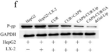
3). Co-Delivery of Curcumin and Capsaicin by Dual-Targeting Liposomes for Inhibition of aHSC-Induced Drug Resistance and Metastasis. ACS Applied Materials & Interfaces, 2021 (PubMed: 33819006) [IF=9.5]

Application: WB    Species: Human    Sample: HepG2 cells

Figure 4. Cytotoxicity of different drug formulations. (a) MTT assay of HepG2 cells and the cocultured model. (b) Cell viability of HepG2 cells. (c) Cell viability of the cocultured system. Scale bars represent 100 μm. (d) Live/dead staining of HepG2 cells, where green- and red-dyed cells represent live and dead HepG2 cells, respectively. (e) Live/dead staining of the cocultured model, where the yellow-, red-, and green-colored cells denote the dead LX-2, dead HepG2, and live LX-2 cells, respectively. Scale bars represent 100 μm. (f) Western blotting assay for the expression of P-gp. (g) Quantitative analysis of P-gp. *P < 0.05, **P < 0.01, ***P < 0.001.

4). Increased autophagy in EOC re-ascites cells can inhibit cell death and promote drug resistance. Cell Death & Disease, 2018 (PubMed: 29549251) [IF=9.0]

Application: WB    Species: human    Sample: EOC ascites cells

Fig. 2| Autophagy is increased and apoptosis is reduced in the EOC re-ascites group. a IHC analysis of LC-3, Belin-1, CASP-9, and c-CASP-3 expression in EOC ascites cells (scale bar =30µm). b, c Western blot analysis of autophagy/apoptosis-related proteins expressed in the chemosensitive group and re-ascites group (***p<0.001, **p<0.01, *p<0.05). d Immunofluorescence analysis of the chemosensitive group and re-ascites group with LC3 (green) and LAMP2 (red) staining. Nuclei were counterstained with DAPI (blue). e qRT-PCR analysis of the average relative mRNA expression levels of autophagy/apoptosis-related genes in the chemosensitive group and re-ascites group (**p<0.01, *p<0.05)

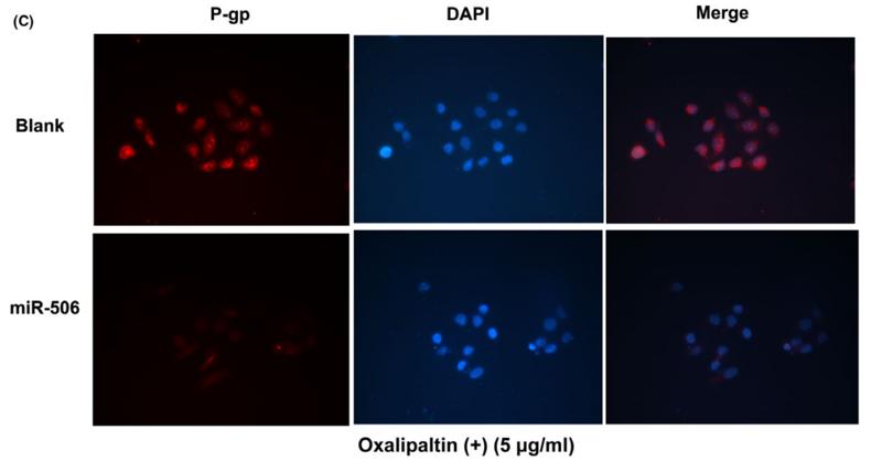
5). miR-506 enhances the sensitivity of human colorectal cancer cells to oxaliplatin by suppressing MDR1/P-gp expression. CELL PROLIFERATION, 2017 (PubMed: 28217977) [IF=8.5]

Application: IF/ICC    Species: human    Sample:

FIGURE 3  miR-506 down-regulated MDR1/P-gp expression in HCT116-OxR. A, The mRNA level of MDR1 was decreased after transfection with the miR-506 mimic of the relative chemoresistant genes as demonstrated by qRT-PCR. B, The protein level of MDR1 was decreased after transfection with the miR-506 mimic of the relative chemoresistant proteins as demonstrated by Western blot. C, Expression of P-gp detected by immunofluorescence staining.HCT116-OxR-miR-506 cells showed low levels of fluorescent staining of P-gp, whereas maximal staining of P-gp was observed in HCT116-OxR cells, readily distinguished from background. Zoom: 200×. *P<.05)

6). d-Borneol enhances cisplatin sensitivity via p21/p27-mediated S-phase arrest and cell apoptosis in non-small cell lung cancer cells and a murine xenograft model. Cellular & Molecular Biology Letters, 2022 (PubMed: 35883026) [IF=8.3]

7). Metformin modified chitosan as a multi-functional adjuvant to enhance cisplatin-based tumor chemotherapy efficacy. International Journal of Biological Macromolecules, 2023 (PubMed: 36283555) [IF=8.2]

8). Efficient Sequential Co-Delivery Nanosystem for Inhibition of Tumor and Tumor-Associated Fibroblast-Induced Resistance and Metastasis. International journal of nanomedicine, 2024 (PubMed: 38414527) [IF=8.0]

Application: WB    Species: Mouse    Sample:

Figure 6 Western blotting against α-SMA, Bcl-2, and P-gp in total soluble protein fractions from (1) control 4T1 cells in monoculture, (2) control co-cultures of 4T1 cells and NIH 3T3 fibroblasts, or co-cultures treated with (3) free CEL, (4) free CEL+BA, (5) CL, (6) CL@BM or (7) F/CL@BM. (A) Western blotting assay for the expression of protein. (B) Quantitative analysis. ***p < 0.001, ****p < 0.0001.

9). Mesenchymal Stem Cells Overexpressing ACE2 Favorably Ameliorate LPS-Induced Inflammatory Injury in Mammary Epithelial Cells. Frontiers in Immunology, 2022 (PubMed: 35095873) [IF=7.3]

10). DHW-221, a Dual PI3K/mTOR Inhibitor, Overcomes Multidrug Resistance by Targeting P-Glycoprotein (P-gp/ABCB1) and Akt-Mediated FOXO3a Nuclear Translocation in Non-small Cell Lung Cancer. Frontiers in Oncology, 2022 (PubMed: 35646704) [IF=4.7]

Application: WB    Species: Human    Sample: A549 and A549/Taxol cells

Figure 2 DHW-221 accelerates intracellular Rho-123 accumulations and inhibits P-gp expression by binding to P-gp. (A) Comparison of P-gp in A549 and A549/Taxol cells. Statistical comparisons were performed with unpaired Student’s t-test (n = 3). **p < 0.01 versus A549. (B, C) After treatment of DHW-221 or verapamil for 48 h, Rho-123 (15 µM) was added into the mediums to co-incubate with DHW-221 or verapamil for 3 h in dark. The intracellular accumulation of Rho-123 was evaluated by flow cytometry and confocal microscope, respectively. Verapamil (VRP, 10 μM) was used as a positive control. Green fluorescence indicated Rho-123, and blue fluorescence indicated DAPI. Scale bar = 100 μm. Nuclear mean fluorescence intensity of Rho 123 was measured by ImageJ and analyzed using FlowJo 10 software. Statistical comparisons were performed with one-way ANOVA followed by Dunnett’s post-hoc test for multiple comparisons (n = 3). *p < 0.05, **p < 0.01, ***p < 0.001 versus control. (D) Effect of DHW-221 or verapamil to reverse Taxol resistance for 48 h in A549/Taxol cells. (E) Western blot analysis of DHW-221 inhibited P-gp expression in A549/Taxol cells. (F) Ribbon diagram of human ABCB1 (PDB code: 7A6E, purple spiral) bound to drug (membrane region) and predicted binding modes for DHW-221 (blue stick) and tariquidar (orange stick, a known third-generation P-gp inhibitor) with ABCB1. Hydrogen bonds were shown as green dashed lines. Key residues for DHW-221 and tariquidar interaction were highlighted. (G) Effect of DHW-221 on the thermal stability of ABCB1 was quantitatively detected by a cellular thermal shift assay (CETSA). Statistical comparisons were performed with one-way ANOVA followed by Dunnett’s post-hoc test for multiple comparisons (n = 3). Data are presented as mean ± SD. **p < 0.01, ****p < 0.0001versus control.

もっと読みます

Restrictive clause


Affinity Biosciences tests all products strictly. Citations are provided as a resource for additional applications that have not been validated by Affinity Biosciences. Please choose the appropriate format for each application and consult Materials and Methods sections for additional details about the use of any product in these publications.

For Research Use Only.
Not for use in diagnostic or therapeutic procedures. Not for resale. Not for distribution without written consent. Affinity Biosciences will not be held responsible for patent infringement or other violations that may occur with the use of our products. Affinity Biosciences, Affinity Biosciences Logo and all other trademarks are the property of Affinity Biosciences LTD.