製品: RUNX2 Antibody
カタログ: AF5186
タンパク質の説明: Rabbit polyclonal antibody to RUNX2
アプリケーション: WB IHC IF/ICC
反応性: Human, Mouse, Rat
予測: Pig, Bovine, Horse, Sheep, Rabbit, Chicken, Xenopus
分子量: 56kDa,48kDa; 57kD(Calculated).
ユニプロット: Q13950
RRID: AB_2837672

類似製品を見る>>

   サイズ 価格 在庫状況
 100ul $280 在庫あり
 200ul $350 在庫あり

リードタイム: 当日配達

For pricing and ordering contact:
お問い合わせ先

製品説明

ソース:
Rabbit
アプリケーション:
WB 1:500-1:2000, IHC 1:50-1:200, IF/ICC 1:100-1:500
*The optimal dilutions should be determined by the end user.
*Tips:

WB: For western blot detection of denatured protein samples. IHC: For immunohistochemical detection of paraffin sections (IHC-p) or frozen sections (IHC-f) of tissue samples. IF/ICC: For immunofluorescence detection of cell samples. ELISA(peptide): For ELISA detection of antigenic peptide.

反応性:
Human,Mouse,Rat
予測:
Pig(100%), Bovine(100%), Horse(100%), Sheep(100%), Rabbit(100%), Chicken(100%), Xenopus(90%)
クローナリティ:
Polyclonal
特異性:
RUNX2 Antibody detects endogenous levels of total RUNX2.
RRID:
AB_2837672
引用形式: Affinity Biosciences Cat# AF5186, RRID:AB_2837672.
コンジュゲート:
Unconjugated.
精製:
The antiserum was purified by peptide affinity chromatography using SulfoLink™ Coupling Resin (Thermo Fisher Scientific).
保存:
Rabbit IgG in phosphate buffered saline , pH 7.4, 150mM NaCl, 0.02% sodium azide and 50% glycerol. Store at -20 °C. Stable for 12 months from date of receipt.
別名:

折りたたみ/展開

Acute myeloid leukemia 3 protein; Alpha subunit 1; AML3; CBF alpha 1; CBF-alpha-1; CBFA1; CCD; CCD1; Cleidocranial dysplasia 1; Core binding factor; Core binding factor runt domain alpha subunit 1; Core binding factor subunit alpha 1; Core-binding factor subunit alpha-1; MGC120022; MGC120023; Oncogene AML 3; Oncogene AML-3; OSF 2; OSF-2; OSF2; Osteoblast specific transcription factor 2; Osteoblast-specific transcription factor 2; OTTHUMP00000016533; PEA2 alpha A; PEA2-alpha A; PEA2aA; PEBP2 alpha A; PEBP2-alpha A; PEBP2A1; PEBP2A2; PEBP2aA; PEBP2aA1; Polyomavirus enhancer binding protein 2 alpha A subunit; Polyomavirus enhancer-binding protein 2 alpha A subunit; Runt domain; Runt related transcription factor 2; Runt-related transcription factor 2; RUNX2; RUNX2_HUMAN; SL3 3 enhancer factor 1 alpha A subunit; SL3-3 enhancer factor 1 alpha A subunit; SL3/AKV core binding factor alpha A subunit; SL3/AKV core-binding factor alpha A subunit;

免疫原

免疫原:
Uniprot:
遺伝子(ID):
発現特異性:
Q13950 RUNX2_HUMAN:

Specifically expressed in osteoblasts.

タンパク質の説明:
Transcription factor involved in osteoblastic differentiation and skeletal morphogenesis. Essential for the maturation of osteoblasts and both intramembranous and endochondral ossification. CBF binds to the core site, 5'-PYGPYGGT-3', of a number of enhancers and promoters, including murine leukemia virus, polyomavirus enhancer, T-cell receptor enhancers, osteocalcin, osteopontin, bone sialoprotein, alpha 1(I) collagen, LCK, IL-3 and GM-CSF promoters.
タンパク質配列:
MASNSLFSTVTPCQQNFFWDPSTSRRFSPPSSSLQPGKMSDVSPVVAAQQQQQQQQQQQQQQQQQQQQQQQEAAAAAAAAAAAAAAAAAVPRLRPPHDNRTMVEIIADHPAELVRTDSPNFLCSVLPSHWRCNKTLPVAFKVVALGEVPDGTVVTVMAGNDENYSAELRNASAVMKNQVARFNDLRFVGRSGRGKSFTLTITVFTNPPQVATYHRAIKVTVDGPREPRRHRQKLDDSKPSLFSDRLSDLGRIPHPSMRVGVPPQNPRPSLNSAPSPFNPQGQSQITDPRQAQSSPPWSYDQSYPSYLSQMTSPSIHSTTPLSSTRGTGLPAITDVPRRISDDDTATSDFCLWPSTLSKKSQAGASELGPFSDPRQFPSISSLTESRFSNPRMHYPATFTYTPPVTSGMSLGMSATTHYHTYLPPPYPGSSQSQSGPFQTSSTPYLYYGTSSGSYQFPMVPGGDRSPSRMLPPCTTTSNGSTLLNPNLPNQNDGVDADGSHSSSPTVLNSSGRMDESVWRPY

種類予測

種類予測:

Score>80(red) has high confidence and is suggested to be used for WB detection. *The prediction model is mainly based on the alignment of immunogen sequences, the results are for reference only, not as the basis of quality assurance.

Species
Results
Score
Pig
100
Horse
100
Bovine
100
Sheep
100
Chicken
100
Rabbit
100
Xenopus
90
Dog
0
Zebrafish
0
Model Confidence:
High(score>80) Medium(80>score>50) Low(score<50) No confidence

PTMs - Q13950 基板として

Site PTM Type Enzyme
S24 Phosphorylation P27361 (MAPK3)
S28 Phosphorylation P31749 (AKT1) , P27361 (MAPK3)
S32 O-Glycosylation
S33 O-Glycosylation
S33 Phosphorylation
S43 Phosphorylation P27361 (MAPK3)
S118 Phosphorylation P45983 (MAPK8)
K176 Ubiquitination
S196 Phosphorylation P31749 (AKT1)
T198 Phosphorylation P31749 (AKT1)
T200 Phosphorylation P31749 (AKT1)
S237 Phosphorylation
K238 Acetylation
K238 Ubiquitination
S247 Phosphorylation
R258 Methylation
R267 Methylation
S275 Phosphorylation P27361 (MAPK3)
S294 Phosphorylation P27361 (MAPK3)
S312 Phosphorylation P27361 (MAPK3)
S314 Phosphorylation P27361 (MAPK3)
S340 Phosphorylation P27361 (MAPK3)
S347 Phosphorylation
S371 O-Glycosylation
R386 Methylation
S388 Phosphorylation
S451 Phosphorylation
S465 Phosphorylation P06493 (CDK1)
S480 Phosphorylation
S499 Phosphorylation
S503 Phosphorylation P27361 (MAPK3)
Y521 Phosphorylation

研究背景

機能:

Transcription factor involved in osteoblastic differentiation and skeletal morphogenesis. Essential for the maturation of osteoblasts and both intramembranous and endochondral ossification. CBF binds to the core site, 5'-PYGPYGGT-3', of a number of enhancers and promoters, including murine leukemia virus, polyomavirus enhancer, T-cell receptor enhancers, osteocalcin, osteopontin, bone sialoprotein, alpha 1(I) collagen, LCK, IL-3 and GM-CSF promoters. In osteoblasts, supports transcription activation: synergizes with SPEN/MINT to enhance FGFR2-mediated activation of the osteocalcin FGF-responsive element (OCFRE) (By similarity). Inhibits KAT6B-dependent transcriptional activation.

PTMs:

Phosphorylated; probably by MAP kinases (MAPK). Phosphorylation by HIPK3 is required for the SPEN/MINT and FGF2 transactivation during osteoblastic differentiation (By similarity). Phosphorylation at Ser-451 by CDK1 promotes endothelial cell proliferation required for tumor angiogenesis probably by facilitating cell cycle progression. Isoform 3 is phosphorylated on Ser-340.

細胞の位置付け:

Nucleus.

Extracellular region or secreted Cytosol Plasma membrane Cytoskeleton Lysosome Endosome Peroxisome ER Golgi apparatus Nucleus Mitochondrion Manual annotation Automatic computational assertionSubcellular location
組織特異性:

Specifically expressed in osteoblasts.

サブユニット構造:

Heterodimer of an alpha and a beta subunit. The alpha subunit binds DNA as a monomer and through the Runt domain. DNA-binding is increased by heterodimerization. Interacts with XRCC6 (Ku70) and XRCC5 (Ku80). Interacts with HIVEP3. Interacts with IFI204. Interaction with SATB2; the interaction results in enhanced DNA binding and transactivation by these transcription factors. Binds to HIPK3. Interacts (isoform 3) with DDX5. Interacts with FOXO1 (via a C-terminal region); the interaction inhibits RUNX2 transcriptional activity towards BGLAP. This interaction is prevented on insulin or IGF1 stimulation as FOXO1 is exported from the nucleus (By similarity). Interacts with CCNB1, KAT6A and KAT6B. Interacts with FOXP3. Interacts with TMEM119 (By similarity). Interacts with OLFM2 (By similarity).

タンパク質ファミリー:

A proline/serine/threonine rich region at the C-terminus is necessary for transcriptional activation of target genes and contains the phosphorylation sites.

研究領域

· Human Diseases > Cancers: Overview > Transcriptional misregulation in cancer.

参考文献

1). Gut microbial alterations in arginine metabolism determine bone mechanical adaptation. Cell metabolism, 2024 (PubMed: 38718794) [IF=29.0]

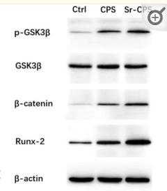
2). Osteoblastic and anti-osteoclastic activities of strontium-substituted silicocarnotite ceramics: In vitro and in vivo studies. Bioactive Materials, 2020 (PubMed: 32280833) [IF=18.9]

Application: WB    Species: Mice    Sample: bone and osteoid tissues

Fig. 4 (A) Expression of the osteogenesis-specific genes including RUNX2, BSP, OPN and OCN; (B) Western blotting showed that Sr-CPS affected the Wnt/β-catenin pathway-related proteins GSK3β, p-GSK3β, β-catenin and RUNX-2; (C) Quantification of the Western blotting. * Compared with the control group; # compared with CPS group.

Application: IHC    Species: Mice    Sample: bone and osteoid tissues

Fig. 9 (A) Masson staining: the region between surrounding bone and scaffold (40x & 100x); (B) TRAP staining: TRAP positive cells (black arrows); (C) IHC staining of RUNX-2; (D) IHC staining of ALP; (E) Quantitative analysis for TRAP staining and IHC staining. * Compared with CPS group; # compared with Sr-5 group.

3). Gold nanoparticles targeting the autophagy–lysosome system to combat the inflammation-compromised osteogenic potential of periodontal ligament stem cells: From mechanism to therapy. Biomaterials, 2022 (PubMed: 36030103) [IF=14.0]

4). Fusion peptide engineered “statically-versatile” titanium implant simultaneously enhancing anti-infection, vascularization and osseointegration. Biomaterials, 2021 (PubMed: 33069134) [IF=14.0]

5). Vascular Derived ECM Improves Therapeutic Index of BMP‐2 and Drives Vascularized Bone Regeneration. Small, 2022 (PubMed: 35218305) [IF=13.3]

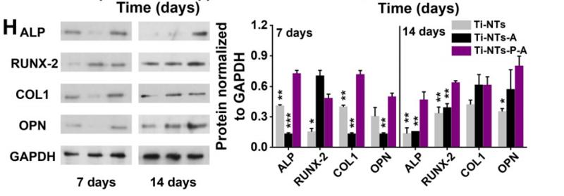
6). On-demand storage and release of antimicrobial peptides using Pandora's box-like nanotubes gated with a bacterial infection-responsive polymer. Theranostics, 2020 (PubMed: 31903109) [IF=12.4]

Application: WB    Species: Human    Sample: Human bone mesenchymal stem cells(hBMSCs)

Figure 5. In vitro biocompatibilities and osteogenic activities of the substrates. (A) CCK-8 assay of hBMSCs on the indicated surfaces after 1, 3 and 7 days of culture. * denotes p < 0.05 and ** denotes p < 0.01 compared to Ti-NTs, # denotes p < 0.05 compared to the corresponding control group without peptides. Error bars denote the standard deviations over quadruplicate measurements with separately implants. (B) Confocal fluorescence microscopy images of hBMSCs stained with vinculin, F-actin and DAPI after being cultured for 24 h. Scale bar, 50 μm. (C-F) qRT-PCR assay of osteogenic gene expression of (C) ALP, (D) RUNX-2, (E) COL1 and (F) OPN of hBMSCs after 7 and 14 days of culture. * denotes p < 0.05, ** denotes p < 0.01 and *** denotes p < 0.001 compared to Ti-NTs-P-A; # denotes p < 0.05, & denotes p < 0.01 and $ denotes p < 0.001 compared to the corresponding control group without peptides. All error bars denote the standard deviations over quadruplicate measurements with separately implants. (G) Immunofluorescence staining of hBMSCs cultured on Ti-NTs-P-A for 7 days (ALP and RUNX-2) and 14 days (OPN). The images were obtained by confocal fluorescence microscopy. Scale bar, 50 μm. (H) Western blotting of hBMSCs cultured on the substrates for 7 and 14 days. At each time point, left lane was Ti-NTs, middle lane was Ti-NTs-A and right lane was Ti-NTs-P-A. * denotes p < 0.05, ** denotes p < 0.01 and *** denotes p < 0.001 compared to Ti-NTs-P-A. Error bars denote the standard deviations over triplicate measurements with separately Western blotting results.

7). Biomimetic hydroxyapatite coating on the 3D-printed bioactive porous composite ceramic scaffolds promoted osteogenic differentiation via PI3K/AKT/mTOR signaling pathways and facilitated bone regeneration in vivo. Journal of Materials Science & Technology, 2023 [IF=10.9]

8). Micro or nano: Evaluation of biosafety and biopotency of magnesium metal organic framework-74 with different particle sizes. Nano Research, 2020 [IF=9.9]

9). Small extracellular vesicles derived from tendon stem cells promote the healing of injured Achilles tendons by regulating miR-145-3p. Acta biomaterialia, 2023 (PubMed: 37806377) [IF=9.7]

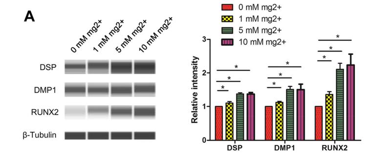
10). Magnesium-enriched microenvironment promotes odontogenic differentiation in human dental pulp stem cells by activating ERK/BMP2/Smads signaling. Stem Cell Research & Therapy, 2019 (PubMed: 31823825) [IF=7.5]

Application: WB    Species: Human    Sample: DPSCs

Fig. 3 Effects of Mg2+-enriched microenvironment on DPSC differentiation. a After 7 days, RUNX2, DMP-1, and DSP protein amounts were markedly elevated in DPSCs treated with 1 mM, 5 mM, and 10 mM Mg2+, compared with the 0 mM Mg2+ group. b Consistently, mineralization nodules were also markedly increased after 14 days. However, there was no difference between the 5 and 10 mM Mg2+ groups

もっと読みます

Restrictive clause


Affinity Biosciences tests all products strictly. Citations are provided as a resource for additional applications that have not been validated by Affinity Biosciences. Please choose the appropriate format for each application and consult Materials and Methods sections for additional details about the use of any product in these publications.

For Research Use Only.
Not for use in diagnostic or therapeutic procedures. Not for resale. Not for distribution without written consent. Affinity Biosciences will not be held responsible for patent infringement or other violations that may occur with the use of our products. Affinity Biosciences, Affinity Biosciences Logo and all other trademarks are the property of Affinity Biosciences LTD.