製品: Claudin 5 Antibody
カタログ: AF5216
タンパク質の説明: Rabbit polyclonal antibody to Claudin 5
アプリケーション: WB IHC IF/ICC
Cited expt.: WB, IHC, IF/ICC
反応性: Human, Mouse, Rat, Pig, Bovine, Monkey
予測: Pig, Bovine, Rabbit, Chicken, Xenopus
分子量: 23 kDa; 23kD(Calculated).
ユニプロット: O00501
RRID: AB_2837702

類似製品を見る>>

   サイズ 価格 在庫状況
 100ul $280 在庫あり
 200ul $350 在庫あり

リードタイム: 当日配達

For pricing and ordering contact:
お問い合わせ先

製品説明

ソース:
Rabbit
アプリケーション:
WB 1:500-1:2000, IHC 1:50-1:200, IF/ICC 1:100-1:500
*The optimal dilutions should be determined by the end user. For optimal experimental results, antibody reuse is not recommended.
*Tips:

WB: For western blot detection of denatured protein samples. IHC: For immunohistochemical detection of paraffin sections (IHC-p) or frozen sections (IHC-f) of tissue samples. IF/ICC: For immunofluorescence detection of cell samples. ELISA(peptide): For ELISA detection of antigenic peptide.

反応性:
Human,Mouse,Rat,Pig,Bovine,Monkey
予測:
Rabbit(93%), Chicken(80%), Xenopus(80%)
クローナリティ:
Polyclonal
特異性:
Claudin 5 Antibody detects endogenous levels of total Claudin 5.
RRID:
AB_2837702
引用形式: Affinity Biosciences Cat# AF5216, RRID:AB_2837702.
コンジュゲート:
Unconjugated.
精製:
The antiserum was purified by peptide affinity chromatography using SulfoLink™ Coupling Resin (Thermo Fisher Scientific).
保存:
Rabbit IgG in phosphate buffered saline , pH 7.4, 150mM NaCl, 0.02% sodium azide and 50% glycerol. Store at -20 °C. Stable for 12 months from date of receipt.
別名:

折りたたみ/展開

Androgen withdrawal and apoptosis induced protein RVP1 like; AWAL; BEC 1; BEC1; Claudin 5 (transmembrane protein deleted in velocardiofacial syndrome); Claudin-5; Claudin5; CLD5_HUMAN; CLDN 5; Cldn5; CPETR L1; CPETRL 1; CPETRL1; TMDVCF; TMVCF; Transmembrane protein deleted in VCFS; Transmembrane protein deleted in velocardiofacial syndrome;

免疫原

免疫原:

A synthesized peptide derived from human Claudin 5, corresponding to a region within C-terminal amino acids.

Uniprot:
遺伝子(ID):
タンパク質の説明:
Plays a major role in tight junction-specific obliteration of the intercellular space.
タンパク質配列:
MGSAALEILGLVLCLVGWGGLILACGLPMWQVTAFLDHNIVTAQTTWKGLWMSCVVQSTGHMQCKVYDSVLALSTEVQAARALTVSAVLLAFVALFVTLAGAQCTTCVAPGPAKARVALTGGVLYLFCGLLALVPLCWFANIVVREFYDPSVPVSQKYELGAALYIGWAATALLMVGGCLLCCGAWVCTGRPDLSFPVKYSAPRRPTATGDYDKKNYV

種類予測

種類予測:

Score>80(red) has high confidence and is suggested to be used for WB detection. *The prediction model is mainly based on the alignment of immunogen sequences, the results are for reference only, not as the basis of quality assurance.

Species
Results
Score
Pig
100
Bovine
93
Rabbit
93
Xenopus
80
Chicken
80
Horse
0
Sheep
0
Dog
0
Zebrafish
0
Model Confidence:
High(score>80) Medium(80>score>50) Low(score<50) No confidence

研究背景

機能:

Plays a major role in tight junction-specific obliteration of the intercellular space.

細胞の位置付け:

Cell junction>Tight junction. Cell membrane>Multi-pass membrane protein.

Extracellular region or secreted Cytosol Plasma membrane Cytoskeleton Lysosome Endosome Peroxisome ER Golgi apparatus Nucleus Mitochondrion Manual annotation Automatic computational assertionSubcellular location
タンパク質ファミリー:

Belongs to the claudin family.

研究領域

· Cellular Processes > Cellular community - eukaryotes > Tight junction.   (View pathway)

· Environmental Information Processing > Signaling molecules and interaction > Cell adhesion molecules (CAMs).   (View pathway)

· Human Diseases > Infectious diseases: Viral > Hepatitis C.

· Organismal Systems > Immune system > Leukocyte transendothelial migration.   (View pathway)

参考文献
1). Migration of CD8 + TSCM cells into intestine via PPBP-CXCR2 axis increases host stress susceptibility by inhibiting gut microbiome-derived homovanillic acid. Nature communications, 2025 (PubMed: 41258106) [IF=16.6]

2). Extracellular vesicle-mediated delivery of circDYM alleviates CUS-induced depressive-like behaviours. Journal of extracellular vesicles, 2022 (PubMed: 35029057) [IF=16.0]

3). Protein palmitoylation-mediated palmitic acid sensing causes blood-testis barrier damage via inducing ER stress. Redox Biology, 2022 (PubMed: 35803125) [IF=10.7]

Application: WB    Species: Mouse    Sample: TM4 Sertoli cells
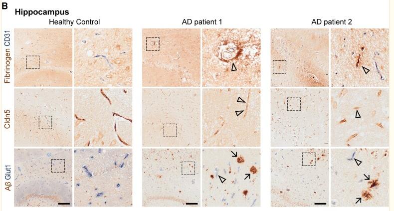
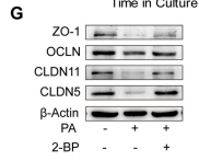
Fig. 4. Inhibition of protein palmitoylation ameliorates PA induced Sertoli cell dysfunction. (A) Analysis of the palmitoylation levels of proteins extracted from the testes of mice administered or not administered the PA injection, with or without gavage of 2-BP (n = 6 for Control, n = 7 for PA and 2-BP + PA). (B) Analysis of the palmitoylation levels of proteins extracted from primary Sertoli cells, Leydig cells and germ cells, which were treated with or without 0.4 mM PA (n = 3). (C) Inhibition of palmitoylation by 2-BP suppressed PA-induced ER stress in Sertoli cells. Translational expression levels of ER stress-related genes were analyzed using Western blotting (n = 3). (D) ROS detection using DCFH-DA staining. The fluorescence densities were calculated using ImageJ (n = 3). Scale bar: 50 μm. (E) TER detection of primary Sertoli cell barriers. The cells were incubated with PA (PA), with PA combined with 2-BP (2-BP + PA), or with the vehicle (Control) for 3 days after barriers were formed on day 4 (n = 5). (F) FITC-dextran permeability assessment of primary Sertoli cell barriers. The cells were treated PA (PA), with PA combined with 2-BP (2-BP + PA), or with the vehicle (Control) for 24 h after cell barriers were formed (n = 5). (G)Tight junction protein levels were examined by western blotting in TM4 Sertoli cells (n = 3). The relative intensities of bands in western blotting results were quantified by ImageJ and normalized to β-actin levels. Data are presented as mean ± SD. n. s., no significant difference vs. Control group. *P < 0.05, **P < 0.01 and ***P < 0.001 vs. Control group. #P < 0.05, ##P < 0.01 and ###P < 0.001 vs. PA group.

4). Activation of Wnt/β-catenin pathway mitigates blood–brain barrier dysfunction in Alzheimer's disease. BRAIN, 2022 (PubMed: 35788280) [IF=10.6]

Application: IHC    Species: Human    Sample: hippocampus

Figure 1 BBB dysfunction and BEC disruption in post-mortem Alzheimer’s disease brains. (A) Fibrinogen/CD31, Cldn5 and Aβ/Glut1 staining in the cortex and (B) hippocampus of post-mortem health control and patients with Alzheimer’s disease. (C) Quantification of Cldn5 and (D) Glut1 intensity, and (E) Aβ plaques in the cortex and hippocampus of healthy controls and patients with Alzheimer’s disease. Black frames in the low-magnification images indicate the location of the high-magnification images to the right. Arrows indicate Aβ plaques, and arrowheads indicate the fibrinogen leakage and decreased Cldn5 and Glut1 in patients with Alzheimer’s disease. Scale bar = 200 µm in A and B. Data are presented as mean ± SEM, n = 5, *P < 0.05, **P < 0.01.

5). A novel IL-1RA-PEP fusion protein alleviates blood-brain barrier disruption after ischemia-reperfusion in male rats. Journal of Neuroinflammation, 2018 (PubMed: 29334965) [IF=9.3]

6). Glyceryl triacetate promotes blood-brain barrier recovery after ischemic stroke through lipogenesis-mediated IL-33 in mice. Journal of neuroinflammation, 2023 (PubMed: 37968698) [IF=9.3]

Application: WB    Species: Mouse    Sample:

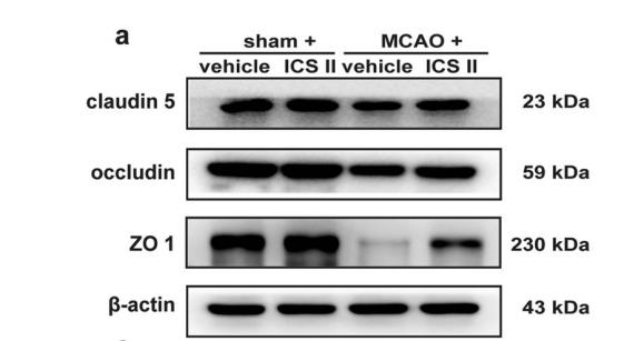
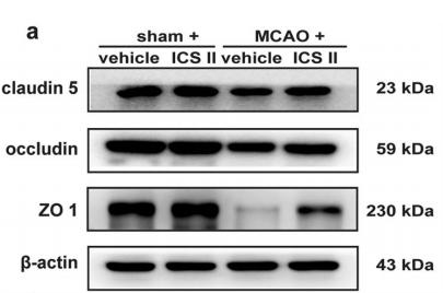
Fig. 3 Treatment with GTA improved BBB permeability in the peri-infarct area on day 7 after cerebral ischemia. A The representative image of extravagated dextran and IgG in the peri-infarct area. B and C Quantification of dextran and IgG intensity based on A. D The representative image of Evans blue leakage. E Quantification of D. F, Western blotting of tight junction proteins in the peri-infarct area. G–I Quantification of ZO-1, Occludin and Claudin-5 based on F. n = 5 for B and C, n = 4 for E–I. Compared with Sham, *P 

7). Robo4 inhibits gamma radiation-induced permeability of a murine microvascular endothelial cell by regulating the junctions. Cellular & Molecular Biology Letters, 2023 (PubMed: 36647012) [IF=9.2]

8). Endothelial delivery of simvastatin by LRP1-targeted nanoparticles ameliorates pathogenesis of alzheimer's disease in a mouse model. Alzheimer's research & therapy, 2025 (PubMed: 40830501) [IF=9.0]

Application: IF/ICC    Species: Mouse    Sample:

Fig. 4 S@A-NPs treatment ameliorates impairment in BBB of APP/PS1 transgenic mice. (a-f) Coronal hippocampal sections were immunostained for CD31 and ZO1 (a), Claudin 5 (b) or Occludin (c), and DAPI. Percentages of ZO1+ (d), Claudin 5+ (e), and Occludin+ (f) area occupied in the total CD31+ area. (g) Representative images in the cortex and hippocampus coronal sections assessed BBB leakage by 10 KDa and 40 KDa FITC-dextran. (h, i) Percentage of 10 KDa (h) or 40 KDa (i) FITC-dextran+ areas in the cortex and hippocampus coronal sections. Data are presented as mean ± SEM. n = 13–15 slices from 3 mice/group (d, e); n = 14–17 slices from 3 mice/group (f); n = 3 mice/group (h, i); * p 

9). Curcumin-loaded milk-derived sEVs fused with platelet membrane attenuate endothelial senescence and promote spinal cord injury recovery in diabetic mice. Materials today. Bio, 2025 (PubMed: 40688662) [IF=8.7]

Application: WB    Species: human    Sample:

Fig. 3. Reduction in HG/IL-1β-induced senescence markers and TJ protein degradation in HUVECs by Cur treatment. (A) Typical images of HUVECs stained with SA-β-gal (blue stain indicates senescent cells). The number of SA-β-gal+ cells decreased in the Cur group compared to the HG/IL-1β + DMSO group, whereas it increased after brusatol treatment. Scale bar = 200 μm. (B) Quantification of SA-β-gal+ cells (n = 5). (C) Typical images of Claudin-5 staining (green, TJ protein). The intensity of Claudin-5 increased in the Cur group compared to the HG/IL-1β + DMSO group, which was decreased by brusatol treatment. Scale bar = 100 μm. (D) Quantification of the relative fluorescence intensity of Claudin-5 (n = 5). (E) Analysis of VE-Cadherin, ZO-1, Occludin and Claudin-5 expression in HUVECs by WB (TJ protein). The level of TJ protein was increased in the Cur group compared to the HG/IL-1β + DMSO group, which was decreased by brusatol treatment. (F–I) Quantification of the level of VE-Cadherin, ZO-1, Claudin-5, Occludin (n = 3). (J) Analysis of NRF2, HO-1, P21 and P16 expression in HUVECs by WB. Note: (1) The levels of NRF2 and HO-1 were upregulated in the Cur group compared to the HG/IL-1β + DMSO group, which was reduced by brusatol treatment. (2) The levels of P21 and P16 were decreased in the Cur group compared to the HG/IL-1β + DMSO group, which were increased by brusatol treatment. (K–N) Quantification of NRF2, HO-1, P21, and P16 levels (n = 3). ∗P < 0.05, ∗∗P < 0.01. (For interpretation of the references to colour in this figure legend, the reader is referred to the Web version of this article.)

10). Endothelial DR6 in blood-brain barrier malfunction in Alzheimer's disease. Cell death & disease, 2024 (PubMed: 38609388) [IF=8.1]

もっと読みます

Restrictive clause


Affinity Biosciences tests all products strictly. Citations are provided as a resource for additional applications that have not been validated by Affinity Biosciences. Please choose the appropriate format for each application and consult Materials and Methods sections for additional details about the use of any product in these publications.

For Research Use Only.
Not for use in diagnostic or therapeutic procedures. Not for resale. Not for distribution without written consent. Affinity Biosciences will not be held responsible for patent infringement or other violations that may occur with the use of our products. Affinity Biosciences, Affinity Biosciences Logo and all other trademarks are the property of Affinity Biosciences LTD.