製品: N Cadherin Antibody
カタログ: AF5239
タンパク質の説明: Rabbit polyclonal antibody to N Cadherin
アプリケーション: WB IHC IF/ICC
Cited expt.: WB, IHC, IF/ICC
反応性: Human, Mouse, Rat
予測: Pig, Zebrafish, Bovine, Horse, Sheep, Rabbit, Dog, Chicken, Xenopus
分子量: 100~150kd; 100kD(Calculated).
ユニプロット: P19022
RRID: AB_2837725

類似製品を見る>>

   サイズ 価格 在庫状況
 100ul $280 在庫あり
 200ul $350 在庫あり

リードタイム: 当日配達

For pricing and ordering contact:
お問い合わせ先

製品説明

ソース:
Rabbit
アプリケーション:
WB 1:500-1:2000, IF/ICC 1:100-1:500, IHC 1:50-1:200
*The optimal dilutions should be determined by the end user. For optimal experimental results, antibody reuse is not recommended.
*Tips:

WB: For western blot detection of denatured protein samples. IHC: For immunohistochemical detection of paraffin sections (IHC-p) or frozen sections (IHC-f) of tissue samples. IF/ICC: For immunofluorescence detection of cell samples. ELISA(peptide): For ELISA detection of antigenic peptide.

反応性:
Human,Mouse,Rat
予測:
Pig(100%), Zebrafish(100%), Bovine(100%), Horse(100%), Sheep(100%), Rabbit(100%), Dog(100%), Chicken(100%), Xenopus(100%)
クローナリティ:
Polyclonal
特異性:
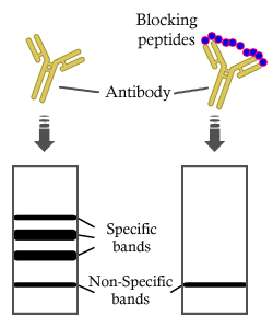
N Cadherin Antibody detects endogenous levels of total N Cadherin.
RRID:
AB_2837725
引用形式: Affinity Biosciences Cat# AF5239, RRID:AB_2837725.
コンジュゲート:
Unconjugated.
精製:
The antiserum was purified by peptide affinity chromatography using SulfoLink™ Coupling Resin (Thermo Fisher Scientific).
保存:
Rabbit IgG in phosphate buffered saline , pH 7.4, 150mM NaCl, 0.02% sodium azide and 50% glycerol. Store at -20 °C. Stable for 12 months from date of receipt.
別名:

折りたたみ/展開

CADH2_HUMAN; Cadherin 2; Cadherin 2 N cadherin neuronal; Cadherin 2 type 1; Cadherin 2 type 1 N cadherin neuronal; Cadherin 2, type 1, N-cadherin (neuronal); Cadherin-2; Cadherin2; Calcium dependent adhesion protein neuronal; CD325; CD325 antigen; CDH2; CDHN; CDw325; CDw325 antigen; N cadherin 1; N-cadherin; NCAD; Neural cadherin; OTTHUMP00000066304; OTTHUMP00000067378;

免疫原

免疫原:

A synthesized peptide derived from human N Cadherin, corresponding to a region within C-terminal amino acids.

Uniprot:
遺伝子(ID):
タンパク質の説明:
Cadherins are calcium-dependent cell adhesion proteins. They preferentially interact with themselves in a homophilic manner in connecting cells; cadherins may thus contribute to the sorting of heterogeneous cell types. CDH2 may be involved in neuronal recognition mechanism. In hippocampal neurons, may regulate dendritic spine density
タンパク質配列:
MCRIAGALRTLLPLLAALLQASVEASGEIALCKTGFPEDVYSAVLSKDVHEGQPLLNVKFSNCNGKRKVQYESSEPADFKVDEDGMVYAVRSFPLSSEHAKFLIYAQDKETQEKWQVAVKLSLKPTLTEESVKESAEVEEIVFPRQFSKHSGHLQRQKRDWVIPPINLPENSRGPFPQELVRIRSDRDKNLSLRYSVTGPGADQPPTGIFIINPISGQLSVTKPLDREQIARFHLRAHAVDINGNQVENPIDIVINVIDMNDNRPEFLHQVWNGTVPEGSKPGTYVMTVTAIDADDPNALNGMLRYRIVSQAPSTPSPNMFTINNETGDIITVAAGLDREKVQQYTLIIQATDMEGNPTYGLSNTATAVITVTDVNDNPPEFTAMTFYGEVPENRVDIIVANLTVTDKDQPHTPAWNAVYRISGGDPTGRFAIQTDPNSNDGLVTVVKPIDFETNRMFVLTVAAENQVPLAKGIQHPPQSTATVSVTVIDVNENPYFAPNPKIIRQEEGLHAGTMLTTFTAQDPDRYMQQNIRYTKLSDPANWLKIDPVNGQITTIAVLDRESPNVKNNIYNATFLASDNGIPPMSGTGTLQIYLLDINDNAPQVLPQEAETCETPDPNSINITALDYDIDPNAGPFAFDLPLSPVTIKRNWTITRLNGDFAQLNLKIKFLEAGIYEVPIIITDSGNPPKSNISILRVKVCQCDSNGDCTDVDRIVGAGLGTGAIIAILLCIIILLILVLMFVVWMKRRDKERQAKQLLIDPEDDVRDNILKYDEEGGGEEDQDYDLSQLQQPDTVEPDAIKPVGIRRMDERPIHAEPQYPVRSAAPHPGDIGDFINEGLKAADNDPTAPPYDSLLVFDYEGSGSTAGSLSSLNSSSSGGEQDYDYLNDWGPRFKKLADMYGGGDD

種類予測

種類予測:

Score>80(red) has high confidence and is suggested to be used for WB detection. *The prediction model is mainly based on the alignment of immunogen sequences, the results are for reference only, not as the basis of quality assurance.

Species
Results
Score
Pig
100
Horse
100
Bovine
100
Sheep
100
Dog
100
Xenopus
100
Zebrafish
100
Chicken
100
Rabbit
100
Model Confidence:
High(score>80) Medium(80>score>50) Low(score<50) No confidence

研究背景

機能:

Calcium-dependent cell adhesion protein; preferentially mediates homotypic cell-cell adhesion by dimerization with a CDH2 chain from another cell. Cadherins may thus contribute to the sorting of heterogeneous cell types. Acts as a regulator of neural stem cells quiescence by mediating anchorage of neural stem cells to ependymocytes in the adult subependymal zone: upon cleavage by MMP24, CDH2-mediated anchorage is affected, leading to modulate neural stem cell quiescence. CDH2 may be involved in neuronal recognition mechanism. In hippocampal neurons, may regulate dendritic spine density.

PTMs:

Cleaved by MMP24. Ectodomain cleavage leads to the generation of a soluble 90 kDa amino-terminal soluble fragment and a 45 kDa membrane-bound carboxy-terminal fragment 1 (CTF1), which is further cleaved by gamma-secretase into a 35 kDa. Cleavage in neural stem cells by MMP24 affects CDH2-mediated anchorage of neural stem cells to ependymocytes in the adult subependymal zone, leading to modulate neural stem cell quiescence (By similarity).

May be phosphorylated by OBSCN.

細胞の位置付け:

Cell membrane>Single-pass type I membrane protein. Cell membrane>Sarcolemma. Cell junction. Cell surface.
Note: Colocalizes with TMEM65 at the intercalated disk in cardiomyocytes. Colocalizes with OBSCN at the intercalated disk and at sarcolemma in cardiomyocytes.

Extracellular region or secreted Cytosol Plasma membrane Cytoskeleton Lysosome Endosome Peroxisome ER Golgi apparatus Nucleus Mitochondrion Manual annotation Automatic computational assertionSubcellular location
タンパク質ファミリー:

Three calcium ions are usually bound at the interface of each cadherin domain and rigidify the connections, imparting a strong curvature to the full-length ectodomain. Calcium-binding sites are occupied sequentially in the order of site 3, then site 2 and site 1.

研究領域

· Environmental Information Processing > Signaling molecules and interaction > Cell adhesion molecules (CAMs).   (View pathway)

· Human Diseases > Cardiovascular diseases > Arrhythmogenic right ventricular cardiomyopathy (ARVC).

参考文献

1). OTUD6A in Airway Epithelial Cells Exacerbates Allergic Asthma by Promoting Airway Inflammation and Airway Remodeling Through Deubiquitination of hResistin/mRELMα. Advanced science (Weinheim, Baden-Wurttemberg, Germany), 2026 (PubMed: 41560298) [IF=15.1]

2). METTL13 Mediates the Translation of Snail in Head and Neck Squamous Cell Carcinoma. International Journal of Oral Science, 2020 (PubMed: 34381012) [IF=10.8]

3). Enhanced oral nanomedicine utilizing biomineralized oncolytic virus for synergistic gastrointestinal cancer therapy. Materials today. Bio, 2025 (PubMed: 41377584) [IF=8.7]

4). Prolyl hydroxylase-2 (EGLN1) mediates TGF-β1 pathway regulated epithelial–mesenchymal transition in yak kidneys. International Journal of Biological Macromolecules, 2025 [IF=8.5]

5). SB431542 partially inhibits high glucose-induced EMT by restoring mitochondrial homeostasis in RPE cells. Cell communication and signaling : CCS, 2024 (PubMed: 38183022) [IF=8.4]

6). BRD7 inhibits enhancer activity and expression of BIRC2 to suppress tumor growth and metastasis in nasopharyngeal carcinoma. Cell Death & Disease, 2023 (PubMed: 36788209) [IF=8.1]

Application: WB    Species: Human    Sample: 5-8 F and HNE1 cells

Fig. 5 Restoration of BIRC2 expression reverses the inhibitory effect of BRD7 on cell migration and invasion. A Scratch wound healing analysis of cell migration in 5-8 F and HNE1 cells stably with BRD7 overexpression, BRD7 and BIRC2 simultaneous overexpression or control. Quantification of the wound recovery rate of the three groups (right). B Matrigel invasion analysis of cell invasive capabilities in 5-8 F and HNE1 cells stably with BRD7 overexpression, BRD7 and BIRC2 simultaneous overexpression or control. C Significantly differently expressed proteins involved in EMT progression (E-cadherin, N-cadherin, Vimentin, ZO-1) in BRD7 overexpression, BIRC2 overexpression and BIRC2 restoration cells, respectively. GAPDH served as an internal control. Error bars represent the mean ± SD. *P 

7). Deprivation of methionine inhibits osteosarcoma growth and metastasis via C1orf112-mediated regulation of mitochondrial functions. Cell death & disease, 2024 (PubMed: 38769167) [IF=8.1]

8). Targeting the MDK/c-Myc complex to overcome temozolomide resistance in glioma. Clinical and translational medicine, 2025 (PubMed: 40468625) [IF=7.9]

Application: WB    Species: Mouse    Sample:

FIGURE 4 MDK affects the ubiquitination modification of c-Myc and the Wnt/β-catenin signalling pathway. (A–C) Results from proteomic analysis indicated that MDK affected the Wnt/β-catenin signalling pathway and protein ubiquitination. The raw data are shown in Table S8. (A) Volcano plot analysis results showed differentially expressed proteins (Table S9). The red dots represent upregulated expression in the siMDK group compared with the control group, and the yellow dots represent downregulated expression in the siMDK group compared with the control group. (B) Cluster analysis results showed similarities and differences among the groups (control vs. siMDK) and the stability of the mass spectrometry results of the three repeated experiments (Table S10). (C) KEGG pathway enrichment analysis revealed the signalling pathway affected by MDK knockdown (Table S11). (D) After treatment with 10 mM MG132 for 4 h, siMDK-U118MG and siMDK-SF126 cell lysates were immunoprecipitated with c-Myc and immunoblotted with ubiquitination antibodies. (E) After treatment with 10 mM MG132 for 4 h, OE-MDK-SHG44 and OE-MDK-U87 cell lysates were immunoprecipitated with c-Myc and immunoblotted with ubiquitination antibodies. (F–G) 50 mg/mL cycloheximide was added (at different time points: 0, 2, 4, 6, and 8 h) to glioma cells transfected with siRNA to block protein synthesis. The expression of c-Myc was then detected by Western blot. (H) MDK knockdown was performed on SF126, U118MG, and U251, and then a Western blot was used to detect the Wnt/β-catenin signalling pathway and EMT pathway markers. (I) MDK was overexpressed in U87, BT325, and SHG44, and Western blot was used to detect the Wnt/β-catenin signalling pathway and EMT pathway markers.

Application: WB    Species: Mouse    Sample:

FIGURE 4 MDK affects the ubiquitination modification of c-Myc and the Wnt/β-catenin signalling pathway. (A–C) Results from proteomic analysis indicated that MDK affected the Wnt/β-catenin signalling pathway and protein ubiquitination. The raw data are shown in Table S8. (A) Volcano plot analysis results showed differentially expressed proteins (Table S9). The red dots represent upregulated expression in the siMDK group compared with the control group, and the yellow dots represent downregulated expression in the siMDK group compared with the control group. (B) Cluster analysis results showed similarities and differences among the groups (control vs. siMDK) and the stability of the mass spectrometry results of the three repeated experiments (Table S10). (C) KEGG pathway enrichment analysis revealed the signalling pathway affected by MDK knockdown (Table S11). (D) After treatment with 10 mM MG132 for 4 h, siMDK-U118MG and siMDK-SF126 cell lysates were immunoprecipitated with c-Myc and immunoblotted with ubiquitination antibodies. (E) After treatment with 10 mM MG132 for 4 h, OE-MDK-SHG44 and OE-MDK-U87 cell lysates were immunoprecipitated with c-Myc and immunoblotted with ubiquitination antibodies. (F–G) 50 mg/mL cycloheximide was added (at different time points: 0, 2, 4, 6, and 8 h) to glioma cells transfected with siRNA to block protein synthesis. The expression of c-Myc was then detected by Western blot. (H) MDK knockdown was performed on SF126, U118MG, and U251, and then a Western blot was used to detect the Wnt/β-catenin signalling pathway and EMT pathway markers. (I) MDK was overexpressed in U87, BT325, and SHG44, and Western blot was used to detect the Wnt/β-catenin signalling pathway and EMT pathway markers.

9). Huaier polysaccharides suppress triple-negative breast cancer metastasis and epithelial-mesenchymal transition by inducing autophagic degradation of Snail. Cell and Bioscience, 2021 (PubMed: 34481526) [IF=7.5]

10). TRIML2 promotes malignant progression of head and neck squamous cell carcinoma via canonical Wnt signaling and tumor immune escape. Journal of Translational Medicine, 2025 [IF=7.5]

Application: WB    Species: human    Sample: HNSC cells

Fig. 8 TRIML2 knockdown inhibits the proliferation, migration, and invasion of HNSC cells. (A, B) Western blot and RT-qPCR were used to validate the knockdown efficiency of TRIML2 in HNSC cell lines. (C-F) The proliferative ability was assessed by CCK-8 and EdU assays. (G-H) Migration and invasion ability were measured by wound-healing and transwell assays. (I) the canonical wnt and EMT-related proteins were detected by Western blot after TRIML2 knockdown. All in vitro experiments were performed in at least three independent replicates. The data are presented as the mean ± SD.

もっと読みます

Restrictive clause


Affinity Biosciences tests all products strictly. Citations are provided as a resource for additional applications that have not been validated by Affinity Biosciences. Please choose the appropriate format for each application and consult Materials and Methods sections for additional details about the use of any product in these publications.

For Research Use Only.
Not for use in diagnostic or therapeutic procedures. Not for resale. Not for distribution without written consent. Affinity Biosciences will not be held responsible for patent infringement or other violations that may occur with the use of our products. Affinity Biosciences, Affinity Biosciences Logo and all other trademarks are the property of Affinity Biosciences LTD.