製品: PPAR alpha Antibody
カタログ: AF5301
タンパク質の説明: Rabbit polyclonal antibody to PPAR alpha
アプリケーション: WB IHC IF/ICC
反応性: Human, Mouse, Rat
予測: Bovine, Horse, Sheep, Rabbit, Dog, Xenopus
分子量: 52 kDa; 52kD(Calculated).
ユニプロット: Q07869
RRID: AB_2837786

類似製品を見る>>

   サイズ 価格 在庫状況
 100ul $280 在庫あり
 200ul $350 在庫あり

リードタイム: 当日配達

For pricing and ordering contact:
お問い合わせ先

製品説明

ソース:
Rabbit
アプリケーション:
WB 1:500-1:2000, IHC 1:50-1:200, IF/ICC 1:100-1:500
*The optimal dilutions should be determined by the end user.
*Tips:

WB: For western blot detection of denatured protein samples. IHC: For immunohistochemical detection of paraffin sections (IHC-p) or frozen sections (IHC-f) of tissue samples. IF/ICC: For immunofluorescence detection of cell samples. ELISA(peptide): For ELISA detection of antigenic peptide.

反応性:
Human,Mouse,Rat
予測:
Bovine(100%), Horse(88%), Sheep(100%), Rabbit(100%), Dog(100%), Xenopus(100%)
クローナリティ:
Polyclonal
特異性:
PPAR alpha Antibody detects endogenous levels of total PPAR alpha.
RRID:
AB_2837786
引用形式: Affinity Biosciences Cat# AF5301, RRID:AB_2837786.
コンジュゲート:
Unconjugated.
精製:
The antiserum was purified by peptide affinity chromatography using SulfoLink™ Coupling Resin (Thermo Fisher Scientific).
保存:
Rabbit IgG in phosphate buffered saline , pH 7.4, 150mM NaCl, 0.02% sodium azide and 50% glycerol. Store at -20 °C. Stable for 12 months from date of receipt.
別名:

折りたたみ/展開

hPPAR; MGC2237; MGC2452; NR1C1; Nuclear receptor subfamily 1 group C member 1; OTTHUMP00000197740; OTTHUMP00000197741; Peroxisome proliferative activated receptor alpha; Peroxisome proliferator activated receptor alpha; Peroxisome proliferator-activated receptor alpha; PPAR; PPAR-alpha; ppara; PPARA_HUMAN; PPARalpha;

免疫原

免疫原:
Uniprot:
遺伝子(ID):
発現特異性:
Q07869 PPARA_HUMAN:

Skeletal muscle, liver, heart and kidney. Expressed in monocytes (PubMed:28167758).

タンパク質の説明:
Ligand-activated transcription factor. Key regulator of lipid metabolism. Activated by the endogenous ligand 1-palmitoyl-2-oleoyl-sn-glycerol-3-phosphocholine (16:0/18:1-GPC). Activated by oleylethanolamide, a naturally occurring lipid that regulates satiety (By similarity). Receptor for peroxisome proliferators such as hypolipidemic drugs and fatty acids. Regulates the peroxisomal beta-oxidation pathway of fatty acids.
タンパク質配列:
MVDTESPLCPLSPLEAGDLESPLSEEFLQEMGNIQEISQSIGEDSSGSFGFTEYQYLGSCPGSDGSVITDTLSPASSPSSVTYPVVPGSVDESPSGALNIECRICGDKASGYHYGVHACEGCKGFFRRTIRLKLVYDKCDRSCKIQKKNRNKCQYCRFHKCLSVGMSHNAIRFGRMPRSEKAKLKAEILTCEHDIEDSETADLKSLAKRIYEAYLKNFNMNKVKARVILSGKASNNPPFVIHDMETLCMAEKTLVAKLVANGIQNKEAEVRIFHCCQCTSVETVTELTEFAKAIPGFANLDLNDQVTLLKYGVYEAIFAMLSSVMNKDGMLVAYGNGFITREFLKSLRKPFCDIMEPKFDFAMKFNALELDDSDISLFVAAIICCGDRPGLLNVGHIEKMQEGIVHVLRLHLQSNHPDDIFLFPKLLQKMADLRQLVTEHAQLVQIIKKTESDAALHPLLQEIYRDMY

種類予測

種類予測:

Score>80(red) has high confidence and is suggested to be used for WB detection. *The prediction model is mainly based on the alignment of immunogen sequences, the results are for reference only, not as the basis of quality assurance.

Species
Results
Score
Bovine
100
Sheep
100
Dog
100
Xenopus
100
Rabbit
100
Horse
88
Pig
0
Zebrafish
0
Chicken
0
Model Confidence:
High(score>80) Medium(80>score>50) Low(score<50) No confidence

PTMs - Q07869 基板として

Site PTM Type Enzyme
S6 Phosphorylation
S12 Phosphorylation P28482 (MAPK1) , P27361 (MAPK3)
S21 Phosphorylation P27361 (MAPK3) , P28482 (MAPK1)
S45 Phosphorylation
S179 Phosphorylation P05771-2 (PRKCB) , P17252 (PRKCA)
K185 Sumoylation
S230 Phosphorylation P05771-2 (PRKCB) , P17252 (PRKCA)

研究背景

機能:

Ligand-activated transcription factor. Key regulator of lipid metabolism. Activated by the endogenous ligand 1-palmitoyl-2-oleoyl-sn-glycerol-3-phosphocholine (16:0/18:1-GPC). Activated by oleylethanolamide, a naturally occurring lipid that regulates satiety. Receptor for peroxisome proliferators such as hypolipidemic drugs and fatty acids. Regulates the peroxisomal beta-oxidation pathway of fatty acids. Functions as transcription activator for the ACOX1 and P450 genes. Transactivation activity requires heterodimerization with RXRA and is antagonized by NR2C2. May be required for the propagation of clock information to metabolic pathways regulated by PER2.

細胞の位置付け:

Nucleus.

Extracellular region or secreted Cytosol Plasma membrane Cytoskeleton Lysosome Endosome Peroxisome ER Golgi apparatus Nucleus Mitochondrion Manual annotation Automatic computational assertionSubcellular location
組織特異性:

Skeletal muscle, liver, heart and kidney. Expressed in monocytes.

サブユニット構造:

Heterodimer; with RXRA. This heterodimerization is required for DNA binding and transactivation activity. Interacts with NCOA3 coactivator. Interacts with CITED2; the interaction stimulates its transcriptional activity. Also interacts with PPARBP in vitro. Interacts with AKAP13, LPIN1, PRDM16 and coactivator NCOA6. Interacts with ASXL1 and ASXL2. Interacts with PER2. Interacts with SIRT1; the interaction seems to be modulated by NAD(+) levels. Interacts with CRY1 and CRY2 (By similarity).

タンパク質ファミリー:

Belongs to the nuclear hormone receptor family. NR1 subfamily.

研究領域

· Environmental Information Processing > Signal transduction > cAMP signaling pathway.   (View pathway)

· Human Diseases > Endocrine and metabolic diseases > Insulin resistance.

· Human Diseases > Endocrine and metabolic diseases > Non-alcoholic fatty liver disease (NAFLD).

· Human Diseases > Infectious diseases: Viral > Hepatitis C.

· Organismal Systems > Endocrine system > PPAR signaling pathway.

· Organismal Systems > Endocrine system > Adipocytokine signaling pathway.

· Organismal Systems > Endocrine system > Glucagon signaling pathway.

参考文献

1). Rescuing SERCA2 pump deficiency improves bone mechano-responsiveness in type 2 diabetes by shaping osteocyte calcium dynamics. Nature communications, 2024 (PubMed: 38291059) [IF=16.6]

Application: WB    Species: Mouse    Sample:

Fig. 9 The nuclear transcription factor PPARα mediates T2D-induced specific reduction in the expression of osteocytic SERCA2 pump. a RNA-seq-based KEGG pathway analysis showing the top 10 enriched KEGG pathways. b, c GSEA analysis showing a significant enrichment of signaling events associated with PPARα and PPARDR1_Q2. d The PPARα and p-PPARα protein expression in osteocytes. e Immunohistochemical staining of osteocytic PPARα in diabetic and non-diabetic tibiae. f The SERCA2 expression in osteocytes treated with antagonists of PPARα (MK886 and GW6471), PPARβ/δ (GSK0660 and GSK3787), and PPARγ (T0070907 and GW9662). g The SERCA2 expression in HGHF-exposed osteocytes treated with agonists of PPARα (Fenofibrate and GW7647), PPARβ/δ (GW0742 and GW501516), and PPARγ (rosiglitazone and pioglitazone). h A schematic representation and the relative luciferase activities of three ATP2A2 promotor regions. i The relative luciferase activity assays of Seg#1, Seg#2 and Seg#3. j The relative luciferase activity in HGHF-treated osteocytes with PPARα silencing. k ChIP assays showing the PPARα enrichment on ATP2A2 promotor in normal and HGHF-treated osteocytes. l EMSA assays confirming the binding of PPARα to the ATP2A2 promoter region (−620 to −608 bp). The nuclear extract were incubated with biotin-labeled wild-type (WT-biotin) probe, unlabeled wild-type (WT) probe, and biotin-labeled mutated (Mut-biotin) probe. Red letters indicate substituted nucleotide sequences in the mutated probes (P1: −620 to −608 bp; P2: −1283 to −1277 bp). m, n Intracellular Ca2+ signaling and protein expression of osteocyte-related cytokines in HGHF-treated osteocytes with PPARα overexpression subjected to FSS. o, p Intracellular Ca2+ signaling and the expression of osteocyte-related cytokines in MLO-Y4 cells with lentiviral silencing of PPARα subjected to FSS. Graphs represent mean ± SD (d, f, g, i–k, m, o n = 6 biologically independent replicates; e n = 8 mice per group; l n = 3 independent replicates; n, p: n = 120 cells per group). a P value was obtained by one-tailed hypergeometric test. b, c P values were obtained by one-tailed permutation test. d–h, n, p ***P 

2). Regulatory effects mediated by ulvan oligosaccharide and its zinc complex on lipid metabolism in high-fat diet-fed mice. Carbohydrate Polymers, 2023 (PubMed: 36372481) [IF=10.7]

3). Yogurt-derived Lactobacillus plantarum Q16 alleviated high-fat diet-induced non-alcoholic fatty liver disease in mice. Food Science and Human Wellness, 2022 [IF=7.0]

Application: WB    Species: Mouse    Sample:

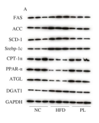
Fig. 5. Effects ofL. plantarum Q16 on key proteins involved in hepatic lipid metabolism in HFD-fed obese mice. Data are presented as mean ± SD (n = 6). Different lowercase alphabet letters were significantly different at the level of P < 0.05.

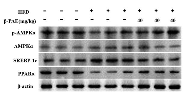
4). β-patchoulene improves lipid metabolism to alleviate non-alcoholic fatty liver disease via activating AMPK signaling pathway. BIOMEDICINE & PHARMACOTHERAPY, 2021 (PubMed: 33341045) [IF=6.9]

Application: WB    Species: Human    Sample: L02 cell

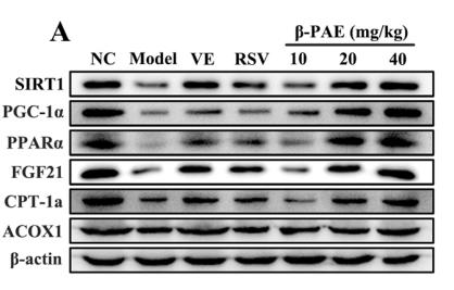
Fig. 6. β-PAE promotes the expression of hepatic lipid oxidation-related proteins and genes in HFD-fed rats. (A–G) Western blot analysis on the expression of SIRT1, PGC-1α, PPARα, FGF21, CPT-1a and ACOX1; (H–K) The mRNA expression of SIRT1, PPARα, CPT-1a and ACOX1. Data are presented as the mean ± SD (n = 6~8). ##p < 0.01 vs. NC group; *p < 0.05, **p < 0.01 vs. Model group.

5). Hepatic NCoR1 deletion exacerbates alcohol-induced liver injury in mice by promoting CCL2-mediated monocyte-derived macrophage infiltration. Acta Pharmacologica Sinica, 2022 (PubMed: 35149852) [IF=6.9]

6). Dietary sanguinarine supplementation recovers the decrease in muscle quality and nutrient composition induced by high-fat diets of grass carp (Ctenopharyngodon idella). Animal nutrition (Zhongguo xu mu shou yi xue hui), 2024 (PubMed: 38800733) [IF=6.1]

Application: WB    Species: fish    Sample:

Fig. 5 The effects of different diets on muscle protein synthesis and lipolysis of grass carp. (A) Protein synthesis related gene expression; (B) lipolysis-related gene expression; (C) Western blot analysis of p-AMPK, AMPK, PPARα, TOR, p70S6K, PI3K, and AKT protein expression; (D) relative quantification of p-AMPK, PPARα, TOR, p70S6K, PI3K, and AKT protein expression. The bars indicate the mean ± standard error (SE). Different superscripts denote significant differences (P < 0.05). CON = a control group (containing 4.95% crude fat); HFD = a high fat diet (containing 10.27% crude fat); HFDS = supplementing 1200 μg/kg sanguinarine to HFD.

7). Effects of polyphenolic maqui (Aristotelia chilensis) extract on the inhibition of NLRP3 inflammasome and activation of mast cells in a mouse model of Crohn's disease-like colitis. Frontiers in immunology, 2024 (PubMed: 38283356) [IF=5.7]

Application: IHC    Species: Mouse    Sample: colon tissue

Figure 6 Impact of Maqui extract rich in anthocyanins on PPAR-α expression in colon tissue from experimental animal model of Crohn’s disease-like colitis. Immuno‐histochemistry analyses for PPAR-α in epithelial cells from colons of each group (n= 6 mice/group): Positive PPAR-α staining in colon epithelium (black arrow) and crypts cells (dashed arrow) in (A) control group, (B) CD group, (C) post-Treatment and (D) pre-Treatment groups. Immuno‐histochemistry analyses for PPAR-α in macrophages cells from colons tissue from 10 representative fields of view for each group: Immune+-PPAR-α staining intensity (black arrow) in (F) control group, (G) CD group, (H) post-Treatment group and (I) pre-Treatment group, (E) Immune positive score (1= 50%) for PPAR−α expression in epithelial cells. (J) Counting cells across 10 fields (400x magnification) for PPAR−α expression in macrophages cells. Values represent mean ± SEM; *Difference at the p < 0.0005 level; Kruskal–Wallis test.

8). Probiotic Yogurt Alleviates High-Fat Diet-Induced Lipid Accumulation and Insulin Resistance in Mice via the Adiponectin Pathway. JOURNAL OF AGRICULTURAL AND FOOD CHEMISTRY, 2023 (PubMed: 36695046) [IF=5.7]

9). D-Mannose Regulates Hepatocyte Lipid Metabolism via PI3K/Akt/mTOR Signaling Pathway and Ameliorates Hepatic Steatosis in Alcoholic Liver Disease. Frontiers in immunology, 2022 (PubMed: 35464439) [IF=5.7]

10). Effects of Poria cocos extract on metabolic dysfunction-associated fatty liver disease via the FXR/PPARα-SREBPs pathway. Frontiers in Pharmacology, 2022 (PubMed: 36278226) [IF=5.6]

Application: WB    Species: Rat    Sample:

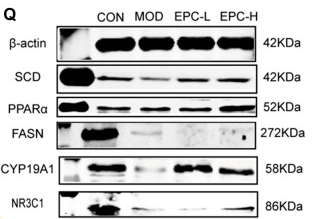
FIGURE 5 EPC regulated the lipid metabolism-related genes and proteins inMAFLD rats. (A) mRNA abundances of FASN. (B) mRNA abundances of PPARα (PPARΑ). (C) mRNA abundances of PPARγ (PPARG). (D) mRNA abundances of RXRα. (E) mRNA abundances of CYP19A1. (F) mRNA abundances of NR3C1. (G) mRNA abundances of SREBP-1c. (H) mRNA abundances of HMGCR. (I) mRNA abundances of SCD. n = 6; (J) Relative expression of protein SCD. (K) Relative expression of protein PPARα. (L) Relative expression of protein FASN. (M) Relative expression of protein p-JNK. (N) Relative expression of protein p-NF-κB. (O) Relative expression of protein CYP19A1. (P) Relative expression of protein NR3C1. (Q-R) Representative immunoblotting images of β-actin, SCD, PPARα, FASN,CYP19A1, NR3C1, JNK, p-JNK, NF-κB, pNF-κB; n = 4, data are presented as mean ± SEM. One-way analysis of variance (ANOVA) was conducted for the group comparison. *p < 0.05, **p < 0.01, ***p < 0.001 vs MOD group. FASN, fatty acid synthase; PPARα/γ, peroxisome proliferator-activated receptor alpha/gamma; RXRa, retinoic acid receptor alpha; SREBP-1c, sterol regulatory element binding protein 1c; HMGCR, 3-hydroxy-3-methylglutaryl-coenzyme A reductase; SCD, Stearoyl-CoA desaturase; p-JNK, phosphorylation (p) -stress-activated protein kinase JNK; NF-κB, nuclear factor kappa B.

もっと読みます

Restrictive clause


Affinity Biosciences tests all products strictly. Citations are provided as a resource for additional applications that have not been validated by Affinity Biosciences. Please choose the appropriate format for each application and consult Materials and Methods sections for additional details about the use of any product in these publications.

For Research Use Only.
Not for use in diagnostic or therapeutic procedures. Not for resale. Not for distribution without written consent. Affinity Biosciences will not be held responsible for patent infringement or other violations that may occur with the use of our products. Affinity Biosciences, Affinity Biosciences Logo and all other trademarks are the property of Affinity Biosciences LTD.