

.
Bands result from membrane strip incubation.")


 and mouse anti-beta tubulin Ab(T0023 1:200) for 1 hour at 37°C. An AlexaFluor594 conjugated goat anti-rabbit IgG(H+L) Ab(Red) and an AlexaFluor488 conjugated goat anti-mouse IgG(H+L) Ab(Green) were used as the secondary antibody.
The nuclear counter stain is DAPI(blue).")

 Oil Red O staining observation of liver (×200, scale bars = 100 μm). (B) H&E staining observation of liver (×200, scale bars = 50 μm). (C) The volume density of quantitation of hepatic steatosis (n = 5). (D) The genes expression involved in fatty acid β-oxidation in liver (n = 5). (E) The mRNA expression of genes involved in lipogenesis and FXR in liver (n = 5). (F) Bands of PPARα,PPARγ, ACOX1, FAS, SREBP-1c, SCD1.")

 PCR analysis of Fas expression (n = 5). (b) The protein
level of Rank and Fas detected by western blotting analyzed by using integrated density (area*mean gray
value) (n = 5). (c) PCR analysis of IL-17, IL-21, RORγt, IFN-γ, Klrd1 and T-bet expression (n = 5). (d) Flow
cytometry analysis of CD4 and IL-17 or IFN-γ positive cells (n = 5). (e) ELISA analysis of IL-17 and IFN-γ
levels in cell culture media. (f) The protein level of SATA1 and STAT3 level by western blotting analyzed by
using integrated density (n = 5). * Indicates that there was a significant difference between two groups,
P < .05; N.S. meant there was no significant difference between two groups. DF: differentiated CD4 + T
cells; DF NC: differentiated negative control siRNA pretreated CD4 + T cells; DF F-ko: differentiated siRNA
of Fas pretreated CD4 + T cells. “n = 5” means the five repeats.")

 293T cells were transfected with FAH-Luc and renila, with or without TNF-α; 48 hours after transfection, cells was performed luciferase assay. The luciferase activity was normalized to Renilla control, and expressed as fold increase (mean ± SD). Experiments were performed 2 times in triplicate. (B and C) SW1353 were co-transfected with TNF-α and FAH or FAH-siRNA; 48 hours after transfection, cells were harvested for immunoblot analysis with indicated antibodies. FAH = fumarylacetoacetase hydrolase; TNF-α = tumor necrosis factor-α.")
 WB analysis of the expression of DR4 and 5 in HepG2 cells that were treated with 2 μM C20/C22 for 0, 6, 12, and 24 h. ImageJ software was used to calculate the gray value (n = 3). (C,D) The immunofluorescence assays used to determine the intracellular and cell surface distribution of DR4/5. (E,F) Annexin V/PI double staining was used to detect the apoptotic cells after the indicated treatments (n = 3). (G,H) WB analysis of the expression of Fas in HepG2 cells treated with C20/C22 for 0, 6, 12, and 24 h. (I) The immunofluorescence assays used to determine the intracellular and cell surface distribution of Fas. (J,K) Flow cytometry analysis of the apoptotic cells after the indicated treatments (n = 3). Scale bars, 10 µm. Data shown correspond to the mean ± SD of three independent experiments. The p-value for all datasets was analyzed by one-way ANOVA followed by Tukey’s test using GraphPad Prism version 8.00, except the data in (B) whose p-value was analyzed by nonparametric Dunnett’s test using GraphPad Prism version 8.00. * p < 0.05, ** p < 0.01, *** p < 0.001, **** p < 0.0001, and ns nonsignificant vs. 0 h/control group.")
 WB analysis of the expression of DR4 and 5 in HepG2 cells that were treated with 2 μM C20/C22 for 0, 6, 12, and 24 h. ImageJ software was used to calculate the gray value (n = 3). (C,D) The immunofluorescence assays used to determine the intracellular and cell surface distribution of DR4/5. (E,F) Annexin V/PI double staining was used to detect the apoptotic cells after the indicated treatments (n = 3). (G,H) WB analysis of the expression of Fas in HepG2 cells treated with C20/C22 for 0, 6, 12, and 24 h. (I) The immunofluorescence assays used to determine the intracellular and cell surface distribution of Fas. (J,K) Flow cytometry analysis of the apoptotic cells after the indicated treatments (n = 3). Scale bars, 10 µm. Data shown correspond to the mean ± SD of three independent experiments. The p-value for all datasets was analyzed by one-way ANOVA followed by Tukey’s test using GraphPad Prism version 8.00, except the data in (B) whose p-value was analyzed by nonparametric Dunnett’s test using GraphPad Prism version 8.00. * p < 0.05, ** p < 0.01, *** p < 0.001, **** p < 0.0001, and ns nonsignificant vs. 0 h/control group.")
 Western blot analysis of PPAR-γ, FAS and p-AMPK. (B) PPAR-γ expressions were normalized that of GAPDH. (C) FAS expressions were normalized that of GAPDH. (D) p-AMPK expressions were normalized that of AMPK. The control or DIO group was treated with Poloxamer 188 aqueous solution. 30 NITyr, 60 NITyr and 100 NITyr were treated with 30 mg/kg NITyr, 60 mg/kg NITyr, 100 mg/kg NITyr, respectively. All values were expressed as means ± SD. (n = 3). * P < 0.05, ** P < 0.01, *** P < 0.001, as compared with the DIO group.")

 Representative images of cell apoptosis in COV362 cells after silencing METTL3 or elevating METTL3 were analyzed by Tunel staining. Scale bar =50 µm. (B) FAS, FADD, p-FADD, Caspase-8, Caspase-3, Bcl-2, and Bax protein expressions were determined in COV362 cells with METTL3 knock-down or overexpression through western blotting. ▴P < 0.05, ▴▴P < 0.01 vs. si-NC group; ∗P < 0.05, ∗∗P < 0.01 vs. PcDNA3.1 group.")
 CPT, (b) FAS and (c) Srebp-1c and key proteins including (d) CPT-1α, (e) FAS and (f) Srebp-1c involved in lipid metabolism in OA-treated HepG2 cells. HepG2 cells (106 cells/mL) were incubated with 100 μL CFE of L. acidophilus NX2-6 (108 CFU/mL) for 24 h, after treated with oleic acid (OA) (0.9 mM) for 24 h. Data are presented as mean ± SD of at least three independent experiments. Different lowercase alphabet letters were significantly different at level of p < 0.05.")
Control Products

製品説明
*The optimal dilutions should be determined by the end user. For optimal experimental results, antibody reuse is not recommended.
*Tips:
WB: For western blot detection of denatured protein samples. IHC: For immunohistochemical detection of paraffin sections (IHC-p) or frozen sections (IHC-f) of tissue samples. IF/ICC: For immunofluorescence detection of cell samples. ELISA(peptide): For ELISA detection of antigenic peptide.
引用形式: Affinity Biosciences Cat# AF5342, RRID:AB_2837827.
折りたたみ/展開
ALPS 1A; ALPS1A; APO 1; Apo 1 antigen; APO 1 cell surface antigen; Apo-1 antigen; APO1; Apo1 antigen; APO1 cell surface antigen; Apoptosis antigen 1; Apoptosis mediating surface antigen FAS; Apoptosis-mediating surface antigen FAS; APT 1; APT1; CD 95; CD 95 antigen; CD95; CD95 antigen; Delta Fas; Delta Fas/APO 1/CD95; Delta Fas/APO1/CD95; Fas (TNF receptor superfamily, member 6); FAS 1; FAS 827dupA; Fas AMA; Fas; FAS Antigen; Fas cell surface death receptor; FAS1; FASLG receptor; FASTM; sFAS; Surface antigen APO1; TNF receptor superfamily, member 6; TNFRSF 6; TNFRSF6; TNR6_HUMAN; Tumor necrosis factor receptor superfamily member 6;
免疫原
A synthesized peptide derived from human FAS, corresponding to a region within C-terminal amino acids.
Isoform 1 and isoform 6 are expressed at equal levels in resting peripheral blood mononuclear cells. After activation there is an increase in isoform 1 and decrease in the levels of isoform 6.
- P25445 TNR6_HUMAN:
- Protein BLAST With
- NCBI/
- ExPASy/
- Uniprot
MLGIWTLLPLVLTSVARLSSKSVNAQVTDINSKGLELRKTVTTVETQNLEGLHHDGQFCHKPCPPGERKARDCTVNGDEPDCVPCQEGKEYTDKAHFSSKCRRCRLCDEGHGLEVEINCTRTQNTKCRCKPNFFCNSTVCEHCDPCTKCEHGIIKECTLTSNTKCKEEGSRSNLGWLCLLLLPIPLIVWVKRKEVQKTCRKHRKENQGSHESPTLNPETVAINLSDVDLSKYITTIAGVMTLSQVKGFVRKNGVNEAKIDEIKNDNVQDTAEQKVQLLRNWHQLHGKKEAYDTLIKDLKKANLCTLAEKIQTIILKDITSDSENSNFRNEIQSLV
研究背景
Receptor for TNFSF6/FASLG. The adapter molecule FADD recruits caspase-8 to the activated receptor. The resulting death-inducing signaling complex (DISC) performs caspase-8 proteolytic activation which initiates the subsequent cascade of caspases (aspartate-specific cysteine proteases) mediating apoptosis. FAS-mediated apoptosis may have a role in the induction of peripheral tolerance, in the antigen-stimulated suicide of mature T-cells, or both. The secreted isoforms 2 to 6 block apoptosis (in vitro).
N- and O-glycosylated. O-glycosylated with core 1 or possibly core 8 glycans.
Cell membrane>Single-pass type I membrane protein.
Secreted.
Secreted.
Secreted.
Secreted.
Secreted.
Isoform 1 and isoform 6 are expressed at equal levels in resting peripheral blood mononuclear cells. After activation there is an increase in isoform 1 and decrease in the levels of isoform 6.
Contains a death domain involved in the binding of FADD, and maybe to other cytosolic adapter proteins.
研究領域
· Cellular Processes > Cell growth and death > p53 signaling pathway. (View pathway)
· Cellular Processes > Cell growth and death > Apoptosis. (View pathway)
· Cellular Processes > Cell growth and death > Necroptosis. (View pathway)
· Environmental Information Processing > Signal transduction > MAPK signaling pathway. (View pathway)
· Environmental Information Processing > Signaling molecules and interaction > Cytokine-cytokine receptor interaction. (View pathway)
· Environmental Information Processing > Signal transduction > TNF signaling pathway. (View pathway)
· Human Diseases > Drug resistance: Antineoplastic > Platinum drug resistance.
· Human Diseases > Endocrine and metabolic diseases > Non-alcoholic fatty liver disease (NAFLD).
· Human Diseases > Endocrine and metabolic diseases > Type I diabetes mellitus.
· Human Diseases > Neurodegenerative diseases > Alzheimer's disease.
· Human Diseases > Infectious diseases: Parasitic > Chagas disease (American trypanosomiasis).
· Human Diseases > Infectious diseases: Parasitic > African trypanosomiasis.
· Human Diseases > Infectious diseases: Viral > Hepatitis B.
· Human Diseases > Infectious diseases: Viral > Measles.
· Human Diseases > Infectious diseases: Viral > Influenza A.
· Human Diseases > Infectious diseases: Viral > Human papillomavirus infection.
· Human Diseases > Infectious diseases: Viral > Herpes simplex infection.
· Human Diseases > Cancers: Overview > Pathways in cancer. (View pathway)
· Human Diseases > Cancers: Overview > Proteoglycans in cancer.
· Human Diseases > Immune diseases > Autoimmune thyroid disease.
· Human Diseases > Immune diseases > Allograft rejection.
· Human Diseases > Immune diseases > Graft-versus-host disease.
· Organismal Systems > Immune system > Natural killer cell mediated cytotoxicity. (View pathway)
参考文献

Application: WB Species: human Sample: HepG2-IR cell

Application: WB Species: Mouse Sample:
Application: WB Species: Human Sample: NB4 cells
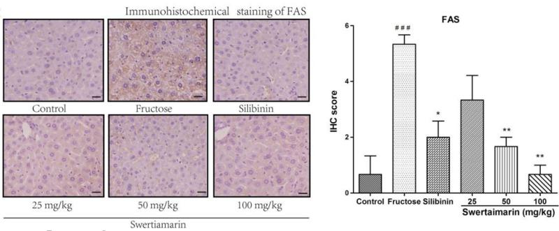
Application: IHC Species: mouse Sample: liver
Application: WB Species: Rat Sample: testes tissue
Restrictive clause
Affinity Biosciences tests all products strictly. Citations are provided as a resource for additional applications that have not been validated by Affinity Biosciences. Please choose the appropriate format for each application and consult Materials and Methods sections for additional details about the use of any product in these publications.
For Research Use Only.
Not for use in diagnostic or therapeutic procedures. Not for resale. Not for distribution without written consent. Affinity Biosciences will not be held responsible for patent infringement or other violations that may occur with the use of our products. Affinity Biosciences, Affinity Biosciences Logo and all other trademarks are the property of Affinity Biosciences LTD.