Cytokeratin 14 Antibody - #AF5370


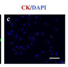
 antibody(Red), diluted at 1/600, was used as secondary antibody.")
製品説明
*The optimal dilutions should be determined by the end user. For optimal experimental results, antibody reuse is not recommended.
*Tips:
WB: For western blot detection of denatured protein samples. IHC: For immunohistochemical detection of paraffin sections (IHC-p) or frozen sections (IHC-f) of tissue samples. IF/ICC: For immunofluorescence detection of cell samples. ELISA(peptide): For ELISA detection of antigenic peptide.
引用形式: Affinity Biosciences Cat# AF5370, RRID:AB_2837855.
折りたたみ/展開
CK 14; CK-14; ck14; Cytokeratin 14; Cytokeratin-14; Cytokeratin14; Dowling Meara; EBS3; EBS4; Epidermolysis bullosa simplex; K14; K1C14_HUMAN; Keratin 14 (epidermolysis bullosa simplex, Dowling-Meara, Koebner); Keratin 14; Keratin; Keratin type I cytoskeletal 14; Keratin, type I cytoskeletal 14; Keratin-14; Keratin14; Koebner; Krt 14; Krt14; NFJ; OTTHUMP00000164624; type I cytoskeletal 14;
免疫原
A synthesized peptide derived from human Cytokeratin 14, corresponding to a region within N-terminal amino acids.
Detected in the basal layer, lowered within the more apically located layers specifically in the stratum spinosum, stratum granulosum but is not detected in stratum corneum. Strongly expressed in the outer root sheath of anagen follicles but not in the germinative matrix, inner root sheath or hair. Found in keratinocytes surrounding the club hair during telogen.
- P02533 K1C14_HUMAN:
- Protein BLAST With
- NCBI/
- ExPASy/
- Uniprot
MTTCSRQFTSSSSMKGSCGIGGGIGGGSSRISSVLAGGSCRAPSTYGGGLSVSSSRFSSGGACGLGGGYGGGFSSSSSSFGSGFGGGYGGGLGAGLGGGFGGGFAGGDGLLVGSEKVTMQNLNDRLASYLDKVRALEEANADLEVKIRDWYQRQRPAEIKDYSPYFKTIEDLRNKILTATVDNANVLLQIDNARLAADDFRTKYETELNLRMSVEADINGLRRVLDELTLARADLEMQIESLKEELAYLKKNHEEEMNALRGQVGGDVNVEMDAAPGVDLSRILNEMRDQYEKMAEKNRKDAEEWFFTKTEELNREVATNSELVQSGKSEISELRRTMQNLEIELQSQLSMKASLENSLEETKGRYCMQLAQIQEMIGSVEEQLAQLRCEMEQQNQEYKILLDVKTRLEQEIATYRRLLEGEDAHLSSSQFSSGSQSSRDVTSSSRQIRTKVMDVHDGKVVSTHEQVLRTKN
種類予測
Score>80(red) has high confidence and is suggested to be used for WB detection. *The prediction model is mainly based on the alignment of immunogen sequences, the results are for reference only, not as the basis of quality assurance.
High(score>80) Medium(80>score>50) Low(score<50) No confidence
研究背景
The nonhelical tail domain is involved in promoting KRT5-KRT14 filaments to self-organize into large bundles and enhances the mechanical properties involved in resilience of keratin intermediate filaments in vitro.
A disulfide bond is formed between rather than within filaments and promotes the formation of a keratin filament cage around the nucleus.
Ubiquitinated by the BCR(KLHL24) E3 ubiquitin ligase complex.
Cytoplasm. Nucleus.
Note: Expressed in both as a filamentous pattern.
Detected in the basal layer, lowered within the more apically located layers specifically in the stratum spinosum, stratum granulosum but is not detected in stratum corneum. Strongly expressed in the outer root sheath of anagen follicles but not in the germinative matrix, inner root sheath or hair. Found in keratinocytes surrounding the club hair during telogen.
Belongs to the intermediate filament family.
研究領域
· Organismal Systems > Endocrine system > Estrogen signaling pathway. (View pathway)
参考文献
Application: IF/ICC Species: mouse Sample: rDFSCs

Application: IF/ICC Species: Human Sample: HDPCs
Restrictive clause
Affinity Biosciences tests all products strictly. Citations are provided as a resource for additional applications that have not been validated by Affinity Biosciences. Please choose the appropriate format for each application and consult Materials and Methods sections for additional details about the use of any product in these publications.
For Research Use Only.
Not for use in diagnostic or therapeutic procedures. Not for resale. Not for distribution without written consent. Affinity Biosciences will not be held responsible for patent infringement or other violations that may occur with the use of our products. Affinity Biosciences, Affinity Biosciences Logo and all other trademarks are the property of Affinity Biosciences LTD.