製品: Integrin beta 1 Antibody
カタログ: AF5379
タンパク質の説明: Rabbit polyclonal antibody to Integrin beta 1
アプリケーション: WB IHC IF/ICC
反応性: Human, Mouse, Rat
予測: Pig, Bovine, Sheep, Rabbit, Dog, Chicken
分子量: 100~150kD; 88kD(Calculated).
ユニプロット: P05556
RRID: AB_2837864

類似製品を見る>>

   サイズ 価格 在庫状況
 100ul $280 在庫あり
 200ul $350 在庫あり

リードタイム: 当日配達

For pricing and ordering contact:
お問い合わせ先

製品説明

ソース:
Rabbit
アプリケーション:
WB 1:500-1:2000, IF/ICC 1:100-1:500, IHC 1:50-1:200
*The optimal dilutions should be determined by the end user.
*Tips:

WB: For western blot detection of denatured protein samples. IHC: For immunohistochemical detection of paraffin sections (IHC-p) or frozen sections (IHC-f) of tissue samples. IF/ICC: For immunofluorescence detection of cell samples. ELISA(peptide): For ELISA detection of antigenic peptide.

反応性:
Human,Mouse,Rat
予測:
Pig(100%), Bovine(100%), Sheep(100%), Rabbit(100%), Dog(100%), Chicken(100%)
クローナリティ:
Polyclonal
特異性:
Integrin beta 1 Antibody detects endogenous levels of total Integrin beta 1.
RRID:
AB_2837864
引用形式: Affinity Biosciences Cat# AF5379, RRID:AB_2837864.
コンジュゲート:
Unconjugated.
精製:
The antiserum was purified by peptide affinity chromatography using SulfoLink™ Coupling Resin (Thermo Fisher Scientific).
保存:
Rabbit IgG in phosphate buffered saline , pH 7.4, 150mM NaCl, 0.02% sodium azide and 50% glycerol. Store at -20 °C. Stable for 12 months from date of receipt.
別名:

折りたたみ/展開

beta1 integrin; CD29; Fibronectin receptor subunit beta; FNRB; Glycoprotein IIa; GP IIa; GPIIA; Integrin beta-1; Integrin subunit beta 1; integrin VLA-4 beta subunit; Integrin, beta 1 (fibronectin receptor, beta polypeptide, antigen CD29 includes MDF2, MSK12); ITB1_HUMAN; ITGB1; MDF2; MSK12; OTTHUMP00000019420; Very late activation protein, beta polypeptide; VLA BETA; VLA-4 subunit beta; VLA-BETA; VLAB; VLAbeta;

免疫原

免疫原:
Uniprot:
遺伝子(ID):
発現特異性:
P05556 ITB1_HUMAN:

Isoform 1 is widely expressed, other isoforms are generally coexpressed with a more restricted distribution. Isoform 2 is expressed in skin, liver, skeletal muscle, cardiac muscle, placenta, umbilical vein endothelial cells, neuroblastoma cells, lymphoma cells, hepatoma cells and astrocytoma cells. Isoform 3 and isoform 4 are expressed in muscle, kidney, liver, placenta, cervical epithelium, umbilical vein endothelial cells, fibroblast cells, embryonal kidney cells, platelets and several blood cell lines. Isoform 4, rather than isoform 3, is selectively expressed in peripheral T-cells. Isoform 3 is expressed in non-proliferating and differentiated prostate gland epithelial cells and in platelets, on the surface of erythroleukemia cells and in various hematopoietic cell lines. Isoform 5 is expressed specifically in striated muscle (skeletal and cardiac muscle).

タンパク質の説明:
Integrins alpha-1/beta-1, alpha-2/beta-1, alpha-10/beta-1 and alpha-11/beta-1 are receptors for collagen. Integrins alpha-1/beta-1 and alpha-2/beta-2 recognize the proline-hydroxylated sequence G-F-P-G-E-R in collagen. Integrins alpha-2/beta-1, alpha-3/beta-1, alpha-4/beta-1, alpha-5/beta-1, alpha-8/beta-1, alpha-10/beta-1, alpha-11/beta-1 and alpha-V/beta-1 are receptors for fibronectin.
タンパク質配列:
MNLQPIFWIGLISSVCCVFAQTDENRCLKANAKSCGECIQAGPNCGWCTNSTFLQEGMPTSARCDDLEALKKKGCPPDDIENPRGSKDIKKNKNVTNRSKGTAEKLKPEDITQIQPQQLVLRLRSGEPQTFTLKFKRAEDYPIDLYYLMDLSYSMKDDLENVKSLGTDLMNEMRRITSDFRIGFGSFVEKTVMPYISTTPAKLRNPCTSEQNCTSPFSYKNVLSLTNKGEVFNELVGKQRISGNLDSPEGGFDAIMQVAVCGSLIGWRNVTRLLVFSTDAGFHFAGDGKLGGIVLPNDGQCHLENNMYTMSHYYDYPSIAHLVQKLSENNIQTIFAVTEEFQPVYKELKNLIPKSAVGTLSANSSNVIQLIIDAYNSLSSEVILENGKLSEGVTISYKSYCKNGVNGTGENGRKCSNISIGDEVQFEISITSNKCPKKDSDSFKIRPLGFTEEVEVILQYICECECQSEGIPESPKCHEGNGTFECGACRCNEGRVGRHCECSTDEVNSEDMDAYCRKENSSEICSNNGECVCGQCVCRKRDNTNEIYSGKFCECDNFNCDRSNGLICGGNGVCKCRVCECNPNYTGSACDCSLDTSTCEASNGQICNGRGICECGVCKCTDPKFQGQTCEMCQTCLGVCAEHKECVQCRAFNKGEKKDTCTQECSYFNITKVESRDKLPQPVQPDPVSHCKEKDVDDCWFYFTYSVNGNNEVMVHVVENPECPTGPDIIPIVAGVVAGIVLIGLALLLIWKLLMIIHDRREFAKFEKEKMNAKWDTGENPIYKSAVTTVVNPKYEGK

種類予測

種類予測:

Score>80(red) has high confidence and is suggested to be used for WB detection. *The prediction model is mainly based on the alignment of immunogen sequences, the results are for reference only, not as the basis of quality assurance.

Species
Results
Score
Pig
100
Bovine
100
Sheep
100
Dog
100
Chicken
100
Rabbit
100
Horse
0
Xenopus
0
Zebrafish
0
Model Confidence:
High(score>80) Medium(80>score>50) Low(score<50) No confidence

PTMs - P05556 基板として

Site PTM Type Enzyme
K71 Ubiquitination
K100 Ubiquitination
K105 Ubiquitination
K107 Ubiquitination
T130 Phosphorylation
K134 Acetylation
K134 Ubiquitination
Y141 Phosphorylation
Y147 Phosphorylation
K163 Ubiquitination
S186 Phosphorylation
K190 Ubiquitination
S197 Phosphorylation
K202 Ubiquitination
N212 N-Glycosylation
K220 Ubiquitination
S224 Phosphorylation
T226 Phosphorylation
K228 Ubiquitination
K238 Ubiquitination
S263 Phosphorylation
N269 N-Glycosylation
T271 Phosphorylation
K289 Acetylation
K346 Ubiquitination
K349 Ubiquitination
T394 O-Glycosylation
K402 Ubiquitination
N406 N-Glycosylation
N481 N-Glycosylation
K540 Ubiquitination
S549 Phosphorylation
K551 Ubiquitination
K575 Ubiquitination
N669 N-Glycosylation
K672 Ubiquitination
K678 Ubiquitination
K692 Ubiquitination
K765 Ubiquitination
K768 Ubiquitination
K774 Ubiquitination
T777 Phosphorylation
Y783 Phosphorylation P42684 (ABL2)
K784 Ubiquitination
S785 Phosphorylation P17252 (PRKCA)
T788 Phosphorylation Q02156 (PRKCE) , Q9UQM7 (CAMK2A)
T789 Phosphorylation Q9UQM7 (CAMK2A) , Q02156 (PRKCE)
K794 Acetylation
K794 Sumoylation
K794 Ubiquitination
Y795 Phosphorylation
K798 Ubiquitination

研究背景

機能:

Integrins alpha-1/beta-1, alpha-2/beta-1, alpha-10/beta-1 and alpha-11/beta-1 are receptors for collagen. Integrins alpha-1/beta-1 and alpha-2/beta-2 recognize the proline-hydroxylated sequence G-F-P-G-E-R in collagen. Integrins alpha-2/beta-1, alpha-3/beta-1, alpha-4/beta-1, alpha-5/beta-1, alpha-8/beta-1, alpha-10/beta-1, alpha-11/beta-1 and alpha-V/beta-1 are receptors for fibronectin. Alpha-4/beta-1 recognizes one or more domains within the alternatively spliced CS-1 and CS-5 regions of fibronectin. Integrin alpha-5/beta-1 is a receptor for fibrinogen. Integrin alpha-1/beta-1, alpha-2/beta-1, alpha-6/beta-1 and alpha-7/beta-1 are receptors for lamimin. Integrin alpha-6/beta-1 (ITGA6:ITGB1) is present in oocytes and is involved in sperm-egg fusion (By similarity). Integrin alpha-4/beta-1 is a receptor for VCAM1. It recognizes the sequence Q-I-D-S in VCAM1. Integrin alpha-9/beta-1 is a receptor for VCAM1, cytotactin and osteopontin. It recognizes the sequence A-E-I-D-G-I-E-L in cytotactin. Integrin alpha-3/beta-1 is a receptor for epiligrin, thrombospondin and CSPG4. Alpha-3/beta-1 may mediate with LGALS3 the stimulation by CSPG4 of endothelial cells migration. Integrin alpha-V/beta-1 is a receptor for vitronectin. Beta-1 integrins recognize the sequence R-G-D in a wide array of ligands. Isoform 2 interferes with isoform 1 resulting in a dominant negative effect on cell adhesion and migration (in vitro). When associated with alpha-7/beta-1 integrin, regulates cell adhesion and laminin matrix deposition. Involved in promoting endothelial cell motility and angiogenesis. Involved in osteoblast compaction through the fibronectin fibrillogenesis cell-mediated matrix assembly process and the formation of mineralized bone nodules. May be involved in up-regulation of the activity of kinases such as PKC via binding to KRT1. Together with KRT1 and RACK1, serves as a platform for SRC activation or inactivation. Plays a mechanistic adhesive role during telophase, required for the successful completion of cytokinesis. Integrin alpha-3/beta-1 provides a docking site for FAP (seprase) at invadopodia plasma membranes in a collagen-dependent manner and hence may participate in the adhesion, formation of invadopodia and matrix degradation processes, promoting cell invasion. ITGA4:ITGB1 binds to fractalkine (CX3CL1) and may act as its coreceptor in CX3CR1-dependent fractalkine signaling. ITGA4:ITGB1 and ITGA5:ITGB1 bind to PLA2G2A via a site (site 2) which is distinct from the classical ligand-binding site (site 1) and this induces integrin conformational changes and enhanced ligand binding to site 1. ITGA5:ITGB1 acts as a receptor for fibrillin-1 (FBN1) and mediates R-G-D-dependent cell adhesion to FBN1. ITGA5:ITGB1 is a receptor for IL1B and binding is essential for IL1B signaling. ITGA5:ITGB3 is a receptor for soluble CD40LG and is required for CD40/CD40LG signaling.

Isoform 5 displaces isoform 1 in striated muscles.

(Microbial infection) Integrin ITGA2:ITGB1 acts as a receptor for Human echoviruses 1 and 8.

(Microbial infection) Acts as a receptor for Cytomegalovirus/HHV-5.

(Microbial infection) Acts as a receptor for Epstein-Barr virus/HHV-4.

(Microbial infection) Integrin ITGA5:ITGB1 acts as a receptor for Human parvovirus B19.

(Microbial infection) Integrin ITGA2:ITGB1 acts as a receptor for Human rotavirus.

(Microbial infection) Acts as a receptor for Mammalian reovirus.

(Microbial infection) In case of HIV-1 infection, integrin ITGA5:ITGB1 binding to extracellular viral Tat protein seems to enhance angiogenesis in Kaposi's sarcoma lesions.

PTMs:

The cysteine residues are involved in intrachain disulfide bonds.

細胞の位置付け:

Cell membrane>Single-pass type I membrane protein. Cell projection>Invadopodium membrane>Single-pass type I membrane protein. Cell projection>Ruffle membrane>Single-pass type I membrane protein. Recycling endosome. Melanosome. Cleavage furrow. Cell projection>Lamellipodium. Cell projection>Ruffle. Cell junction>Focal adhesion. Cell surface.
Note: Isoform 2 does not localize to focal adhesions. Highly enriched in stage I melanosomes. Located on plasma membrane of neuroblastoma NMB7 cells. In a lung cancer cell line, in prometaphase and metaphase, localizes diffusely at the membrane and in a few intracellular vesicles. In early telophase, detected mainly on the matrix-facing side of the cells. By mid-telophase, concentrated to the ingressing cleavage furrow, mainly to the basal side of the furrow. In late telophase, concentrated to the extending protrusions formed at the opposite ends of the spreading daughter cells, in vesicles at the base of the lamellipodia formed by the separating daughter cells. Colocalizes with ITGB1BP1 and metastatic suppressor protein NME2 at the edge or peripheral ruffles and lamellipodia during the early stages of cell spreading on fibronectin or collagen. Translocates from peripheral focal adhesions sites to fibrillar adhesions in a ITGB1BP1-dependent manner. Enriched preferentially at invadopodia, cell membrane protrusions that correspond to sites of cell invasion, in a collagen-dependent manner. Localized at plasma and ruffle membranes in a collagen-independent manner.

Cell membrane>Sarcolemma. Cell junction.
Note: In cardiac muscle, isoform 5 is found in costameres and intercalated disks.

Extracellular region or secreted Cytosol Plasma membrane Cytoskeleton Lysosome Endosome Peroxisome ER Golgi apparatus Nucleus Mitochondrion Manual annotation Automatic computational assertionSubcellular location
組織特異性:

Isoform 1 is widely expressed, other isoforms are generally coexpressed with a more restricted distribution. Isoform 2 is expressed in skin, liver, skeletal muscle, cardiac muscle, placenta, umbilical vein endothelial cells, neuroblastoma cells, lymphoma cells, hepatoma cells and astrocytoma cells. Isoform 3 and isoform 4 are expressed in muscle, kidney, liver, placenta, cervical epithelium, umbilical vein endothelial cells, fibroblast cells, embryonal kidney cells, platelets and several blood cell lines. Isoform 4, rather than isoform 3, is selectively expressed in peripheral T-cells. Isoform 3 is expressed in non-proliferating and differentiated prostate gland epithelial cells and in platelets, on the surface of erythroleukemia cells and in various hematopoietic cell lines. Isoform 5 is expressed specifically in striated muscle (skeletal and cardiac muscle).

サブユニット構造:

Heterodimer of an alpha and a beta subunit. Beta-1 associates with either alpha-1, alpha-2, alpha-3, alpha-4, alpha-5, alpha-6, alpha-7, alpha-8, alpha-9, alpha-10, alpha-11 or alpha-V. ITGA6:ITGB1 is found in a complex with CD9; interaction takes place in oocytes and is involved in sperm-egg fusion (By similarity). Interacts with seprase FAP (seprase); the interaction occurs at the cell surface of invadopodia membrane in a collagen-dependent manner. Binds LGALS3BP and NMRK2, when associated with alpha-7, but not with alpha-5. Interacts with FGR and HCK. Interacts (via the cytoplasmic region) with RAB25 (via the hypervariable C-terminal region). Interacts with RAB21. Interacts with KRT1 in the presence of RACK1 and SRC. Interacts with JAML; integrin alpha-4/beta-1 may regulate leukocyte to endothelial cells adhesion by controlling JAML homodimerization. Interacts with FLNA, FLNB and RANBP9. Isoform 5 interacts with ACE2. Isoform 1 interacts with the C-terminal region of FLNC. Interacts with MYO10. Interacts with DAB2. Interacts with FERMT2; the interaction is inhibited in presence of ITGB1BP1. Interacts with ITGB1BP1 (via C-terminal region); the interaction is a prerequisite for focal adhesion disassembly. Interacts with TLN1; the interaction is prevented by competitive binding of ITGB1BP1. Interacts with ACAP1; required for ITGB1 recycling. Interacts with ASAP3. Isoform 5 interacts with alpha-7A and alpha-7B in adult skeletal muscle. Isoform 5 interacts with alpha-7B in cardiomyocytes of adult heart. Interacts with EMP2; the interaction may be direct or indirect and ITGB1 has a heterodimer form (By similarity). ITGA5:ITGB1 interacts with CCN3. ITGA4:ITGB1 is found in a ternary complex with CX3CR1 and CX3CL1. ITGA5:ITGB1 interacts with FBN1. ITGA5:ITGB1 interacts with IL1B. Interacts with MDK. ITGA4:ITGB1 interacts with MDK; this interaction mediates MDK-induced osteoblast cells migration through PXN phosphorylation. ITGA6:ITGB1 interacts with MDK; this interaction mediates MDK-induced neurite-outgrowth.

(Microbial infection) Integrin ITGA2:ITGB1 interacts with human echoviruses 1 and 8 capsid proteins.

(Microbial infection) Interacts with human cytomegalovirus/HHV-5 envelope glycoprotein B/gB.

(Microbial infection) Interacts with Epstein-Barr virus/HHV-4 gB protein.

(Microbial infection) Integrin ITGA5:ITGB1 interacts with human parvovirus B19 capsid protein.

(Microbial infection) Integrin ITGA2:ITGB1 interacts with human rotavirus VP4 protein.

(Microbial infection) Interacts with mammalian reovirus capsid proteins.

(Microbial infection) Integrin ITGA5:ITGB1 interacts with HIV-1 Tat.

タンパク質ファミリー:

Belongs to the integrin beta chain family.

研究領域

· Cellular Processes > Transport and catabolism > Phagosome.   (View pathway)

· Cellular Processes > Cellular community - eukaryotes > Focal adhesion.   (View pathway)

· Cellular Processes > Cellular community - eukaryotes > Tight junction.   (View pathway)

· Cellular Processes > Cell motility > Regulation of actin cytoskeleton.   (View pathway)

· Environmental Information Processing > Signal transduction > Rap1 signaling pathway.   (View pathway)

· Environmental Information Processing > Signal transduction > PI3K-Akt signaling pathway.   (View pathway)

· Environmental Information Processing > Signaling molecules and interaction > ECM-receptor interaction.   (View pathway)

· Environmental Information Processing > Signaling molecules and interaction > Cell adhesion molecules (CAMs).   (View pathway)

· Human Diseases > Infectious diseases: Bacterial > Bacterial invasion of epithelial cells.

· Human Diseases > Infectious diseases: Bacterial > Pathogenic Escherichia coli infection.

· Human Diseases > Infectious diseases: Bacterial > Shigellosis.

· Human Diseases > Infectious diseases: Bacterial > Pertussis.

· Human Diseases > Infectious diseases: Parasitic > Leishmaniasis.

· Human Diseases > Infectious diseases: Parasitic > Toxoplasmosis.

· Human Diseases > Infectious diseases: Viral > Human papillomavirus infection.

· Human Diseases > Cancers: Overview > Pathways in cancer.   (View pathway)

· Human Diseases > Cancers: Overview > Proteoglycans in cancer.

· Human Diseases > Cancers: Specific types > Small cell lung cancer.   (View pathway)

· Human Diseases > Cardiovascular diseases > Hypertrophic cardiomyopathy (HCM).

· Human Diseases > Cardiovascular diseases > Arrhythmogenic right ventricular cardiomyopathy (ARVC).

· Human Diseases > Cardiovascular diseases > Dilated cardiomyopathy (DCM).

· Organismal Systems > Development > Axon guidance.   (View pathway)

· Organismal Systems > Immune system > Platelet activation.   (View pathway)

· Organismal Systems > Immune system > Leukocyte transendothelial migration.   (View pathway)

参考文献

1). Abnormally activated OPN/integrin αVβ3/FAK signalling is responsible for EGFR-TKI resistance in EGFR mutant non-small-cell lung cancer. Journal of hematology & oncology, 2020 (PubMed: 33287873) [IF=28.5]

Application: WB    Species: Human    Sample:

Additional file 11: Fig. S7. Under gefitinib treatment, p-FAK in PC9 cells transfected with si-ITGB1 was detected by western blot analysis.

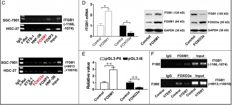
2). hTERT promotes the invasion of gastric cancer cells by enhancing FOXO3a ubiquitination and subsequent ITGB1 upregulation. GUT, 2017 (PubMed: 26370108) [IF=23.0]

Application: WB    Species: human    Sample: human gastric cancer

Figure 3 Human telomerase reverse transcriptase (hTERT) promotes integrin β1 (ITGB1) expression by modulating the binding of both forkhead box M1 (FOXM1) and forkhead box O3 (FOXO3a) to the ITGB1 gene. (A) Chromatin immunoprecipitation (ChIP)-qPCR analysis of the binding sites of hTERT in the regulatory region of the ITGB1 gene. ChIP assays were performed using chromatin isolated from SGC-7901 and HGC-27 cells, and final DNA extractions were qPCR amplified by 49 pairs of primers covering the regulatory region of ITGB1 (Chr10: 33 253 528∼Chr10: 33 226 885). (B) Influence of hTERT on the luciferase activity of the pGL3-promoter constructs. The recombinant constructs (named pGL3-p1-6 (Left) and pGL3-I1-6 (Right)) were separately cotransfected with pIRES2-hTERT plasmid (or control plasmid) into U2OS cells, and cells were harvested 24 h later for dual luciferase activity assays. (C) ChIP analysis of the binding of transcription factors to the (−1166∼−1074) or (+9913∼+10 010) region of ITGB1 in both SGC-7901 and HGC-27 cells. (D) ITGB1 expression after FOXM1 or FOXO3a overexpression in SGC-7901 cells. Forty-eight hours after the transient transfection of pEGFP-FOXM1 or pcDNA-FOXO3a plasmids, together with the corresponding controls, SGC-7901 cells were harvested for qPCR and western blot analysis. (E) Effect of FOXM1 or FOXO3a on the activity of pGL3-P5 or pGL3-I5, respectively. SGC-7901 cells in 24-well plates were transiently transfected with the luciferase reporters, together with pEGFP-FOXM1 or pcDNA-FOXO3a plasmids. Twenty-four hours later, the luciferase activity was measured using luciferase assay substrate. (F) ChIP assays were performed using chromatin isolated from gastric cancer cells infected with lenti-hTERT or lenti-Control. Final DNA extractions were PCR amplified using primers that cover FOXM1 binding element (F1BE) (−1104∼−1109) or FOXO3a binding element (F3BE) (+9972∼+9978) within the ITGB1 gene. *p<0.05, **p<0.01.

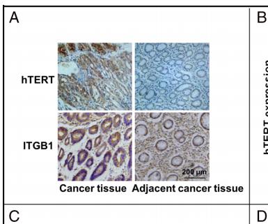
Application: IHC    Species: human    Sample: human gastric cancer

Figure 2 Human telomerase reverse transcriptase (hTERT) expression parallels that of integrin β1 (ITGB1) in gastric cancer tissues, and both are correlated with poor prognosis. (A) Representative immunohistochemical staining of hTERT and ITGB1 in gastric cancer tissues and corresponding adjacent non-cancerous tissues. (B) The association between hTERT/ITGB1 expression and TNM stage/lymphatic metastasis in patients with gastric cancer (p<0.01). (C) The correlations between the protein levels of hTERT and ITGB1 (p<0.01). (D) The receiver operating characteristic (ROC) curves for predicting patients’ survival time using hTERT or ITGB1 expression. (E) Kaplan–Meier analyses of overall survival according to hTERT or ITGB1 expression levels (p<0.01). (F) Kaplan–Meier analyses of overall survival according to the combination of the above two indices (p<0.01). AUC, area under the curve; TNM, tumour node metastases.

3). Mussel-Derived and Bioclickable Peptide Mimic for Enhanced Interfacial Osseointegration via Synergistic Immunomodulation and Vascularized Bone Regeneration. Advanced science (Weinheim, Baden-Wurttemberg, Germany), 2024 (PubMed: 38922775) [IF=15.1]

4). ZIF-8 Modified Multifunctional Bone-Adhesive Hydrogels Promoting Angiogenesis and Osteogenesis for Bone Regeneration. ACS Applied Materials & Interfaces, 2020 (PubMed: 32814397) [IF=8.3]

5). Rab34 regulates adhesion, migration, and invasion of breast cancer cells. Oncogene, 2018 (PubMed: 29622794) [IF=6.9]

Application: WB    Species: human    Sample: MDA-MB-231 cells

Fig. 6 Rab34 phosphorylation contributes to accumulation of inter-nalized ITGB3. c MDA-MB-231 cells stably expressing pSuper.GFP-shRNA-Rab34 or shRNA-Ctrl were lysed and the lysates were subjected for Western blot assay to analyze the protein level of ITGB3 and ITGB1, showing two independent shRNAs targeting Rab34 decrease the protein levels of ITGB3.

6). Jinfukang regulates integrin/Src pathway and anoikis mediating circulating lung cancer cells migration. JOURNAL OF ETHNOPHARMACOLOGY, 2021 (PubMed: 33068649) [IF=4.8]

Application: WB    Species: human    Sample: CTC-TJH-01 cells

Fig. 5. |Jinfukang inhibited the cell migration via Integrin/Src axis in lung cancer cells. Cells were incubated with Jinfukang (100 μg/mL), ATN-161 (10 μmol/L) or a combination of both for 24 h. A Western blot assay was used to measure the protein expression levels of Integrin β1, Src, and p-Src in CTC-TJH-01 (A) and H1975 (B)cells. GAPDH was used as an internal standard.

7). SCO-spondin-derived peptide NX210 rescues neurons from cerebral ischemia/reperfusion injury through modulating the Integrin-β1 mediated PI3K/Akt pathway. International Immunopharmacology, 2022 (PubMed: 35930911) [IF=4.8]

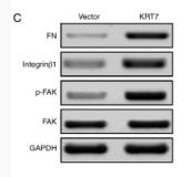
8). KRT7 promotes epithelial‑mesenchymal transition in ovarian cancer via the TGF‑β/Smad2/3 signaling pathway. ONCOLOGY REPORTS, 2021 (PubMed: 33416175) [IF=3.8]

Application: WB    Species: Human    Sample: HEY cells

Figure 5. - KRT7 expression affects the integrin-β1-FAK signaling and TGF-β signaling pathways. (A and B) Expression of proliferation- and migration-associated genes (PCNA, MMP9 and TIMP-1) were evaluated using western blotting in HEY cells. (C and D) Western blotting of proteins involved in integrin-β1-FAK signaling pathway in the KRT7-overexpressing HEY cells. (E) Expression of MMPs after knockdown of KRT7 in OVCAR433 cells. (F and G) Expression of the TGF-β signaling pathway-related proteins was evaluated by western blotting in KRT7-overexpressing HEY cells and KRT7-knockdown OVCAR433 cells. All experiments were performed at least three times. Results are presented as the mean ± standard deviation. **P<0.01. FAK, focal adhesion kinase; PCNA, proliferating cell nuclear antigen; FN, fibronectin; TIMP-1, TIMP metallopeptidase inhibitor 1; p-, phosphorylated; MMP, matrix metalloproteinase; KRT7, keratin 7; sh, short hairpin RNA; NC, negative control.

9). Downregulation of glycoprotein non-metastatic melanoma protein B prevents high glucose-induced angiogenesis in diabetic retinopathy. MOLECULAR AND CELLULAR BIOCHEMISTRY, 2023 (PubMed: 36036335) [IF=3.5]

10). Identification of prothymosin alpha (PTMA) as a biomarker for esophageal squamous cell carcinoma (ESCC) by label-free quantitative proteomics and Quantitative Dot Blot (QDB). Clinical Proteomics, 2019 (PubMed: 30988666) [IF=2.8]

Application: WB    Species: human    Sample: ESCC tissues

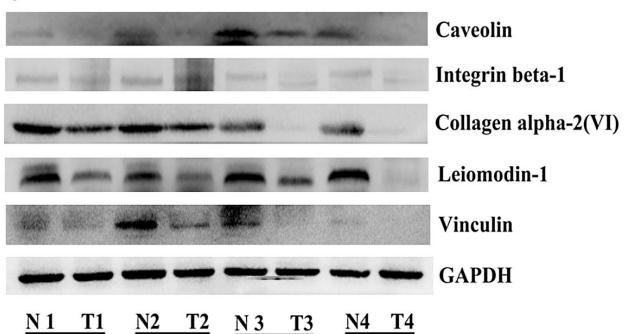
Fig. 5|The diferentially expressed proteins were validated by Western Blot. Compared with adjacent normal tissues, the protein expression of PTMA, PAK2, PPP1CA, HMGB2 were up-regulated (a, b), and the protein expression of Caveolin, Integrin beta-1, Collagen alpha-2(VI), Leiomodin-1, Vinculin were down-regulated in ESCC tissues from four pairs of samples (c, d). Representative immunoblot images (a, c) and histograms (mean ± SD; b, d).The experiments were repeated at least three times, N represented normal tissues and T represented tumor tissues

もっと読みます

Restrictive clause


Affinity Biosciences tests all products strictly. Citations are provided as a resource for additional applications that have not been validated by Affinity Biosciences. Please choose the appropriate format for each application and consult Materials and Methods sections for additional details about the use of any product in these publications.

For Research Use Only.
Not for use in diagnostic or therapeutic procedures. Not for resale. Not for distribution without written consent. Affinity Biosciences will not be held responsible for patent infringement or other violations that may occur with the use of our products. Affinity Biosciences, Affinity Biosciences Logo and all other trademarks are the property of Affinity Biosciences LTD.