製品: Nanog Antibody
カタログ: AF5388
タンパク質の説明: Rabbit polyclonal antibody to Nanog
アプリケーション: WB IHC IF/ICC
反応性: Human, Mouse, Rat
予測: Bovine, Horse, Sheep, Rabbit
分子量: 35 kDa; 35kD(Calculated).
ユニプロット: Q9H9S0
RRID: AB_2814953

類似製品を見る>>

   サイズ 価格 在庫状況
 100ul $280 在庫あり
 200ul $350 在庫あり

リードタイム: 当日配達

For pricing and ordering contact:
お問い合わせ先

製品説明

ソース:
Rabbit
アプリケーション:
WB 1:500-1:2000, IF/ICC 1:100-1:500, IHC 1:50-1:200
*The optimal dilutions should be determined by the end user.
*Tips:

WB: For western blot detection of denatured protein samples. IHC: For immunohistochemical detection of paraffin sections (IHC-p) or frozen sections (IHC-f) of tissue samples. IF/ICC: For immunofluorescence detection of cell samples. ELISA(peptide): For ELISA detection of antigenic peptide.

反応性:
Human,Mouse,Rat
予測:
Bovine(82%), Horse(91%), Sheep(82%), Rabbit(100%)
クローナリティ:
Polyclonal
特異性:
Nanog Antibody detects endogenous levels of total Nanog.
RRID:
AB_2814953
引用形式: Affinity Biosciences Cat# AF5388, RRID:AB_2814953.
コンジュゲート:
Unconjugated.
精製:
The antiserum was purified by peptide affinity chromatography using SulfoLink™ Coupling Resin (Thermo Fisher Scientific).
保存:
Rabbit IgG in phosphate buffered saline , pH 7.4, 150mM NaCl, 0.02% sodium azide and 50% glycerol. Store at -20 °C. Stable for 12 months from date of receipt.
別名:

折りたたみ/展開

Embryonic stem cell specific homeobox protein (Nanog); ENK; FLJ12581; hNanog; Homeobox protein NANOG; Homeobox transcription factor Nanog; homeobox transcription factor Nanog-delta 48; NANOG; Nanog homeobox; NANOG_HUMAN;

免疫原

免疫原:
Uniprot:
遺伝子(ID):
発現特異性:
Q9H9S0 NANOG_HUMAN:

Expressed in testicular carcinoma and derived germ cell tumors (at protein level). Expressed in fetal gonads, ovary and testis. Also expressed in ovary teratocarcinoma cell line and testicular embryonic carcinoma. Not expressed in many somatic organs and oocytes.

タンパク質の説明:
Transcription regulator involved in inner cell mass and embryonic stem (ES) cells proliferation and self-renewal. Imposes pluripotency on ES cells and prevents their differentiation towards extraembryonic endoderm and trophectoderm lineages.
タンパク質配列:
MSVDPACPQSLPCFEASDCKESSPMPVICGPEENYPSLQMSSAEMPHTETVSPLPSSMDLLIQDSPDSSTSPKGKQPTSAEKSVAKKEDKVPVKKQKTRTVFSSTQLCVLNDRFQRQKYLSLQQMQELSNILNLSYKQVKTWFQNQRMKSKRWQKNNWPKNSNGVTQKASAPTYPSLYSSYHQGCLVNPTGNLPMWSNQTWNNSTWSNQTQNIQSWSNHSWNTQTWCTQSWNNQAWNSPFYNCGEESLQSCMQFQPNSPASDLEAALEAAGEGLNVIQQTTRYFSTPQTMDLFLNYSMNMQPEDV

種類予測

種類予測:

Score>80(red) has high confidence and is suggested to be used for WB detection. *The prediction model is mainly based on the alignment of immunogen sequences, the results are for reference only, not as the basis of quality assurance.

Species
Results
Score
Rabbit
100
Horse
91
Bovine
82
Sheep
82
Pig
0
Dog
0
Xenopus
0
Zebrafish
0
Chicken
0
Model Confidence:
High(score>80) Medium(80>score>50) Low(score<50) No confidence

PTMs - Q9H9S0 基板として

Site PTM Type Enzyme
S2 Phosphorylation
Y35 Phosphorylation Q05397 (PTK2)
T50 Phosphorylation
S52 Phosphorylation P28482 (MAPK1)
S56 Phosphorylation
S57 Phosphorylation
S65 Phosphorylation
T70 Phosphorylation
S71 Phosphorylation P28482 (MAPK1) , P06493 (CDK1)
T78 Phosphorylation Q02156 (PRKCE) , P28482 (MAPK1)
S79 Phosphorylation Q02156 (PRKCE)
S135 Phosphorylation Q02156 (PRKCE)
Y174 Phosphorylation Q05397 (PTK2)
T200 Phosphorylation Q02156 (PRKCE)
S258 Phosphorylation
T280 Phosphorylation Q02156 (PRKCE)
T286 Phosphorylation
T289 Phosphorylation

研究背景

機能:

Transcription regulator involved in inner cell mass and embryonic stem (ES) cells proliferation and self-renewal. Imposes pluripotency on ES cells and prevents their differentiation towards extraembryonic endoderm and trophectoderm lineages. Blocks bone morphogenetic protein-induced mesoderm differentiation of ES cells by physically interacting with SMAD1 and interfering with the recruitment of coactivators to the active SMAD transcriptional complexes. Acts as a transcriptional activator or repressor. Binds optimally to the DNA consensus sequence 5'-TAAT[GT][GT]-3' or 5'-[CG][GA][CG]C[GC]ATTAN[GC]-3'. Binds to the POU5F1/OCT4 promoter. Able to autorepress its expression in differentiating (ES) cells: binds to its own promoter following interaction with ZNF281/ZFP281, leading to recruitment of the NuRD complex and subsequent repression of expression. When overexpressed, promotes cells to enter into S phase and proliferation.

細胞の位置付け:

Nucleus.

Extracellular region or secreted Cytosol Plasma membrane Cytoskeleton Lysosome Endosome Peroxisome ER Golgi apparatus Nucleus Mitochondrion Manual annotation Automatic computational assertionSubcellular location
組織特異性:

Expressed in testicular carcinoma and derived germ cell tumors (at protein level). Expressed in fetal gonads, ovary and testis. Also expressed in ovary teratocarcinoma cell line and testicular embryonic carcinoma. Not expressed in many somatic organs and oocytes.

サブユニット構造:

Interacts with SMAD1 and SALL4. Interacts with ZNF281/ZFP281 (By similarity). Interacts with PCGF1. Interacts with ESRRB; reciprocally modulates their transcriptional activities (By similarity).

タンパク質ファミリー:

Belongs to the Nanog homeobox family.

研究領域

· Cellular Processes > Cellular community - eukaryotes > Signaling pathways regulating pluripotency of stem cells.   (View pathway)

· Human Diseases > Cancers: Overview > Proteoglycans in cancer.

参考文献

1). Exploration of the mechanisms of Ge Gen Decoction against influenza A virus infection. Chinese Journal of Natural Medicines, 2019 (PubMed: 31526500) [IF=4.6]

2). CX43 down-regulation promotes cell aggressiveness and 5-fluorouracil-resistance by attenuating cell stiffness in colorectal carcinoma. Cancer Biology & Therapy, 2023 (PubMed: 37342072) [IF=3.6]

Application: WB    Species: Human    Sample: CRC cells

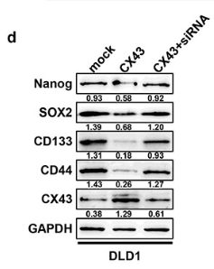
Figure 5. CX43 increased cell stiffness by polymerizing CRC cytoskeleton. (a and b) Confocal microscopy analysis of α-tubulin polymerization and F-actin cytoskeletal remodeling in CRC cells with CX43 overexpression and knock-down. (c) The young’s modulus of CX43 overexpressed DLD1 cells was measured by atomic mechanics microscope. Mechanical curve (left) and quantitative analysis (right) are displayed. (d) Western blot analyses expression of stem cell characteristic related proteins in CRC cells with CX43 overexpression. (e) The tumor cell spheroidizing ability of CX43 overexpression cells was detected by tumor sphere formation assays. (f) Western blot was used to survey the relative expression of apoptosis pathway related proteins in CX43 overexpression cells. The downregulation of CX43 could rescue the above phenotypes (d, e and f).

3). The adenosine-A2a receptor regulates the radioresistance of gastric cancer via PI3K-AKT-mTOR pathway. International Journal of Clinical Oncology, 2022 (PubMed: 35122587) [IF=3.3]

4). Lycorine attenuates lipopolysaccharide-induced acute lung injury through the HMGB1/TLRs/NF-κB pathway. 3 Biotech, 2020 (PubMed: 32818131) [IF=2.8]

5). Emodin reverses resistance to gemcitabine in pancreatic cancer by suppressing stemness through regulation of the epithelial‑mesenchymal transition. Experimental and Therapeutic Medicine, 2023 (PubMed: 36545274) [IF=2.7]

Application: WB    Species: Human    Sample: SW1990/GZ cells

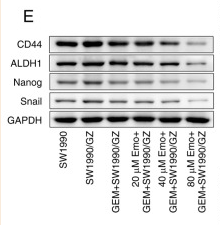
Figure 2 Emo enhances inhibitory effects of GEM on self-renewal of SW1990/GZ cells. (A) Morphology of cells observed under an inverted light microscope (scale bar, 100 µm). (B) The ultrastructure of cells observed under a transmission electron microscope (x10,000 magnification). (C) Cell sphere-forming rate evaluated using the sphere formation assay (x100 magnification). (D) Number of spheres measured using the colony-formation assay (x200 magnification). (E) Expression levels of CD44, ALDH1, Nanog and Snail determined using western blotting assays. *P

Application: IHC    Species: Human    Sample: SW1990/GZ cells

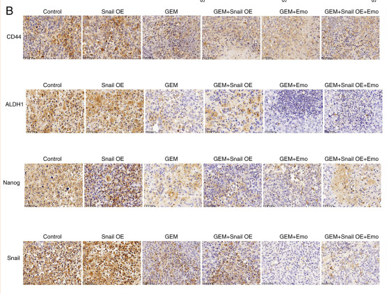
Figure 7 Growth of stem cells and EMT progression in SW1990/GZ xenograft model is inhibited by Emo by inactivating Snail. (A) Expression levels of CD44, ALDH1, Nanog and Snail as determined via western blotting. (B) Expression level of CD44, ALDH1, Nanog and Snail in tumor tissues measured via immunohistochemistry (x200 magnification). (C) Expression levels of vimentin and E-cadherin as determined via western blotting. (D) Expression levels of vimentin and E-cadherin in tumor tissues measured via immunohistochemistry (x200 magnification). *P

6). Sigma-1 receptor and binding immunoglobulin protein interacts with ulinastatin contributing to a protective effect on rat cerebral ischemia-reperfusion. World Neurosurgery, 2022 (PubMed: 34767993) [IF=2.0]

7). EFNA4 deletion suppresses the migration, invasion, stemness, and angiogenesis of gastric cancer cells through the inactivation of Pygo2/Wnt signaling. Histology and histopathology, 2024 (PubMed: 38953488) [IF=2.0]

8). Generation of a human induced pluripotent stem cell line (SDUBMSi001-A) from a hereditary spastic paraplegia patient carrying kif1a c.773C>T missense mutation. Stem Cell Research, 2020 (PubMed: 32045731) [IF=1.2]

9). Non-muscle myosin II knockdown improves survival and therapeutic effects of implanted bone marrow-derived mesenchymal stem cells in lipopolysaccharide-induced acute lung injury. Annals of Translational Medicine, 2021 (PubMed: 33708889)

Application: WB    Species: Rat    Sample: BMSCs

Figure 7 NM-II knockdown inhibited the decreased of BMSCs viability. Nanog and Ang-1 were significantly upregulated in BMSCs upon NM-II knockdown, while the activity of Caspase-3 was decreased in BMSCs upon NM-II knockdown. Cell viability was measured by CCK8 assay (A). Expression of self-renewal and apoptosis-associated proteins were evaluated by western blotting (B). Representative of three independent experiments. Relative protein levels were quantified by densitometry and expressed as optical density ratio with GAPDH serving as internal standards (C). *, P<0.05, **, P<0.01 vs. control.

Restrictive clause


Affinity Biosciences tests all products strictly. Citations are provided as a resource for additional applications that have not been validated by Affinity Biosciences. Please choose the appropriate format for each application and consult Materials and Methods sections for additional details about the use of any product in these publications.

For Research Use Only.
Not for use in diagnostic or therapeutic procedures. Not for resale. Not for distribution without written consent. Affinity Biosciences will not be held responsible for patent infringement or other violations that may occur with the use of our products. Affinity Biosciences, Affinity Biosciences Logo and all other trademarks are the property of Affinity Biosciences LTD.