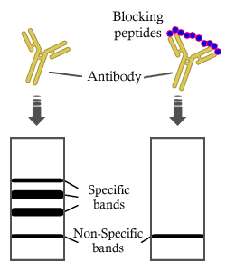
製品: CDKN2A/p16INK4a Antibody
カタログ: AF5484
タンパク質の説明: Rabbit polyclonal antibody to CDKN2A/p16INK4a
アプリケーション: WB IF/ICC
Cited expt.: WB, IF/ICC
反応性: Human, Mouse
予測: Pig, Bovine, Horse, Rabbit
分子量: 16 kDa; 17kD(Calculated).
ユニプロット: P42771
RRID: AB_2837964

類似製品を見る>>

   サイズ 価格 在庫状況
 100ul $280 在庫あり
 200ul $350 在庫あり

リードタイム: 当日配達

For pricing and ordering contact:
お問い合わせ先

製品説明

ソース:
Rabbit
アプリケーション:
WB 1:500-1:2000, IF/ICC 1:100-1:500
*The optimal dilutions should be determined by the end user. For optimal experimental results, antibody reuse is not recommended.
*Tips:

WB: For western blot detection of denatured protein samples. IHC: For immunohistochemical detection of paraffin sections (IHC-p) or frozen sections (IHC-f) of tissue samples. IF/ICC: For immunofluorescence detection of cell samples. ELISA(peptide): For ELISA detection of antigenic peptide.

反応性:
Human,Mouse
予測:
Pig(85%), Bovine(82%), Horse(85%), Rabbit(100%)
クローナリティ:
Polyclonal
特異性:
CDKN2A/p16INK4a Antibody detects endogenous levels of total CDKN2A/p16INK4a.
RRID:
AB_2837964
引用形式: Affinity Biosciences Cat# AF5484, RRID:AB_2837964.
コンジュゲート:
Unconjugated.
精製:
The antiserum was purified by peptide affinity chromatography using SulfoLink™ Coupling Resin (Thermo Fisher Scientific).
保存:
Rabbit IgG in phosphate buffered saline , pH 7.4, 150mM NaCl, 0.02% sodium azide and 50% glycerol. Store at -20 °C. Stable for 12 months from date of receipt.
別名:

折りたたみ/展開

Cyclin-dependent kinase inhibitor 2A;Cyclin-dependent kinase 4 inhibitor A;CDK4I;Multiple tumor suppressor 1;MTS-1;p16-INK4a;p16-INK4;p16INK4A;CDKN2A;CDKN2;MTS1;

免疫原

免疫原:

A synthesized peptide derived from human CDKN2A/p16INK4a, corresponding to a region within N-terminal amino acids.

Uniprot:
遺伝子(ID):
発現特異性:
P42771 CDN2A_HUMAN:

Widely expressed but not detected in brain or skeletal muscle. Isoform 3 is pancreas-specific.

タンパク質の説明:
Acts as a negative regulator of the proliferation of normal cells by interacting strongly with CDK4 and CDK6. This inhibits their ability to interact with cyclins D and to phosphorylate the retinoblastoma protein.
タンパク質配列:
MEPAAGSSMEPSADWLATAAARGRVEEVRALLEAGALPNAPNSYGRRPIQVMMMGSARVAELLLLHGAEPNCADPATLTRPVHDAAREGFLDTLVVLHRAGARLDVRDAWGRLPVDLAEELGHRDVARYLRAAAGGTRGSNHARIDAAEGPSDIPD

種類予測

種類予測:

Score>80(red) has high confidence and is suggested to be used for WB detection. *The prediction model is mainly based on the alignment of immunogen sequences, the results are for reference only, not as the basis of quality assurance.

Species
Results
Score
Rabbit
100
Pig
85
Horse
85
Bovine
82
Sheep
0
Dog
0
Xenopus
0
Zebrafish
0
Chicken
0
Model Confidence:
High(score>80) Medium(80>score>50) Low(score<50) No confidence

研究背景

機能:

Acts as a negative regulator of the proliferation of normal cells by interacting strongly with CDK4 and CDK6. This inhibits their ability to interact with cyclins D and to phosphorylate the retinoblastoma protein.

PTMs:

Phosphorylation seems to increase interaction with CDK4.

細胞の位置付け:

Cytoplasm. Nucleus.

Extracellular region or secreted Cytosol Plasma membrane Cytoskeleton Lysosome Endosome Peroxisome ER Golgi apparatus Nucleus Mitochondrion Manual annotation Automatic computational assertionSubcellular location
組織特異性:

Widely expressed but not detected in brain or skeletal muscle. Isoform 3 is pancreas-specific.

タンパク質ファミリー:

Belongs to the CDKN2 cyclin-dependent kinase inhibitor family.

研究領域

· Cellular Processes > Cell growth and death > Cell cycle.   (View pathway)

· Cellular Processes > Cell growth and death > p53 signaling pathway.   (View pathway)

· Cellular Processes > Cell growth and death > Cellular senescence.   (View pathway)

· Human Diseases > Drug resistance: Antineoplastic > Endocrine resistance.

· Human Diseases > Drug resistance: Antineoplastic > Platinum drug resistance.

· Human Diseases > Infectious diseases: Viral > HTLV-I infection.

· Human Diseases > Cancers: Overview > Pathways in cancer.   (View pathway)

· Human Diseases > Cancers: Overview > Viral carcinogenesis.

· Human Diseases > Cancers: Overview > MicroRNAs in cancer.

· Human Diseases > Cancers: Specific types > Pancreatic cancer.   (View pathway)

· Human Diseases > Cancers: Specific types > Glioma.   (View pathway)

· Human Diseases > Cancers: Specific types > Melanoma.   (View pathway)

· Human Diseases > Cancers: Specific types > Bladder cancer.   (View pathway)

· Human Diseases > Cancers: Specific types > Chronic myeloid leukemia.   (View pathway)

· Human Diseases > Cancers: Specific types > Non-small cell lung cancer.   (View pathway)

· Human Diseases > Cancers: Specific types > Hepatocellular carcinoma.   (View pathway)

参考文献

1). Senescent-like macrophages mediate angiogenesis for endplate sclerosis via IL-10 secretion in male mice. Nature communications, 2024 (PubMed: 38580630) [IF=16.6]

Application: IF/ICC    Species: Mouse    Sample:

Fig. 6: Knockout of cdkn2a (p16) in Lyz2+ cells abrogates LSI-induced endplate sclerosis. a Representative immunofluorescent images of p16+ (red), F4/80+ (green) cells and DAPI (blue) staining of nuclei in mouse caudal endplates of L4/5 in Cdkn2aΔLyz2 or Cdkn2af/f mice at 8 weeks after LSI or sham surgery. Scale bars, 50 μm. b Representative immunofluorescent images of CD31 (green), Emcn (red) and DAPI (blue) staining of nuclei in the endplates of Cdkn2aΔLyz2 or Cdkn2af/f mice after LSI or sham surgery. Scale bars, 50 μm. c Permillage of CD31+Emcn+ area in the endplates of b. d Representative µCT images of the caudal endplates of L4/5 (coronal view) in Cdkn2aΔLyz2 or Cdkn2af/f mice at 8 weeks after LSI or sham surgery. Scale bars, 500 μm. e Quantitative analysis of the total porosity of d. f Representative images of safranin O and fast green staining of endplates in Cdkn2aΔLyz2 or Cdkn2af/f mice at 8 weeks after LSI or sham surgery. Scale bars, 50 μm. g Endplate scores of the endplates of f. h Representative immunohistochemical images of Osterix (Osx) in the endplates of Cdkn2aΔLyz2 or Cdkn2af/f mice at 8 weeks after LSI or sham surgery. Scale bars, 50 μm. i Quantitative analysis of the number of Osx+ cells in the endplates of h. j Representative images of immunofluorescent analysis of CGRP+ sensory nerves (red) and DAPI (blue) staining of nuclei in the endplates of Cdkn2aΔLyz2 or Cdkn2af/f mice after LSI or sham surgery. Scale bars, 50 μm. k Permillage of CGRP+ area in the endplates of l. n = 6 per group. Data are represented as means ± standard deviations, as determined by One-way ANOVA. Source data are provided as a Source Data file.

2). Morusin Alleviates Aortic Valve Calcification by Inhibiting Valve Interstitial Cell Senescence Through Ccnd1/Trim25/Nrf2 Axis. Advanced science (Weinheim, Baden-Wurttemberg, Germany), 2024 (PubMed: 38502885) [IF=15.1]

Application: IHC    Species: human    Sample: VICs

Figure 1 Cellular senescence is associated with aortic valve calcification. A) GSEA plot showing differential expression of signature genes in the cellular senescence pathway in calcific valve and normal valvular tissue based on the RNA-seq data sets from the GEO database. B) DEG plot showing the differential expression genes between the calcific valve and normal valvular tissue. C) Violin plots representing the expression of P21, P16, and P53 in the aortic valve from CAVD patients, elder and young individuals. D) Representative Vonkossa staining, Alizarin red staining, and immunohistochemical staining of P16, P21, and P53 in aortic valves from CAVD patients and controls. Scale bar 200 µm. E) Immunofluorescent staining of NQO-1 (red), CCND1 (red), and DAPI (blue) in the aortic valve from CAVD patients and controls. Scale bar 100 µm. F) Protein expression of ALP, Runx2, NQO1, and P21 in the aortic valve from CAVD patients (n = 10) and controls (n = 10). Bar plots representing the fold change of specific protein expression over control. Data are means ± SD. **p < 0.01; ***p < 0.001 (unpaired two-tailed Student's t-test).

3). Complement C4b as a key mediator of synaptic loss and cognitive decline in brain aging. Molecular therapy : the journal of the American Society of Gene Therapy, 2025 (PubMed: 40931518) [IF=12.1]

4). A sonoelectric niche for noninvasive intervertebral disc regeneration via targeted cell cycle modulation. Science advances, 2025 (PubMed: 40779637) [IF=11.7]

Application: WB    Species: goat    Sample: NPCs
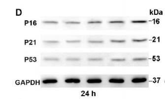
Fig. 3. The effects of TCPP@PPPy-PPy/PVA gel on NPCs’ viability and anabolic/catabolic metabolism. (A) The TCPP@PPy-PPy/PVA gel’s impact on NPC viability within a 3D culture system was evaluated through live/dead fluorescence staining on days 1, 3, and 7. (B) The MTT assay was used to determine the impact of varying US intensity on cell viability, with the culture time plotted as a line graph. Data shown are means ± SD of n = 3 biological replicates. (C) NPCs from Control, TBHP, TBHP + US, TBHP + Gel, and TBHP + Gel + US groups were performed live/dead and phalloidin fluorescence staining. (D) The PCR assay results from the Control-, TBHP-, TBHP + US–, TBHP + Gel–, and TBHP + Gel + US–treated NPCs were normalized and subsequently displayed using a heatmap. Data shown are means ± SD of n = 3 biological replicates. (E) WB analysis of inflammatory factor expression in NPCs following treatment with Control, TBHP, TBHP + US, TBHP + Gel, and TBHP + Gel + US experimental conditions. (F) The WB assay band images of ECM metabolism–related protein in NPCs following treatment with Control, TBHP, TBHP + US, TBHP + Gel, and TBHP + Gel + US. (G) Control-, TBHP-, TBHP + US–, TBHP + Gel–, and TBHP + Gel + US–treated NPCs were stained by Alcian. The darker the blue staining represents the better the catabolism of proteoglycan in NPCs.

5). Sirt3-mediated mitophagy regulates AGEs-induced BMSCs senescence and senile osteoporosis. Redox Biology, 2021 (PubMed: 33662874) [IF=10.7]

Application: WB    Species: mice    Sample: bone marrow mesenchymal stem (BMSCs)

Fig. 1. Effects of AGEs in different concentrations on the senescence of BMSCs. The BMSCs were treated with AGEs (50–200 μg/mL) or BSA for 24–72 h. (A) SA-β-gal assay for detection of BMSCs senescence. Scale bar: 100 μm. (B) Detection of H3K9me3 by immunofluorescence in BMSCs. Scale bar: 100 μm. (C) Detection of γ-H2AX by immunofluorescence in BMSCs. Scale bar: 100 μm. (D–I) Representative Western blotting assay and quantitation of the level of P16, P21, P53. **p < 0.01 versus BSA.

6). Targeted Inhibition of cGAS/STING signaling induced by aberrant R-Loops in the nucleus pulposus to alleviate cellular senescence and intervertebral disc degeneration. Journal of nanobiotechnology, 2025 (PubMed: 40660286) [IF=10.2]

Application: IHC    Species: Rat    Sample:

Fig. 1 NPC senescence is accompanied by DNA damage and the activation of the cGAS-STING axis during IVDD. A: Overview of the overall workflow of the integrated single-cell analysis. B: UMAP visualizing human NPCs as five different subclusters after unsupervised clustering. Each point indicates a single cell, colored according to different cell subclusters. C: Relative proportion of each NPC cluster between the control group and IVDD group. D: Differentially expressed genes in each NPC cluster. E: Cell cycle analysis of all NPC subclusters or that between the control group and IVDD group. F: Senescence score based on single-cell sequencing of each NPC cluster between the control group and IVDD group. G: Representative T2-weighted MRI images and SOFG staining of NP tissues with varying degrees of degeneration. Scale bar = 20 μm. H and I: Representative images and quantitative results of immunohistochemistry for p16INK4a in NP tissues with different degrees of degeneration (n = 3). Scale bar = 20 μm. J and K: Representative images and quantitative results of IF staining for γH2AX in NP tissues with varying degrees of degeneration (n = 3). Scale bar = 20 μm. L and M: Representative images and quantitative results of IF staining for γH2AX and p16INK4a in primary rat NPCs treated with or without H2O2 for 24 h (n = 3). Scale bar = 20 μm. N and O: Representative images and quantitative results of IF staining for S-β-Gal in primary rat NPCs treated with or without H2O2 for 24 h (n = 3). P: Neutral comet assay in primary rat NPCs treated with or without H2O2 for 24 h. Q: Quantification of the percentage of DNA in the tail (Tail DNA) (n = 3). Significance was calculated by (I, K, M, O, and Q) one-way ANOVA. *p 

Application: IF/ICC    Species: Rat    Sample:

Fig. 1 NPC senescence is accompanied by DNA damage and the activation of the cGAS-STING axis during IVDD. A: Overview of the overall workflow of the integrated single-cell analysis. B: UMAP visualizing human NPCs as five different subclusters after unsupervised clustering. Each point indicates a single cell, colored according to different cell subclusters. C: Relative proportion of each NPC cluster between the control group and IVDD group. D: Differentially expressed genes in each NPC cluster. E: Cell cycle analysis of all NPC subclusters or that between the control group and IVDD group. F: Senescence score based on single-cell sequencing of each NPC cluster between the control group and IVDD group. G: Representative T2-weighted MRI images and SOFG staining of NP tissues with varying degrees of degeneration. Scale bar = 20 μm. H and I: Representative images and quantitative results of immunohistochemistry for p16INK4a in NP tissues with different degrees of degeneration (n = 3). Scale bar = 20 μm. J and K: Representative images and quantitative results of IF staining for γH2AX in NP tissues with varying degrees of degeneration (n = 3). Scale bar = 20 μm. L and M: Representative images and quantitative results of IF staining for γH2AX and p16INK4a in primary rat NPCs treated with or without H2O2 for 24 h (n = 3). Scale bar = 20 μm. N and O: Representative images and quantitative results of IF staining for S-β-Gal in primary rat NPCs treated with or without H2O2 for 24 h (n = 3). P: Neutral comet assay in primary rat NPCs treated with or without H2O2 for 24 h. Q: Quantification of the percentage of DNA in the tail (Tail DNA) (n = 3). Significance was calculated by (I, K, M, O, and Q) one-way ANOVA. *p 

7). Heme oxygenase-1 prevents heart against myocardial infarction by attenuating ischemic injury-induced cardiomyocytes senescence. EBioMedicine, 2019 (PubMed: 30527623) [IF=9.7]

8). The brain-protective mechanism of fecal microbiota transplantation from young donor mice in the natural aging process via exosome, gut microbiota, and metabolomics analyses. Pharmacological research, 2024 (PubMed: 39053865) [IF=9.1]

9). DLK/JNK3 Upregulation Aggravates Hair Cell Senescence in Mice Cochleae via Excessive Autophagy. Aging cell, 2025 (PubMed: 40356329) [IF=8.0]

Application: WB    Species: Mouse    Sample:

FIGURE 5 Autophagy was excessively activated, and hair cell senescence was aggravated by DLK overexpression. (a, c–e) Western blot showing that DLK, p-JNK, and JNK3 were upregulated after DLK overexpression. (b, f–h) Western blot showing that the expression of ULK1, Beclin-1, and LC3B increased after DLK overexpression. (i–l) Western blot showing that the expression of p21, p16, and p53 increased after DLK overexpression. (m–p) Immunofluorescence showing that the expression of p21, p16, and p53 increased in HEI-OC1 cells following DLK overexpression. (q) Transmission electron microscopy image showing that autophagosomes (blue arrows) and autolysosomes (red arrows) increased in HEI-OC1 cells after DLK overexpression. (r) Number of autophagosomes and autolysosomes in a single cell in (p). Data are presented as mean ± SD

Application: IF/ICC    Species: Mouse    Sample:

FIGURE 5 Autophagy was excessively activated, and hair cell senescence was aggravated by DLK overexpression. (a, c–e) Western blot showing that DLK, p-JNK, and JNK3 were upregulated after DLK overexpression. (b, f–h) Western blot showing that the expression of ULK1, Beclin-1, and LC3B increased after DLK overexpression. (i–l) Western blot showing that the expression of p21, p16, and p53 increased after DLK overexpression. (m–p) Immunofluorescence showing that the expression of p21, p16, and p53 increased in HEI-OC1 cells following DLK overexpression. (q) Transmission electron microscopy image showing that autophagosomes (blue arrows) and autolysosomes (red arrows) increased in HEI-OC1 cells after DLK overexpression. (r) Number of autophagosomes and autolysosomes in a single cell in (p). Data are presented as mean ± SD

10). VDR activation attenuates osteoblastic ferroptosis and senescence by stimulating the Nrf2/GPX4 pathway in age-related osteoporosis. Free Radical Biology and Medicine, 2022 (PubMed: 36402439) [IF=7.1]

もっと読みます

Restrictive clause


Affinity Biosciences tests all products strictly. Citations are provided as a resource for additional applications that have not been validated by Affinity Biosciences. Please choose the appropriate format for each application and consult Materials and Methods sections for additional details about the use of any product in these publications.

For Research Use Only.
Not for use in diagnostic or therapeutic procedures. Not for resale. Not for distribution without written consent. Affinity Biosciences will not be held responsible for patent infringement or other violations that may occur with the use of our products. Affinity Biosciences, Affinity Biosciences Logo and all other trademarks are the property of Affinity Biosciences LTD.