製品: FGF2 Antibody
カタログ: DF6038
タンパク質の説明: Rabbit polyclonal antibody to FGF2
アプリケーション: WB IHC IF/ICC
反応性: Human, Mouse, Rat
分子量: 21kDa; 31kD(Calculated).
ユニプロット: P09038
RRID: AB_2838011

類似製品を見る>>

   サイズ 価格 在庫状況
 100ul $280 在庫あり
 200ul $350 在庫あり

リードタイム: 当日配達

For pricing and ordering contact:
お問い合わせ先

製品説明

ソース:
Rabbit
アプリケーション:
WB 1:500-1:2000, IHC 1:50-1:200, IF/ICC 1:100-1:500
*The optimal dilutions should be determined by the end user.
*Tips:

WB: For western blot detection of denatured protein samples. IHC: For immunohistochemical detection of paraffin sections (IHC-p) or frozen sections (IHC-f) of tissue samples. IF/ICC: For immunofluorescence detection of cell samples. ELISA(peptide): For ELISA detection of antigenic peptide.

反応性:
Human,Mouse,Rat
クローナリティ:
Polyclonal
特異性:
FGF2 Antibody detects endogenous levels of total FGF2.
RRID:
AB_2838011
引用形式: Affinity Biosciences Cat# DF6038, RRID:AB_2838011.
コンジュゲート:
Unconjugated.
精製:
The antiserum was purified by peptide affinity chromatography using SulfoLink™ Coupling Resin (Thermo Fisher Scientific).
保存:
Rabbit IgG in phosphate buffered saline , pH 7.4, 150mM NaCl, 0.02% sodium azide and 50% glycerol. Store at -20 °C. Stable for 12 months from date of receipt.
別名:

折りたたみ/展開

Basic fibroblast growth factor; Basic fibroblast growth factor bFGF; BFGF; FGF 2; FGF B; FGF-2; Fgf2; FGF2 basic; FGF2_HUMAN; FGFB; Fibroblast growth factor 2 (basic); Fibroblast growth factor 2; Fibroblast growth factor, basic; HBGF 2; HBGF-2; HBGF2; HBGH 2; HBGH2; Heparin binding growth factor 2 precursor; Heparin-binding growth factor 2; Prostatropin;

免疫原

免疫原:
Uniprot:
遺伝子(ID):
発現特異性:
P09038 FGF2_HUMAN:

Expressed in granulosa and cumulus cells. Expressed in hepatocellular carcinoma cells, but not in non-cancerous liver tissue.

タンパク質の説明:
Fibroblast growth factors are a family of broad-spectrum growth factors influencing a plethora of cellular activities. The interaction of at least 23 ligands, 4 receptors and multiple coreceptors provides a dramatic complexity to a signaling system capable of effecting a multitude of responses (1,2). Basic fibroblast growth factor (bFGF or FGF2), initially identified as a mitogen with prominent angiogenic properties, is now recognized as a multifunctional growth factor (3). It is clear that bFGF produces its biological effects in target cells by signaling through cell-surface FGF receptors. bFGF binds to all four FGF receptors. Ligand binding induces receptor dimerization and autophosphorylation, allowing binding and activation of cytoplasmic downstream target proteins, including FRS-2, PLC and Crk (4,5). The FGF signaling pathway appears to play a significant role not only in normal cell growth regulation but also in tumor development and progression (6).Acidic FGF (aFGF or FGF1) is another extensively investigated protein of the FGF family. aFGF shares 55% DNA sequence homology with bFGF. These two growth factors are ubiquitously expressed and exhibit a wide spectrum of similiar biological activities with quantitative differences likely due to variation in receptor affinity or binding (7).
タンパク質配列:
MVGVGGGDVEDVTPRPGGCQISGRGARGCNGIPGAAAWEAALPRRRPRRHPSVNPRSRAAGSPRTRGRRTEERPSGSRLGDRGRGRALPGGRLGGRGRGRAPERVGGRGRGRGTAAPRAAPAARGSRPGPAGTMAAGSITTLPALPEDGGSGAFPPGHFKDPKRLYCKNGGFFLRIHPDGRVDGVREKSDPHIKLQLQAEERGVVSIKGVCANRYLAMKEDGRLLASKCVTDECFFFERLESNNYNTYRSRKYTSWYVALKRTGQYKLGSKTGPGQKAILFLPMSAKS

PTMs - P09038 基板として

Site PTM Type Enzyme
S62 Phosphorylation
T65 Phosphorylation
R82 Methylation
R84 Methylation
R86 Methylation
R96 Methylation
R98 Methylation
R108 Methylation
R110 Methylation
R112 Methylation
R118 Methylation
R124 Methylation
S138 Phosphorylation
T141 Phosphorylation
K168 Acetylation
S206 Phosphorylation
Y215 Phosphorylation P42680 (TEC)
K228 Sumoylation
K228 Ubiquitination
S242 Phosphorylation
Y245 Phosphorylation
S250 Phosphorylation
Y253 Phosphorylation
T254 Phosphorylation P17612 (PRKACA)
Y257 Phosphorylation P42680 (TEC)
K261 Acetylation
K267 Acetylation
K267 Ubiquitination
K271 Acetylation
K271 Ubiquitination
K277 Acetylation
S285 Phosphorylation
S288 Phosphorylation

研究背景

機能:

Acts as a ligand for FGFR1, FGFR2, FGFR3 and FGFR4. Also acts as an integrin ligand which is required for FGF2 signaling. Binds to integrin ITGAV:ITGB3. Plays an important role in the regulation of cell survival, cell division, cell differentiation and cell migration. Functions as a potent mitogen in vitro. Can induce angiogenesis.

PTMs:

Phosphorylation at Tyr-215 regulates FGF2 unconventional secretion.

Several N-termini starting at positions 94, 125, 126, 132, 143 and 162 have been identified by direct sequencing.

細胞の位置付け:

Secreted. Nucleus.
Note: Exported from cells by an endoplasmic reticulum (ER)/Golgi-independent mechanism. Unconventional secretion of FGF2 occurs by direct translocation across the plasma membrane. Binding of exogenous FGF2 to FGFR facilitates endocytosis followed by translocation of FGF2 across endosomal membrane into the cytosol. Nuclear import from the cytosol requires the classical nuclear import machinery, involving proteins KPNA1 and KPNB1, as well as CEP57.

Extracellular region or secreted Cytosol Plasma membrane Cytoskeleton Lysosome Endosome Peroxisome ER Golgi apparatus Nucleus Mitochondrion Manual annotation Automatic computational assertionSubcellular location
組織特異性:

Expressed in granulosa and cumulus cells. Expressed in hepatocellular carcinoma cells, but not in non-cancerous liver tissue.

サブユニット構造:

Monomer. Homodimer. Interacts with FGFR1, FGFR2, FGFR3 and FGFR4. Affinity between fibroblast growth factors (FGFs) and their receptors is increased by heparan sulfate glycosaminoglycans that function as coreceptors. Interacts with CSPG4, FGFBP1 and TEC. Found in a complex with FGFBP1, FGF1 and FGF2. Interacts with FGFBP3. Interacts with integrin ITGAV:ITGB3; the interaction is required for FGF2 signaling. Interacts with SNORC (via the extracellular domain) (By similarity). Interacts with glypican GPC3 (By similarity).

タンパク質ファミリー:

Belongs to the heparin-binding growth factors family.

研究領域

· Cellular Processes > Cellular community - eukaryotes > Signaling pathways regulating pluripotency of stem cells.   (View pathway)

· Cellular Processes > Cell motility > Regulation of actin cytoskeleton.   (View pathway)

· Environmental Information Processing > Signal transduction > MAPK signaling pathway.   (View pathway)

· Environmental Information Processing > Signal transduction > Ras signaling pathway.   (View pathway)

· Environmental Information Processing > Signal transduction > Rap1 signaling pathway.   (View pathway)

· Environmental Information Processing > Signal transduction > PI3K-Akt signaling pathway.   (View pathway)

· Human Diseases > Drug resistance: Antineoplastic > EGFR tyrosine kinase inhibitor resistance.

· Human Diseases > Cancers: Overview > Pathways in cancer.   (View pathway)

· Human Diseases > Cancers: Overview > Proteoglycans in cancer.

· Human Diseases > Cancers: Specific types > Melanoma.   (View pathway)

· Human Diseases > Cancers: Specific types > Breast cancer.   (View pathway)

· Human Diseases > Cancers: Specific types > Gastric cancer.   (View pathway)

参考文献

1). Adipose-derived stem cell/FGF19-loaded microfluidic hydrogel microspheres for synergistic restoration of critical ischemic limb. Bioactive Materials, 2023 (PubMed: 37122899) [IF=18.9]

Application: WB    Species: Mouse    Sample: ADSCs

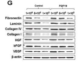
Fig. 3. Proliferation, migration and angiogenesis performance of ADSCs on GelMA microspheres. A) The proliferation curves of ADSCs seeded on culture plate and GelMA microspheres. B) ADSCs grown on indicated substrates were subjected to a transwell migration assay and C) the results of statistical evaluation results. D) ADSCs grown on indicated substrates were used for an in vitro scratching wound healing assay, and E) the results of statistical evaluation results. F) qRT-PCR assay was used to evaluate the gene expression of extracellular matrix (fibronectin, laminin, collagen I and collagen IV) and angiogenic factors (HGF, bFGF and VEGF). Western blot and qRT-PCR outcomes are shown relative to β-actin. (units: cells/10 mg microspheres) G) Western blotting was used to assess fibronectin, laminin, collagen I, collagen IV, HGF, bFGF and VEGF protein expression in the 6 different groups (Group1-3: FGF19-loading microspheres with high, medium and low densities of ADSCs; Group 4–6: Blank microspheres with high, medium and low densities of ADSCs). H) Western blotting data of the levels of fibronectin, laminin, collagen I, collagen IV, HGF, bFGF and VEGF in different groups. Experiments were repeated 3 times. *p < 0.05

2). Dihydroartemisinin inhibited vasculogenic mimicry in gastric cancer through the FGF2/FGFR1 signaling pathway. Phytomedicine : international journal of phytotherapy and phytopharmacology, 2024 (PubMed: 39214017) [IF=7.9]

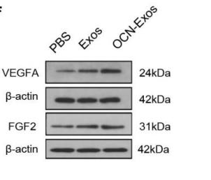
3). Exosomes secreted from osteocalcin-overexpressed endothelial progenitor cells promoted endothelial cell angiogenesis. AMERICAN JOURNAL OF PHYSIOLOGY-CELL PHYSIOLOGY, 2019 (PubMed: 31411920) [IF=5.5]

Application: WB    Species: rat    Sample: RAOECs

Figure 3 |OCN-Exos were more effective than Exos in promoting endothelial cell angiogenesis. RAOECs treated by PBS, Exos and OCN-Exos for 24 hours were seeded on Matrigel. The representative photomicrographs of Matrigel tube formation angiogenesis assays were taken after 4, and 8 hours incubated in Matrigel-coated culture plates (A). Number (B and C) and length (D and E) of master segments were counted and measured. Protein levels of VEGFA and FGF2 were analysed by western blot. (F, G and H) Data were from four independent experiments and expressed as means ± SEM. *p<0.05, **p<0.01 vs. PBS; #p<0.05, ## p<0.05 vs. Exos. Scale bars=100 μm.

4). Exploring the Efficacy of Hydroxybenzoic Acid Derivatives in Mitigating Jellyfish Toxin-Induced Skin Damage: Insights into Protective and Reparative Mechanisms. Marine drugs, 2024 (PubMed: 38786596) [IF=5.4]

Application: IHC    Species: Mouse    Sample:

Figure 3 PCA and DHB enhance collagen remodeling and stimulate growth factor expression in NnNV-induced wounds. (A–D) Picrosirius red staining of the dorsal skin wound site in mice observed using polarized light microscopy after NnNV injection and 7-day treatment with PCA (green) or DHB (yellow) (n = 4). (A) Scale bar, 50 μm. (B) Quantitative data showing the percentage of type I collagen fibers in the wound bed field for each skin tissue group. (C) Quantitative data showing the percentage of type III collagen fibers in the wound bed field for each skin tissue group. (D) Proportion of type III/type I collagen fibers in the wound bed domain. (E) Immunohistochemical staining showing the expression of vascular endothelial growth factor (VEGF) and basic fibroblast growth factor (bFGF) at the margins of dorsal skin wounds in mice 7 days after NnNV injection and treatment with PCA (green) or DHB (yellow). Scale bar, 100 μm. Blank groups received injections of an equal volume of PBS buffer (PBS, black). The NnNV control group received only NnNV injection without cream application (NnNV, blue), while the Cream control group received NnNV injection with cream application but without drug loading (Cream, grey). Data are mean  ±  s.e.m. n  =  biological replicates.

5). Newly generated neurons at 2 months post-status epilepticus are functionally integrated into neuronal circuitry in mouse hippocampus. EXPERIMENTAL NEUROLOGY, 2015 (PubMed: 26384773) [IF=5.3]

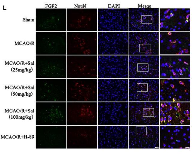
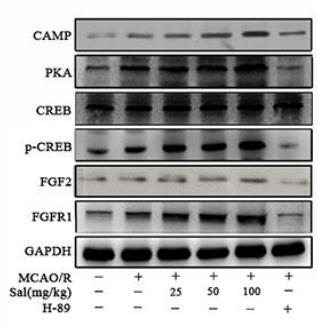
6). Fibroblast growth factor 2 contributes to the effect of salidroside on dendritic and synaptic plasticity after cerebral ischemia/reperfusion injury. Aging (Albany NY), 2020 (PubMed: 32518214) [IF=5.2]

Application: IF/ICC    Species: rat    Sample:

Figure 6. | Sal upregulates the FGF2-mediated cAMP/PKA/CREB signaling pathway following MCAO/R.(A–G) Representative western blot bands of cAMP, PKA, CREB, p-CREB, FGF2 and FGFR1 in each group. (H–K) QPCR analysis of PKA, p-CREB, FGF2 and FGFR1 mRNA expression at 7 days after MCAO/R in different groups. (I) Double staining for FGF2-positive (green) and NeuN-positive neurons (red) neurons(the scale bars are 20 μm and 10 μm). Values are expressed as the mean ± SD. #p < 0.05, ##p < 0.01 vs. sham; *p < 0.05, **p < 0.01 vs. MCAO/R.

Application: WB    Species: rat    Sample:

Figure 6. |Sal upregulates the FGF2-mediated cAMP/PKA/CREB signaling pathway following MCAO/R. (A–G) Representative western blot bands of cAMP, PKA, CREB, p-CREB, FGF2 and FGFR1 in each group.

7). FGFR inhibitors combined with nab-paclitaxel - A promising strategy to treat non-small cell lung cancer and overcome resistance. Frontiers in Oncology, 2023 (PubMed: 36845692) [IF=4.7]

Application: WB    Species: Human    Sample:

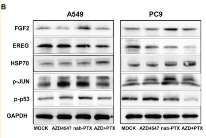
Figure 6 Effects of AZD4547 and nab-PTX on the MAPK signaling pathway. (A) The expression of FGF2, EREG, and HSP70 was assessed by qRT−PCR. (B) Western blot analysis of the protein expression of FGF2, EREG, and HSP70 and the phosphorylation of JUN and p53. (C) AZD4547 combined with nab-PTX inhibited phosphorylation of the JNK, ERK, and p38 proteins. *: p< 0.05, **: p< 0.01, ****: p< 0.0001.

8). Forced Physical Training Increases Neuronal Proliferation and Maturation with Their Integration into Normal Circuits in Pilocarpine Induced Status Epilepticus Mice. NEUROCHEMICAL RESEARCH, 2019 (PubMed: 31560103) [IF=4.4]

9). Hair growth predicts a depression-like phenotype in rats as a mirror of stress traceability. NEUROCHEMISTRY INTERNATIONAL, 2021 (PubMed: 34166749) [IF=4.2]

10). Fabrication of porous bovine pericardium scaffolds incorporated with bFGF for tissue engineering applications. XENOTRANSPLANTATION, 2020 (PubMed: 31693254) [IF=3.9]

もっと読みます

Restrictive clause


Affinity Biosciences tests all products strictly. Citations are provided as a resource for additional applications that have not been validated by Affinity Biosciences. Please choose the appropriate format for each application and consult Materials and Methods sections for additional details about the use of any product in these publications.

For Research Use Only.
Not for use in diagnostic or therapeutic procedures. Not for resale. Not for distribution without written consent. Affinity Biosciences will not be held responsible for patent infringement or other violations that may occur with the use of our products. Affinity Biosciences, Affinity Biosciences Logo and all other trademarks are the property of Affinity Biosciences LTD.