製品: WNT3A Antibody
カタログ: DF6113
タンパク質の説明: Rabbit polyclonal antibody to WNT3A
アプリケーション: WB IHC IF/ICC
反応性: Human, Mouse, Rat
予測: Pig, Zebrafish, Bovine, Rabbit, Dog, Chicken, Xenopus
分子量: 39kDa; 39kD(Calculated).
ユニプロット: P56704
RRID: AB_2838080

類似製品を見る>>

   サイズ 価格 在庫状況
 100ul $280 在庫あり
 200ul $350 在庫あり

リードタイム: 当日配達

For pricing and ordering contact:
お問い合わせ先

製品説明

ソース:
Rabbit
アプリケーション:
WB 1:500-1:2000, IHC 1:50-1:200, IF/ICC 1:100-500
*The optimal dilutions should be determined by the end user.
*Tips:

WB: For western blot detection of denatured protein samples. IHC: For immunohistochemical detection of paraffin sections (IHC-p) or frozen sections (IHC-f) of tissue samples. IF/ICC: For immunofluorescence detection of cell samples. ELISA(peptide): For ELISA detection of antigenic peptide.

反応性:
Human,Mouse,Rat
予測:
Pig(88%), Zebrafish(88%), Bovine(88%), Rabbit(88%), Dog(88%), Chicken(88%), Xenopus(88%)
クローナリティ:
Polyclonal
特異性:
WNT3A Antibody detects endogenous levels of total WNT3A.
RRID:
AB_2838080
引用形式: Affinity Biosciences Cat# DF6113, RRID:AB_2838080.
コンジュゲート:
Unconjugated.
精製:
The antiserum was purified by peptide affinity chromatography using SulfoLink™ Coupling Resin (Thermo Fisher Scientific).
保存:
Rabbit IgG in phosphate buffered saline , pH 7.4, 150mM NaCl, 0.02% sodium azide and 50% glycerol. Store at -20 °C. Stable for 12 months from date of receipt.
別名:

折りたたみ/展開

Protein Wnt 3a Precursor; Protein Wnt-3a; Wingless type MMTV integration site family member 3A; Wnt 3a; wnt3a; WNT3A protein; WNT3A_HUMAN;

免疫原

免疫原:
Uniprot:
遺伝子(ID):
発現特異性:
P56704 WNT3A_HUMAN:

Moderately expressed in placenta and at low levels in adult lung, spleen, and prostate.

タンパク質の説明:
The Wnt family includes several secreted glycoproteins that play important roles in animal development (1). There are 19 Wnt genes in the human genome that encode functionally distinct Wnt proteins (2). Wnt members bind to the Frizzled family of seven-pass transmembrane proteins and activate several signaling pathways (3). The canonical Wnt/β-catenin pathway also requires a coreceptor from the low-density lipoprotein receptor family (4). Aberrant activation of Wnt signaling pathways is involved in several types of cancers (5).
タンパク質配列:
MAPLGYFLLLCSLKQALGSYPIWWSLAVGPQYSSLGSQPILCASIPGLVPKQLRFCRNYVEIMPSVAEGIKIGIQECQHQFRGRRWNCTTVHDSLAIFGPVLDKATRESAFVHAIASAGVAFAVTRSCAEGTAAICGCSSRHQGSPGKGWKWGGCSEDIEFGGMVSREFADARENRPDARSAMNRHNNEAGRQAIASHMHLKCKCHGLSGSCEVKTCWWSQPDFRAIGDFLKDKYDSASEMVVEKHRESRGWVETLRPRYTYFKVPTERDLVYYEASPNFCEPNPETGSFGTRDRTCNVSSHGIDGCDLLCCGRGHNARAERRREKCRCVFHWCCYVSCQECTRVYDVHTCK

種類予測

種類予測:

Score>80(red) has high confidence and is suggested to be used for WB detection. *The prediction model is mainly based on the alignment of immunogen sequences, the results are for reference only, not as the basis of quality assurance.

Species
Results
Score
Pig
88
Bovine
88
Dog
88
Xenopus
88
Zebrafish
88
Chicken
88
Rabbit
88
Horse
0
Sheep
0
Model Confidence:
High(score>80) Medium(80>score>50) Low(score<50) No confidence

PTMs - P56704 基板として

Site PTM Type Enzyme
S65 Phosphorylation
N87 N-Glycosylation
Y235 Phosphorylation
S237 Phosphorylation
N298 N-Glycosylation

研究背景

機能:

Ligand for members of the frizzled family of seven transmembrane receptors (Probable). Functions in the canonical Wnt signaling pathway that results in activation of transcription factors of the TCF/LEF family. Required for normal embryonic mesoderm development and formation of caudal somites. Required for normal morphogenesis of the developing neural tube (By similarity). Mediates self-renewal of the stem cells at the bottom on intestinal crypts (in vitro).

PTMs:

Palmitoleoylation by PORCN is required for efficient binding to frizzled receptors. Palmitoleoylation is required for proper trafficking to cell surface, vacuolar acidification is critical to release palmitoleoylated WNT3A from WLS in secretory vesicles. Depalmitoleoylated by NOTUM, leading to inhibit Wnt signaling pathway, possibly by promoting disulfide bond formation and oligomerization.

Proteolytic processing by TIKI1 and TIKI2 promotes oxidation and formation of large disulfide-bond oligomers, leading to inactivation of WNT3A.

Disulfide bonds have critical and distinct roles in secretion and activity. Loss of each conserved cysteine in WNT3A results in high molecular weight oxidized Wnt oligomers, which are formed through inter-Wnt disulfide bonding.

細胞の位置付け:

Secreted>Extracellular space>Extracellular matrix. Secreted.

Extracellular region or secreted Cytosol Plasma membrane Cytoskeleton Lysosome Endosome Peroxisome ER Golgi apparatus Nucleus Mitochondrion Manual annotation Automatic computational assertionSubcellular location
組織特異性:

Moderately expressed in placenta and at low levels in adult lung, spleen, and prostate.

サブユニット構造:

Forms a soluble 1:1 complex with AFM; this prevents oligomerization and is required for prolonged biological activity. The complex with AFM may represent the physiological form in body fluids. Homooligomer; disulfide-linked, leading to inactivation (By similarity). Interacts with PORCN. Interacts with APCDD1 and WLS. Component of the Wnt-Fzd-LRP5-LRP6 signaling complex that contains a WNT protein, a FZD protein and LRP5 or LRP6. Interacts directly in the complex with LRP6. Interacts with glypican GPC3. Interacts with PKD1 (via extracellular domain).

タンパク質ファミリー:

Belongs to the Wnt family.

研究領域

· Cellular Processes > Cellular community - eukaryotes > Signaling pathways regulating pluripotency of stem cells.   (View pathway)

· Environmental Information Processing > Signal transduction > mTOR signaling pathway.   (View pathway)

· Environmental Information Processing > Signal transduction > Wnt signaling pathway.   (View pathway)

· Environmental Information Processing > Signal transduction > Hippo signaling pathway.   (View pathway)

· Human Diseases > Infectious diseases: Viral > Human papillomavirus infection.

· Human Diseases > Infectious diseases: Viral > HTLV-I infection.

· Human Diseases > Cancers: Overview > Pathways in cancer.   (View pathway)

· Human Diseases > Cancers: Overview > Proteoglycans in cancer.

· Human Diseases > Cancers: Overview > MicroRNAs in cancer.

· Human Diseases > Cancers: Specific types > Basal cell carcinoma.   (View pathway)

· Human Diseases > Cancers: Specific types > Breast cancer.   (View pathway)

· Human Diseases > Cancers: Specific types > Hepatocellular carcinoma.   (View pathway)

· Human Diseases > Cancers: Specific types > Gastric cancer.   (View pathway)

· Organismal Systems > Endocrine system > Melanogenesis.

参考文献

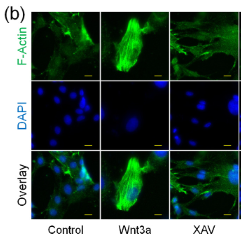
1). Regulation of hepatic stellate cell contraction and cirrhotic portal hypertension by Wnt/β-catenin signalling via interaction with Gli1. British Journal of Pharmacology, 2021 (PubMed: 33085791) [IF=6.8]

Application: IF/ICC    Species: Human    Sample: HSCs

FIGURE 2 Wnt/β-catenin signalling promotes HSC contraction. LX2 human HSCs were treated with Wnt3a (50 ng·ml−1), XAV (10 μM), or IWR (5 μM) for 24 h or transfected with β-catenin siRNA with or without Wnt3a (50 ng·ml−1) for 48 h. (a) Collagen gel contraction; n = 5. (b) Cytoskeleton immunofluorescence (400× magnification, scale bars: 10 μm); n = 3. (c) Western blotting of phospho-MLC2; n = 5. (d) Immunofluorescence of phospho-MLC2 (400× magnification, scale bars: 10 μm); n = 3. In (a) and (c), *P 

2). Quercetin induces MGMT+ glioblastoma cells apoptosis via dual inhibition of Wnt3a/β-Catenin and Akt/NF-κB signaling pathways. Phytomedicine, 2023 (PubMed: 37451151) [IF=6.7]

3). YAP and Wnt3a independently promote AECIIs proliferation and differentiation by increasing nuclear β‑catenin expression in experimental bronchopulmonary dysplasia. INTERNATIONAL JOURNAL OF MOLECULAR MEDICINE, 2021 (PubMed: 33236141) [IF=5.7]

Application: WB    Species: Rat    Sample: lung tissues

Figure 2 YAP, Wnt3a and β-catenin expression is decreased in lung tissue affected by BPD. (A) YAP mRNA and (C) protein expression in the lungs. (B) Wnt3a mRNA and (C) protein expression in the lungs. (D) β-catenin protein expression in lung nucleoli. Data are expressed as the means ± SD (n=6). *P<0.05, **P<0.01. (E) YAP and (F) β-catenin expression in IHC-stained lung sections. Scale bar, 40 µm. Expression of (G) SPC and (G) YAP, as well as (H) AQP5 and YAP detected by double IF staining. Scale bar, 40 µm. (I and J) Expression of (I) SPC and β-catenin, as well as (J) AQP5 and β-catenin detected by double IF staining. Scale bar, 40 µm. N, normoxia-exposed rats; H, hyperoxia-exposed rats; SPC, surfactant protein C; AQP5, aquaporin 5; BPD, bronchopulmonary dysplasia.

4). Interaction between the Neuroprotective and Hyperglycemia Mitigation Effects of Walnut-Derived Peptide LVRL via the Wnt3a/β-Catenin/GSK-3β Pathway in a Type 2 Diabetes Mellitus Model. Journal of agricultural and food chemistry, 2024 (PubMed: 38984968) [IF=5.7]

5). DEPDC1B-mediated USP5 deubiquitination of β-catenin promotes breast cancer metastasis by activating the wnt/β-catenin pathway. American Journal of Physiology-Cell Physiology, 2023 (PubMed: 37642235) [IF=5.0]

Application: WB    Species: Human    Sample: breast cancer cells

Figure 6. The expression of DEP domain-containing protein 1B (DEPDC1B) can activate the wnt/β-catenin signaling pathway. A: functional enrichment analysis of the results of protein spectrum identification. B: the results of KEGG analysis showed that the signal pathway related to the DEPDC1B interacting proteins. C and D: DEPDC1B is involved in the nuclear translocation of β-catenin. Hs578T cells (C) were transfected with negative control (NC) and DEPDC1B siRNA, MDA-MB-157 cells (D) were transfected with vector and DEPDC1B overexpression plasmids, and the expression of β-catenin was detected by immunofluorescence after 48 h. Scale bars, 20 μm. E: the results of the statistical analysis of C and D. Analysis between 2 groups was conducted by unpaired Student’s t test. *P < 0.05. F: the expression levels of wnt3a, phospho (p-)GSK-3β (ser9), GSK-3β, and β-catenin were detected by Western blot, and GAPDH was used as the internal control. G: Western blot results of the expression of DEPDC1B, wnt3a, and β-catenin in MDA-MB-157 cells transfected with DEPDC1B overexpression plasmid alone or in combination with XAV-939, with GAPDH as the internal control.

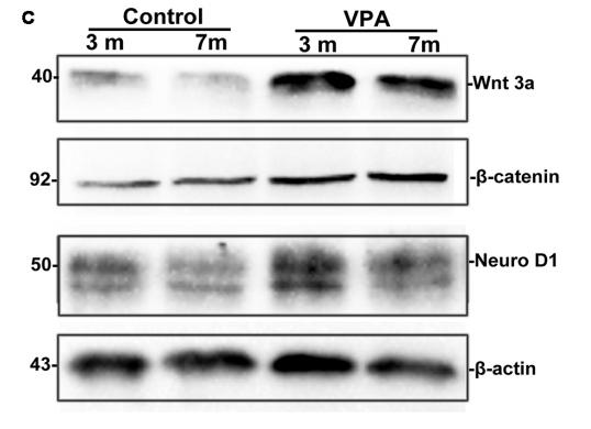
6). Valproic Acid Stimulates Hippocampal Neurogenesis via Activating the Wnt/β-Catenin Signaling Pathway in the APP/PS1/Nestin-GFP Triple Transgenic Mouse Model of Alzheimer's Disease. Frontiers in Aging Neuroscience, 2019 (PubMed: 30971911) [IF=4.1]

Application: WB    Species: mouse    Sample: hippocampus

FIGURE 6 | VPA inhibits glycogen synthase kinase-3β (GSK-3β) activity and stimulates the Wnt/β-catenin signaling pathway. (A) Western blot analysis of total GSK-3β and p-GSK-3βS9(pSer9). GAPDH was used as an internal control. (B) Densitometric analysis of total GSK-3β and p-GSK-3βS9(pSer9). VPA increased pSer9-GSK-3β levels but not total GSK-3β levels in AD mice. (C) Immunoblots of β-catenin, Wnt3a and NeuroD1; β-actin was used as an internal control.

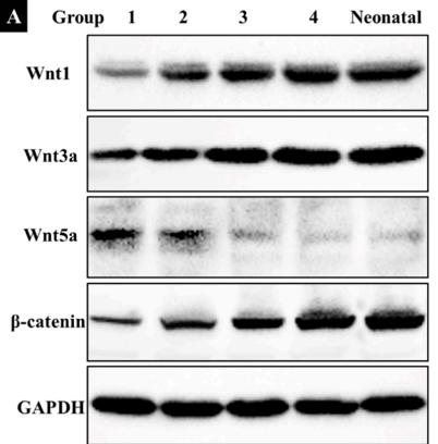
7). Exercise Promotes the Osteoinduction of HA/β-TCP Biomaterials via the Wnt Signaling Pathway. Metabolites, 2020 (PubMed: 32151083) [IF=4.1]

Application: WB    Species: mouse    Sample: bone

Figure 5. |(A) Western blot showing the expression of wingless/integrated 1 (Wnt1), Wnt3a, Wnt5a, β‐catenin, and glyceraldehyde‐3 phosphate dehydrogenase (GAPDH) in five groups (n = 3). G1: negative control group; G2: running group; G3: weight‐bearing group; G4: positive control group; Neonatal: the neonatal bone, control.

8). Potential actions of capsaicin for preventing vascular calcification of vascular smooth muscle cells in vitro and in vivo. Heliyon, 2024 (PubMed: 38524547) [IF=4.0]

Application: WB    Species: Mouse    Sample: VSMCs

Fig. 4 Cap inhibits Pi-induced calcification of VSMCs by inhibiting the Wnt/β-catenin signaling pathway by upregulation of TRPV1 receptor expression. A) RT-qPCR showing the mRNA levels of Wnt3a and β-catenin; B) Western blot illustrating the protein expression levels of Wnt3a and β-catenin; C) Alizarin Red staining assay; D) Calcium content in VSMCs assessed using a calcium colorimetric assay kit. E) RT-qPCR showing the expression of osteogenesis-specific and phenotypic markers in VSMC calcification. F) Western blot depicting the protein expression levels of osteogenesis-specific and phenotypic markers in VSMC calcification; G) Western blot showing the protein expression of β-catenin after treatment with DKK1. VSMCs cultured in the complete and pro-calcifying medium were defined as the control and Pi group, respectively; Pi + Cap: VSMCs cultured in the pro-calcifying medium with 20 μM Cap; Pi + Cap + CPZ: VSMCs cultured in the pro-calcifying medium with 20 μM Cap and 10 μM CPZ; Pi + Cap + DKK1: VSMCs cultured in the pro-calcifying medium with 20 μM Cap and 20 ng/mL DKK1; Data are mean ± SD (n = 3); #: p < 0.05, ##: p < 0.01, ###: p < 0.001 vs Control; *: p < 0.05, **: p < 0.01, ***: p < 0.001 vs Pi; ※: p < 0.05, ※※: p < 0.01, ※※※: p < 0.001 vs Pi + Cap; ns: p > 0.05 vs Control; p > 0.05 vs Pi; p > 0.05 vs Pi + Cap. (For interpretation of the references to color in this figure legend, the reader is referred to the Web version of this article.)

9). Wnt3a/YTHDF1 Regulated Oxaliplatin-Induced Neuropathic Pain Via TNF-α/IL-18 Expression in the Spinal Cord. Cellular and Molecular Neurobiology, 2022 (PubMed: 35939138) [IF=3.6]

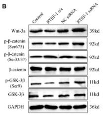
10). RTEF-1 Inhibits Vascular Smooth Muscle Cell Calcification through Regulating Wnt/β-Catenin Signaling Pathway. CALCIFIED TISSUE INTERNATIONAL, 2021 (PubMed: 33713163) [IF=3.3]

Application: WB    Species: Human    Sample: VSMCs

Fig. 4 RTEF-1 inhibits β-GP-activated Wnt/β‐catenin signaling pathway in human VSMCs. RTEF-1-overexpressed VSMCs, RTEF-1-siRNA VSMCs, or control VSMCs were treated in the presence of 10 mM β-GP for 9 days (n = 3). A Wnt3a mRNA levels were analyzed by qRT-PCR. B Wnt3a, p-β‐catenin (Ser675), p-β‐catenin (Ser33/37), β‐catenin, p-GSK-3β (Ser9), and GSK-3β protein expression were analyzed by Western blot and quantified by densitometry. C GSK-3β mRNA expression was detected by qRT-PCR. a Western blotting analysis of Wnt3a, p-β‐catenin (Ser675), p-β‐catenin (Ser33/37), p-GSK-3β (Ser9), and GSK-3β. b Western blotting analysis of the ratio of p-β‐catenin (Ser33/37) to β‐catenin protein, c Western blotting analysis of the ratio of p-GSK-3β to GSK-3β protein. Statistical significance was assessed using t-test. All graphs show mean + SD. *P < 0.05 vs. Control. #P < 0.05 vs. NC siRNA

もっと読みます

Restrictive clause


Affinity Biosciences tests all products strictly. Citations are provided as a resource for additional applications that have not been validated by Affinity Biosciences. Please choose the appropriate format for each application and consult Materials and Methods sections for additional details about the use of any product in these publications.

For Research Use Only.
Not for use in diagnostic or therapeutic procedures. Not for resale. Not for distribution without written consent. Affinity Biosciences will not be held responsible for patent infringement or other violations that may occur with the use of our products. Affinity Biosciences, Affinity Biosciences Logo and all other trademarks are the property of Affinity Biosciences LTD.