, using BIRC2 Antibody at 1/1000 dilution.")
, using BIRC2 Antibody at 1/1000 dilution.")


製品説明
*The optimal dilutions should be determined by the end user. For optimal experimental results, antibody reuse is not recommended.
*Tips:
WB: For western blot detection of denatured protein samples. IHC: For immunohistochemical detection of paraffin sections (IHC-p) or frozen sections (IHC-f) of tissue samples. IF/ICC: For immunofluorescence detection of cell samples. ELISA(peptide): For ELISA detection of antigenic peptide.
引用形式: Affinity Biosciences Cat# DF6167, RRID:AB_2838134.
折りたたみ/展開
API 1; API1; Apoptosis inhibitor 1; Baculoviral IAP repeat containing 2; Baculoviral IAP repeat containing protein 2; Baculoviral IAP repeat-containing protein 2; BIRC 2; BIRC2; BIRC2_HUMAN; C IAP1; C-IAP1; cIAP 1; cIAP1; HIAP 2; HIAP-2; HIAP2; IAP 2; IAP homolog B; IAP-2; IAP2; Inhibitor of apoptosis protein 2; MIHB; NFR2 TRAF signalling complex protein; RING finger protein 48; RNF 48; RNF48; TNFR2 TRAF signaling complex protein 2; TNFR2-TRAF-signaling complex protein 2;
免疫原
A synthesized peptide derived from human BIRC2, corresponding to a region within C-terminal amino acids.
Present in many fetal and adult tissues. Mainly expressed in adult skeletal muscle, thymus, testis, ovary, and pancreas, low or absent in brain and peripheral blood leukocytes.
- Q13490 BIRC2_HUMAN:
- Protein BLAST With
- NCBI/
- ExPASy/
- Uniprot
MHKTASQRLFPGPSYQNIKSIMEDSTILSDWTNSNKQKMKYDFSCELYRMSTYSTFPAGVPVSERSLARAGFYYTGVNDKVKCFCCGLMLDNWKLGDSPIQKHKQLYPSCSFIQNLVSASLGSTSKNTSPMRNSFAHSLSPTLEHSSLFSGSYSSLSPNPLNSRAVEDISSSRTNPYSYAMSTEEARFLTYHMWPLTFLSPSELARAGFYYIGPGDRVACFACGGKLSNWEPKDDAMSEHRRHFPNCPFLENSLETLRFSISNLSMQTHAARMRTFMYWPSSVPVQPEQLASAGFYYVGRNDDVKCFCCDGGLRCWESGDDPWVEHAKWFPRCEFLIRMKGQEFVDEIQGRYPHLLEQLLSTSDTTGEENADPPIIHFGPGESSSEDAVMMNTPVVKSALEMGFNRDLVKQTVQSKILTTGENYKTVNDIVSALLNAEDEKREEEKEKQAEEMASDDLSLIRKNRMALFQQLTCVLPILDNLLKANVINKQEHDIIKQKTQIPLQARELIDTILVKGNAAANIFKNCLKEIDSTLYKNLFVDKNMKYIPTEDVSGLSLEEQLRRLQEERTCKVCMDKEVSVVFIPCGHLVVCQECAPSLRKCPICRGIIKGTVRTFLS
種類予測
Score>80(red) has high confidence and is suggested to be used for WB detection. *The prediction model is mainly based on the alignment of immunogen sequences, the results are for reference only, not as the basis of quality assurance.
High(score>80) Medium(80>score>50) Low(score<50) No confidence
研究背景
Multi-functional protein which regulates not only caspases and apoptosis, but also modulates inflammatory signaling and immunity, mitogenic kinase signaling, and cell proliferation, as well as cell invasion and metastasis. Acts as an E3 ubiquitin-protein ligase regulating NF-kappa-B signaling and regulates both canonical and non-canonical NF-kappa-B signaling by acting in opposite directions: acts as a positive regulator of the canonical pathway and suppresses constitutive activation of non-canonical NF-kappa-B signaling. The target proteins for its E3 ubiquitin-protein ligase activity include: RIPK1, RIPK2, RIPK3, RIPK4, CASP3, CASP7, CASP8, TRAF2, DIABLO/SMAC, MAP3K14/NIK, MAP3K5/ASK1, IKBKG/NEMO, IKBKE and MXD1/MAD1. Can also function as an E3 ubiquitin-protein ligase of the NEDD8 conjugation pathway, targeting effector caspases for neddylation and inactivation. Acts as an important regulator of innate immune signaling via regulation of Toll-like receptors (TLRs), Nodlike receptors (NLRs) and RIG-I like receptors (RLRs), collectively referred to as pattern recognition receptors (PRRs). Protects cells from spontaneous formation of the ripoptosome, a large multi-protein complex that has the capability to kill cancer cells in a caspase-dependent and caspase-independent manner. Suppresses ripoptosome formation by ubiquitinating RIPK1 and CASP8. Can stimulate the transcriptional activity of E2F1. Plays a role in the modulation of the cell cycle.
Auto-ubiquitinated and degraded by the proteasome in apoptotic cells.
Upon stimulation of death receptors, including TNFRSF10B, recruited to receptors and cleaved by caspases. Proteolytic fragments remain associated with the receptors. This cleavage presumably inactivates the protein.
Cytoplasm. Nucleus.
Note: Agents that induce either the extrinsic or intrinsic apoptotic pathways promote its redistribution from the nuclear compartment to the cytoplasmic compartment. Associated with the midbody in telophase cells, and found diffusely in the nucleus of interphase cells.
Present in many fetal and adult tissues. Mainly expressed in adult skeletal muscle, thymus, testis, ovary, and pancreas, low or absent in brain and peripheral blood leukocytes.
The BIR domains mediate nuclear localization.
The CARD domain is necessary to stabilize the protein and inhibit the activation of E3 ubiquitin-protein ligase activity of BIRC2/c-IAP1 by preventing RING domain dimerization and E2 ubiquitin donor binding and activation.
Belongs to the IAP family.
研究領域
· Cellular Processes > Cell growth and death > Apoptosis. (View pathway)
· Cellular Processes > Cell growth and death > Apoptosis - multiple species. (View pathway)
· Cellular Processes > Cell growth and death > Necroptosis. (View pathway)
· Cellular Processes > Cellular community - eukaryotes > Focal adhesion. (View pathway)
· Environmental Information Processing > Signal transduction > NF-kappa B signaling pathway. (View pathway)
· Environmental Information Processing > Signal transduction > Hippo signaling pathway. (View pathway)
· Environmental Information Processing > Signal transduction > TNF signaling pathway. (View pathway)
· Genetic Information Processing > Folding, sorting and degradation > Ubiquitin mediated proteolysis. (View pathway)
· Human Diseases > Drug resistance: Antineoplastic > Platinum drug resistance.
· Human Diseases > Infectious diseases: Parasitic > Toxoplasmosis.
· Human Diseases > Cancers: Overview > Pathways in cancer. (View pathway)
· Human Diseases > Cancers: Specific types > Small cell lung cancer. (View pathway)
· Organismal Systems > Immune system > NOD-like receptor signaling pathway. (View pathway)
参考文献

Application: IHC Species: human Sample:

Application: IHC Species: Human Sample: Ameloblastoma
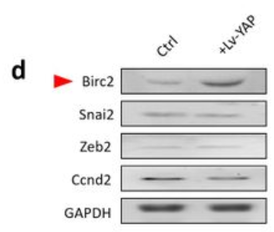
Application: WB Species: Human Sample: CSPCs
Restrictive clause
Affinity Biosciences tests all products strictly. Citations are provided as a resource for additional applications that have not been validated by Affinity Biosciences. Please choose the appropriate format for each application and consult Materials and Methods sections for additional details about the use of any product in these publications.
For Research Use Only.
Not for use in diagnostic or therapeutic procedures. Not for resale. Not for distribution without written consent. Affinity Biosciences will not be held responsible for patent infringement or other violations that may occur with the use of our products. Affinity Biosciences, Affinity Biosciences Logo and all other trademarks are the property of Affinity Biosciences LTD.