製品: CD44 Antibody
カタログ: DF6392
タンパク質の説明: Rabbit polyclonal antibody to CD44
アプリケーション: WB IHC IF/ICC
Cited expt.: WB, IHC, IF/ICC
反応性: Human, Mouse, Rat
予測: Pig, Bovine, Horse, Sheep, Rabbit, Dog
分子量: 82kDa; 82kD(Calculated).
ユニプロット: P16070
RRID: AB_2838355

類似製品を見る>>

   サイズ 価格 在庫状況
 100ul $280 在庫あり
 200ul $350 在庫あり

リードタイム: 当日配達

For pricing and ordering contact:
お問い合わせ先

製品説明

ソース:
Rabbit
アプリケーション:
WB 1:500-1:2000, IHC 1:50-1:200, IF/ICC 1:100-1:500
*The optimal dilutions should be determined by the end user. For optimal experimental results, antibody reuse is not recommended.
*Tips:

WB: For western blot detection of denatured protein samples. IHC: For immunohistochemical detection of paraffin sections (IHC-p) or frozen sections (IHC-f) of tissue samples. IF/ICC: For immunofluorescence detection of cell samples. ELISA(peptide): For ELISA detection of antigenic peptide.

反応性:
Human,Mouse,Rat
予測:
Pig(82%), Bovine(82%), Horse(82%), Sheep(82%), Rabbit(82%), Dog(91%)
クローナリティ:
Polyclonal
特異性:
CD44 Antibody detects endogenous levels of total CD44.
RRID:
AB_2838355
引用形式: Affinity Biosciences Cat# DF6392, RRID:AB_2838355.
コンジュゲート:
Unconjugated.
精製:
The antiserum was purified by peptide affinity chromatography using SulfoLink™ Coupling Resin (Thermo Fisher Scientific).
保存:
Rabbit IgG in phosphate buffered saline , pH 7.4, 150mM NaCl, 0.02% sodium azide and 50% glycerol. Store at -20 °C. Stable for 12 months from date of receipt.
別名:

折りたたみ/展開

LHR; BA-1; CD 44; CD44; CD44 antigen; CD44 molecule (Indian blood group); CD44 molecule; CD44_HUMAN; CDw44; Cell surface glycoprotein CD44; chondroitin sulfate proteoglycan 8; CSPG8; ECMR-III; Epican; Extracellular matrix receptor III; GP90 lymphocyte homing/adhesion receptor; HCELL; hematopoietic cell E- and L-selectin ligand; Heparan sulfate proteoglycan; Hermes antigen; homing function and Indian blood group system; HSA; HUTCH-I; HUTCH1; Hyaluronate receptor; IN; INLU-related p80 Glycoprotein; MC56; MDU2; MDU3; MGC10468; MIC4; MUTCH1; PGP-1; PGP-I; PGP1; Phagocytic glycoprotein 1; Phagocytic glycoprotein I; Soluble CD44;

免疫原

免疫原:

A synthesized peptide derived from human CD44, corresponding to a region within the internal amino acids.

Uniprot:
遺伝子(ID):
発現特異性:
P16070 CD44_HUMAN:

Isoform 10 (epithelial isoform) is expressed by cells of epithelium and highly expressed by carcinomas. Expression is repressed in neuroblastoma cells.

タンパク質の説明:
CD44 is a type I transmembrane glycoprotein that mediates cell-cell and cell-matrix interaction through its affinity for hyaluronic acid (HA) and possibly through other parts of the extracellular matrix (ECM). CD44 is highly polymorphic, possesses a number of alternative splice variants and undergoes extensive post-translational modifications (1,2). Increased surface levels of CD44 are characteristic of T cell activation, and expression of the protein is upregulated during the inflammatory response. Interactions between CD44 and HER2 have been linked to an increase in ovarian carcinoma cell growth (1-3). CD44 interacts with ezrin, radixin and moesin (ERM), linking the actin cytoskeleton to the plasma membrane and the ECM (4-6). CD44 is constitutively phosphorylated at Ser325 in resting cells. Activation of PKC results in phosphorylation of Ser291, dephosphorylation of Ser325, disassociation of ezrin from CD44, and directional motility (4).
タンパク質配列:
MDKFWWHAAWGLCLVPLSLAQIDLNITCRFAGVFHVEKNGRYSISRTEAADLCKAFNSTLPTMAQMEKALSIGFETCRYGFIEGHVVIPRIHPNSICAANNTGVYILTSNTSQYDTYCFNASAPPEEDCTSVTDLPNAFDGPITITIVNRDGTRYVQKGEYRTNPEDIYPSNPTDDDVSSGSSSERSSTSGGYIFYTFSTVHPIPDEDSPWITDSTDRIPATTLMSTSATATETATKRQETWDWFSWLFLPSESKNHLHTTTQMAGTSSNTISAGWEPNEENEDERDRHLSFSGSGIDDDEDFISSTISTTPRAFDHTKQNQDWTQWNPSHSNPEVLLQTTTRMTDVDRNGTTAYEGNWNPEAHPPLIHHEHHEEEETPHSTSTIQATPSSTTEETATQKEQWFGNRWHEGYRQTPKEDSHSTTGTAAASAHTSHPMQGRTTPSPEDSSWTDFFNPISHPMGRGHQAGRRMDMDSSHSITLQPTANPNTGLVEDLDRTGPLSMTTQQSNSQSFSTSHEGLEEDKDHPTTSTLTSSNRNDVTGGRRDPNHSEGSTTLLEGYTSHYPHTKESRTFIPVTSAKTGSFGVTAVTVGDSNSNVNRSLSGDQDTFHPSGGSHTTHGSESDGHSHGSQEGGANTTSGPIRTPQIPEWLIILASLLALALILAVCIAVNSRRRCGQKKKLVINSGNGAVEDRKPSGLNGEASKSQEMVHLVNKESSETPDQFMTADETRNLQNVDMKIGV

種類予測

種類予測:

Score>80(red) has high confidence and is suggested to be used for WB detection. *The prediction model is mainly based on the alignment of immunogen sequences, the results are for reference only, not as the basis of quality assurance.

Species
Results
Score
Dog
91
Pig
82
Horse
82
Bovine
82
Sheep
82
Rabbit
82
Xenopus
0
Zebrafish
0
Chicken
0
Model Confidence:
High(score>80) Medium(80>score>50) Low(score<50) No confidence

研究背景

機能:

Cell-surface receptor that plays a role in cell-cell interactions, cell adhesion and migration, helping them to sense and respond to changes in the tissue microenvironment. Participates thereby in a wide variety of cellular functions including the activation, recirculation and homing of T-lymphocytes, hematopoiesis, inflammation and response to bacterial infection. Engages, through its ectodomain, extracellular matrix components such as hyaluronan/HA, collagen, growth factors, cytokines or proteases and serves as a platform for signal transduction by assembling, via its cytoplasmic domain, protein complexes containing receptor kinases and membrane proteases. Such effectors include PKN2, the RhoGTPases RAC1 and RHOA, Rho-kinases and phospholipase C that coordinate signaling pathways promoting calcium mobilization and actin-mediated cytoskeleton reorganization essential for cell migration and adhesion.

PTMs:

Proteolytically cleaved in the extracellular matrix by specific proteinases (possibly MMPs) in several cell lines and tumors.

N-glycosylated.

O-glycosylated. O-glycosylation contains more-or-less-sulfated chondroitin sulfate glycans, whose number may affect the accessibility of specific proteinases to their cleavage site(s). It is uncertain if O-glycosylation occurs on Thr-637 or Thr-638.

Phosphorylated; activation of PKC results in the dephosphorylation of Ser-706 (constitutive phosphorylation site), and the phosphorylation of Ser-672.

細胞の位置付け:

Cell membrane>Single-pass type I membrane protein. Cell projection>Microvillus.
Note: Colocalizes with actin in membrane protrusions at wounding edges. Co-localizes with RDX, EZR and MSN in microvilli. Localizes to cholesterol-rich membrane-bound lipid raft domains.

Extracellular region or secreted Cytosol Plasma membrane Cytoskeleton Lysosome Endosome Peroxisome ER Golgi apparatus Nucleus Mitochondrion Manual annotation Automatic computational assertionSubcellular location
組織特異性:

Isoform 10 (epithelial isoform) is expressed by cells of epithelium and highly expressed by carcinomas. Expression is repressed in neuroblastoma cells.

タンパク質ファミリー:

The lectin-like LINK domain is responsible for hyaluronan binding.

研究領域

· Environmental Information Processing > Signaling molecules and interaction > ECM-receptor interaction.   (View pathway)

· Human Diseases > Infectious diseases: Bacterial > Shigellosis.

· Human Diseases > Infectious diseases: Viral > Epstein-Barr virus infection.

· Human Diseases > Cancers: Overview > Proteoglycans in cancer.

· Human Diseases > Cancers: Overview > MicroRNAs in cancer.

· Organismal Systems > Immune system > Hematopoietic cell lineage.   (View pathway)

参考文献

1). The heterogeneous immune landscape between lung adenocarcinoma and squamous carcinoma revealed by single-cell RNA sequencing. Signal transduction and targeted therapy, 2022 (PubMed: 36008393) [IF=40.8]

2). Metabolic reprogramming of proinflammatory macrophages by target delivered roburic acid effectively ameliorates rheumatoid arthritis symptoms. Signal Transduction and Targeted Therapy, 2023 (PubMed: 37500654) [IF=40.8]

Application: IF/ICC    Species: Mouse    Sample: RAW264.7 cells

Fig. 2 RBA-NPs display targeting capability in vitro. a, b Confocal micrographs showed that DiD-NPs (red) enjoyed significantly improved cell uptake compared with free DiD in LPS + IFN-γ activated RAW264.7 cells. Scale bar = 10 μm. c Flow cytometry analysis showed that the fluorescence intensity of DiD-NPs in LPS + IFN-γ activated RAW264.7 cells was higher than that of free DiD in RAW264.7. d The expression of CD44 and folate receptors increased significantly on the surface of LPS + IFN-γ activated RAW264.7 cells (d). Scale bar = 10 μm. e LPS + IFN-γ activated macrophages were pretreated with HA or FA or both HA and FA to compete for the overexpressed CD44 or folate receptors, and cellular uptake of DiD-NPs was reduced. f Confocal micrographs showed co-localization of DiD-NPs (orange) with CD44 receptor (red) and folate receptor (green). Scale bar = 20 μm. Cell nuclei was stained with DAPI (blue). All results are shown as mean ± SD. *P 

3). Mas Signaling Potentiates Neutrophil Extracellular Traps Formation Induced by Endothelial Cells Derived S1P in Mice with Acute Liver Failure. Advanced science (Weinheim, Baden-Wurttemberg, Germany), 2025 (PubMed: 40285622) [IF=15.1]

Application: IF/ICC    Species: Mouse    Sample: liver
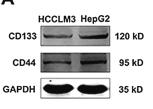
Figure 4 Col4a1+ ECs trigger NETs formation in Cldn1+CD177+ neutrophils via Col4a1-CD44 interaction. A,B) Heatmap shows the number of interactions between neutrophil clusters and other cell types in the WT-LG (A) and Mas1−/−-L/G (B) groups. C) Bubble plots display the likelihood of ligand-receptor pairs for ECs signaling to Neu2 in both WT-LG and Mas1−/−-L/G mice. D) KEGG enrichment of DEGs in ECs. Top 20 downregulated are shown in Mas1−/−-L/G versus WT-L/G. E) Violin plots illustrate the score of sphingolipid signaling pathway for EC clusters. F) Dot plots show scaled expression of the gene signature for EC cluster 2 (EC2). WT-L/G mice were pre-treated with CD44 mAb or IgG control (n = 6 per group, G–J). G) Representative liver photographs and immunohistochemical staining with the quantification (below) of H&E, TUNEL, and Ly6G (two-sided Student's t-test, p = 7.82 × 10⁻⁴, p = 4.51 × 10⁻³ and p = 6.06 × 10⁻³ from left to right). Scale bars are shown as indicated. H) Serum levels of ALT and TBA (two-sided Student's t-test, p = 3.98 × 10⁻⁴ and p = 6.15 × 10⁻⁴ from left to right). I) Serum concentrations of S1P (two-sided Mann–Whitney U test, p = 8.21 × 10⁻⁴). J) Plasma levels of cell-free DNA (two-sided Student's t-test, p = 4.31 × 10⁻⁴). K) Representative mIHC staining of intrahepatic Col4a1+ ECs and Cldn1+CD44+ neutrophils in WT-LG and Mas1−/−-L/G mice. Scale bar: 20 µm. Data are presented as mean ± SD or median ± IQR (***p < 0.001, **p < 0.01). See also related Figures S4–S6 (Supporting Information).

4). HYD-PEP06 suppresses hepatocellular carcinoma metastasis, epithelial–mesenchymal transition and cancer stem cell-like properties by inhibiting PI3K/AKT and WNT/β-catenin signaling activation. Acta Pharmaceutica Sinica B, 2021 (PubMed: 34221870) [IF=14.7]

Application: WB    Species: Human    Sample: HepG2 cells

Figure 5 Tumor spheres isolated from hepatocellular carcinoma HepG2 cells obtained the characteristics of cancer stem cells. (A) Comparison of the protein levels of CD133 and CD44 in HCCLM3 and HepG2 cells. The protein expression of (B) CD133 and (C) CD44 was dramatically increased in HepG2 cells compared with those in HCCLM3 cells. ∗P < 0.05, compared with the HCCLM3 cells. (D) The images of 3D tumor spheroids were captured using the fluorescence microscope on Day 0 in attachment surface plates and Days 0, 3, 5, 7, and 10 in ultra-low attachment surface plates with CSC medium (magnification 200 ×); scale bar: 20 μm. (E) The protein expression of CD133 and CD44 was analyzed by Western blot in HepG2 and CSC-like cells. The protein expression of (F) CD133 and (G) CD44 was dramatically increased in CSC-like cells compared with HepG2 cells. Data are presented as mean ± SEM; ∗P < 0.05, ∗∗P < 0.01. (H) The expression of CD133 and CD44 were assessed by immunofluorescence staining in HepG2 and tumor sphere cells. Blue represents DAPI; green represents CD44; red represents CD133. Magnification: 400 ×. (I) CSC augments tumor-initiating in vivo. Representative xenograft tumors derived from CSC-like cells compared to HepG2 cells after two weeks subcutaneous injection in nude mice (n = 3). Left panel: HepG2 cells; right: CSCs. (J) The protein levels of CD133 and CD44 were analyzed by Western blot in the tumor tissues isolated from xenograft models. GAPDH served as a sample loading control. The protein levels of CSC markers (K) CD133 and (L) CD44 were dramatically upregulated in right tissues compared with left tissues. Data are presented as mean ± SEM; ∗P < 0.05, ∗∗∗P < 0.001.

5). Exosomes of human adipose stem cells mitigate irradiation injury to salivary glands by inhibiting epithelial-mesenchymal transition through miR-199a-3p targeting Twist1 and regulating TGFβ1/Smad3 pathway. Theranostics, 2025 (PubMed: 39897544) [IF=12.4]

6). CD44‐targeting Drug Delivery System of Exosomes Loading Forsythiaside A Combats Liver Fibrosis via Regulating NLRP3‐mediated Pyroptosis. Advanced Healthcare Materials, 2023 (PubMed: 36603210) [IF=10.0]

7). ZIF-8 Modified Multifunctional Bone-Adhesive Hydrogels Promoting Angiogenesis and Osteogenesis for Bone Regeneration. ACS Applied Materials & Interfaces, 2020 (PubMed: 32814397) [IF=8.3]

8). Targeting TNFAIP2 with NIR-II CRISPR-Cas9 nanosystem to overcome cisplatin resistance in laryngeal cancer. NPJ precision oncology, 2025 (PubMed: 40731142) [IF=7.9]

9). GSH-responsive targeted copper-based nanoparticle improve ovarian cancer treatment via induction of apoptosis-ferroptosis-cuproptosis. MATERIALS & DESIGN, 2025 [IF=7.6]

Application: WB    Species: human    Sample:

Fig. 3. Targeting ability of HTC@DOX. (A) Boxplot of CD44 expression difference between OC tumour and normal tissues using GEPIA (http://gepia.cancer-pku.cn/). The red and gray box respectively represent tumour and normal tissues. (B) CD44 protein expression level of ID8, NRK-49F, AML-12 and H9c2 cells, *compared to the ID8 group (n = 3). (C) Fluorescent inverted microscope images and quantitative of ID8 cells incubated with HTC@DOX and HA + HTC@DOX for 6 h (scale = 20 μm), *compared to the HA + HTC@DOX group (n = 3). (D) Fluorescent images of tumour-bearing mice at different time points and various organs and tumours at 24 h after tail vein injection of fluorescent dye DIR and DIR-loaded nanoparticle HT@DIR (n = 5). (E) Quantitative graph of fluorescence intensity of tumour from tumour-bearing mice at different time points (n = 5), *compared to the DIR group.

10). Ginsenoside Rg1 prevents bone marrow mesenchymal stem cell senescence via NRF2 and PI3K/Akt signaling. Free radical biology & medicine, 2021 (PubMed: 34364981) [IF=7.1]

もっと読みます

Restrictive clause


Affinity Biosciences tests all products strictly. Citations are provided as a resource for additional applications that have not been validated by Affinity Biosciences. Please choose the appropriate format for each application and consult Materials and Methods sections for additional details about the use of any product in these publications.

For Research Use Only.
Not for use in diagnostic or therapeutic procedures. Not for resale. Not for distribution without written consent. Affinity Biosciences will not be held responsible for patent infringement or other violations that may occur with the use of our products. Affinity Biosciences, Affinity Biosciences Logo and all other trademarks are the property of Affinity Biosciences LTD.