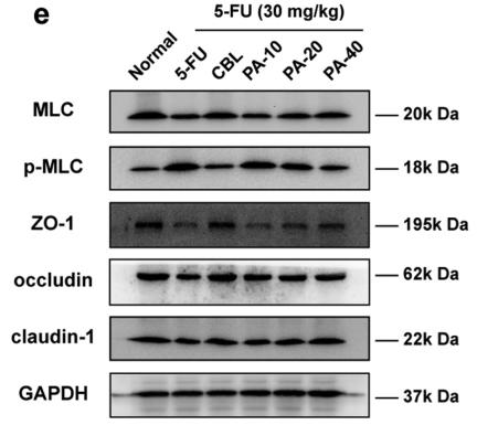
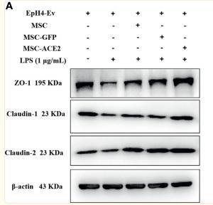
製品: Claudin 1 Antibody
カタログ: AF0127
タンパク質の説明: Rabbit polyclonal antibody to Claudin 1
アプリケーション: WB IHC
Cited expt.: WB, IHC
反応性: Human, Mouse, Rat
予測: Pig, Bovine, Horse, Sheep, Rabbit, Chicken
分子量: 22kDa; 23kD(Calculated).
ユニプロット: O95832
RRID: AB_2833311

類似製品を見る>>

   サイズ 価格 在庫状況
 100ul $280 在庫あり
 200ul $350 在庫あり

リードタイム: 当日配達

For pricing and ordering contact:
お問い合わせ先

製品説明

ソース:
Rabbit
アプリケーション:
WB 1:500-1:3000, IHC 1:50-1:200
*The optimal dilutions should be determined by the end user. For optimal experimental results, antibody reuse is not recommended.
*Tips:

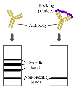
WB: For western blot detection of denatured protein samples. IHC: For immunohistochemical detection of paraffin sections (IHC-p) or frozen sections (IHC-f) of tissue samples. IF/ICC: For immunofluorescence detection of cell samples. ELISA(peptide): For ELISA detection of antigenic peptide.

反応性:
Human,Mouse,Rat
予測:
Pig(100%), Bovine(100%), Horse(100%), Sheep(100%), Rabbit(100%), Chicken(83%)
クローナリティ:
Polyclonal
特異性:
Claudin 1 Antibody detects endogenous levels of total Claudin 1.
RRID:
AB_2833311
引用形式: Affinity Biosciences Cat# AF0127, RRID:AB_2833311.
コンジュゲート:
Unconjugated.
精製:
The antiserum was purified by peptide affinity chromatography using SulfoLink™ Coupling Resin (Thermo Fisher Scientific).
保存:
Rabbit IgG in phosphate buffered saline , pH 7.4, 150mM NaCl, 0.02% sodium azide and 50% glycerol. Store at -20 °C. Stable for 12 months from date of receipt.
別名:

折りたたみ/展開

Claudin-1; Claudin1; CLD 1; CLD1; CLD1_HUMAN; CLDN 1; Cldn1; ILVASC; SEMP 1; SEMP1; Senescence associated epithelial membrane protein 1; Senescence associated epithelial membrane protein; Senescence-associated epithelial membrane protein;

免疫原

免疫原:

A synthesized peptide derived from human Claudin 1, corresponding to a region within C-terminal amino acids.

Uniprot:
遺伝子(ID):
発現特異性:
O95832 CLD1_HUMAN:

Strongly expressed in liver and kidney. Expressed in heart, brain, spleen, lung and testis.

タンパク質の説明:
claudin 1 Plays a major role in tight junction-specific obliteration of the intercellular space, through calcium- independent cell-adhesion activity. Acts as a co- receptor for HCV entry into hepatic cells. Belongs to the claudin family. Can form homo- and heteropolymers with other CLDN. Homopolymers interact with CLDN3, but not CLDN2, homopolymers. Directly interacts with TJP1/ZO-1, TJP2/ZO-2 and TJP3/ZO-3. Interacts with MPDZ and INADL. May interact with HCV E1 and E2 proteins.
タンパク質配列:
MANAGLQLLGFILAFLGWIGAIVSTALPQWRIYSYAGDNIVTAQAMYEGLWMSCVSQSTGQIQCKVFDSLLNLSSTLQATRALMVVGILLGVIAIFVATVGMKCMKCLEDDEVQKMRMAVIGGAIFLLAGLAILVATAWYGNRIVQEFYDPMTPVNARYEFGQALFTGWAAASLCLLGGALLCCSCPRKTTSYPTPRPYPKPAPSSGKDYV

種類予測

種類予測:

Score>80(red) has high confidence and is suggested to be used for WB detection. *The prediction model is mainly based on the alignment of immunogen sequences, the results are for reference only, not as the basis of quality assurance.

Species
Results
Score
Pig
100
Horse
100
Bovine
100
Sheep
100
Rabbit
100
Chicken
83
Dog
0
Xenopus
0
Zebrafish
0
Model Confidence:
High(score>80) Medium(80>score>50) Low(score<50) No confidence

研究背景

機能:

Claudins function as major constituents of the tight junction complexes that regulate the permeability of epithelia. While some claudin family members play essential roles in the formation of impermeable barriers, others mediate the permeability to ions and small molecules. Often, several claudin family members are coexpressed and interact with each other, and this determines the overall permeability. CLDN1 is required to prevent the paracellular diffusion of small molecules through tight junctions in the epidermis and is required for the normal barrier function of the skin. Required for normal water homeostasis and to prevent excessive water loss through the skin, probably via an indirect effect on the expression levels of other proteins, since CLDN1 itself seems to be dispensable for water barrier formation in keratinocyte tight junctions.

(Microbial infection) Acts as a receptor for hepatitis C virus (HCV) in hepatocytes. Associates with CD81 and the CLDN1-CD81 receptor complex is essential for HCV entry into host cell. Acts as a receptor for dengue virus.

細胞の位置付け:

Cell junction>Tight junction. Cell membrane>Multi-pass membrane protein. Basolateral cell membrane.
Note: Associates with CD81 and the CLDN1-CD81 complex localizes to the basolateral cell membrane.

Extracellular region or secreted Cytosol Plasma membrane Cytoskeleton Lysosome Endosome Peroxisome ER Golgi apparatus Nucleus Mitochondrion Manual annotation Automatic computational assertionSubcellular location
組織特異性:

Strongly expressed in liver and kidney. Expressed in heart, brain, spleen, lung and testis.

タンパク質ファミリー:

Belongs to the claudin family.

研究領域

· Cellular Processes > Cellular community - eukaryotes > Tight junction.   (View pathway)

· Environmental Information Processing > Signaling molecules and interaction > Cell adhesion molecules (CAMs).   (View pathway)

· Human Diseases > Infectious diseases: Bacterial > Pathogenic Escherichia coli infection.

· Human Diseases > Infectious diseases: Viral > Hepatitis C.

· Organismal Systems > Immune system > Leukocyte transendothelial migration.   (View pathway)

参考文献

1). Ectoparasites enhance survival by suppressing host exploration and limiting dispersal. Nature communications, 2025 (PubMed: 40346081) [IF=16.6]

Application: WB    Species: Mouse    Sample:
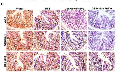
Fig. 5: Flea bites affect neural function through the skin-brain axis. a GSEA result on upregulated genes from the mice skin transcriptome. Functions related to immunoglobulin and hemoglobin genes are significantly enriched. b Cytokine levels in mice skin presented in boxplot where red is Flea− (n = 6/12) and blue is Flea+ (n = 6/12). Two-sided t test was used. Asterisks indicate the significance. All p-values were below 0.001. c Cytokine levels in mice serum presented in boxplot where red is Flea− (n = 12) and blue is Flea+ (n = 12). Two-sided t test was used. Thick bars indicate the interquartile range (IQR) around the median, and whiskers represent 1.5 times the interquartile range (maxima: Q3 + 1.5 × IQR, minima: Q1 − 1.5 × IQR). Asterisks indicate the significance. All p-values were below 0.001. d Cytokine levels in mice PFC presented in boxplot where red is Flea− (n = 10) and blue is Flea+ (n = 10). Two-sided t test was used. Asterisks indicate the significance. All p-values were below 0.001. e Representative HE plots of the PFC. In the Flea+ group, cortical tissue shows evident cellular damage, with minor mononuclear cell infiltration (yellow arrow) at the injury site. Additionally, widespread nuclear pyknosis and condensation (red arrow) are observed in the parenchyma. The infected group exhibits a larger area of damage; Scale 50 µm. f Western blot analysis of Claudin1 markers of tight junction protein from the PFC brain region (n = 5 samples each group). g Comparison of Claudin1 in WB between the Flea− (red, n = 5) and Flea+ (blue, n = 5) groups of mice. Two-sided t test was used. Each black dot represents an individual. Asterisks indicates brain regions with significant differences. Data are presented as mean ± SEM. Source data are provided as a Source Data file. h Validation by qPCR of select BBB genes altered between Flea− (n = 3) and Flea+ (n = 3) group. Two-sided t test was used. Thick bars indicate the interquartile range (IQR) around the median, and whiskers represent 1.5 times the interquartile range (maxima: Q3 + 1.5 × IQR, minima: Q1 − 1.5 × IQR). Each black dot represents pooled sample of 3 decapitated individuals. Asterisks indicate the significance.

2). Extracellular vesicles of Fusobacterium nucleatum compromise intestinal barrier through targeting RIPK1-mediated cell death pathway. Gut Microbes, 2021 (PubMed: 33769187) [IF=12.2]

Application: IHC    Species: mouse    Sample: colon

Figure 5. |FnEVs increase gut barrier leakage in experimental colitis models. (a) Representative images and ex vivo imaging with the intestine, liver, heart, spleen and kidney of mice. (b) Relative fluorescence intensity of translocated EGFP-labeled E.coli in every tissues. (c) Representative images of immunohistochemical stainings of ZO-1, claudin-1 and occludin in the colon on day 3 after colitis induction. Scale bar = 50 um.

3). Gut flora-derived succinate exacerbates Allergic Airway Inflammation by promoting protein succinylation. Redox biology, 2025 (PubMed: 40174477) [IF=10.7]

Application: WB    Species: Mouse    Sample:
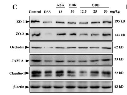
Fig. 3. The mRNA level, protein expression level and Western blotting analysis of tight junction proteins in intestinal and lung tissues of AAI mice. A, Relative mRNA expression levels of Occludin, Claudin-1 and ZO1 in lung and gut tissues. B, Representative Western-blot images of Occludin, Claudin1, and ZO1 proteins in gut and lung tissues with β-Actin as the internal reference. Statistical significance was determined by Student's t-test.

4). Stem cell membrane-coated isotretinoin for acne treatment. JOURNAL OF NANOBIOTECHNOLOGY, 2020 (PubMed: 32723398) [IF=10.2]

5). Oxyberberine, a novel gut microbiota-mediated metabolite of berberine, possesses superior anti-colitis effect: impact on intestinal epithelial barrier, gut microbiota profile and TLR4-MyD88-NF-κB pathway. PHARMACOLOGICAL RESEARCH, 2020 (PubMed: 31863867) [IF=9.1]

Application: WB    Species: Mice    Sample: colonic tissues
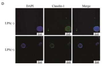
Fig. 4. OBB protected intestinal epithelial barrier by modulating TJs proteins. Effect of OBB on the mRNA levels of mucin-1(A) and mucin-2 (B). (C) Representative Western blotting images of TJs protein, and the relative protein expressions were normalized to β-actin. (D-H) Changes in the relative protein expression levels of ZO-1, ZO-2, occludin, JAM-A, and claudin-1 were measured respectively. Data are shown as mean ± SEM (n = 3). # P < 0.05, ## P < 0.01 vs. Control group, * P < 0.05, ** P < 0.01 vs. DSS group, & P < 0.05, && P < 0.01 vs. BBR group.

6). Lipopolysaccharide induced intestinal epithelial injury: a novel organoids-based model for sepsis in vitro. CHINESE MEDICAL JOURNAL, 2022 (PubMed: 36355867) [IF=7.5]

Application: IF/ICC    Species: Mouse    Sample:

Figure 2 The immunofluorescence of organoids with or without LPS stimulation. The images were captured by Opera Phenix™ high-content imaging system and compressed by Harmony. (A) The expression of Ki-67, LPS 100 μg/mL, 24 h. (B) The expression of ZO-1, LPS 100 μg/mL, 24 h. (C) The expression of occludins, LPS 100 μg/mL, 24 h. (D) The expression of claudin-1, LPS 100 μg/mL, 24 h. (E) Quantitative analysis of Ki-67 with different concentrations of LPS stimulated for 8 h or 24 h. (F) Quantitative analysis of ZO-1 with different concentrations of LPS stimulated for 8 h or 24 h. (G) Quantitative analysis of occludins with different concentrations of LPS stimulated for 8 h or 24 h. (H) Quantitative analysis of claudin-1 with different concentrations of LPS stimulated for 8 h or 24 h. Data are presented as means ± SD, compared with LPS 0 μg/mL 8 h or 24 h group accordingly using Student's t-tests. ∗ is used for 24 h. SD: Standard deviation; LPS: Lipopolysaccharide; ZO-1: Zonula occludens-1.

7). SIRT1 Stabilizes β-TrCP1 to Inhibit Snail1 Expression in Maintaining Intestinal Epithelial Integrity to Alleviate Colitis. Cellular and molecular gastroenterology and hepatology, 2024 (PubMed: 38729522) [IF=7.1]

Application: WB    Species: human    Sample: Caco2 cells
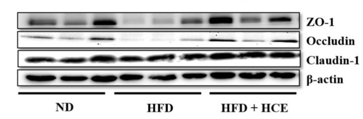
Figure 3. SIRT1 deacetylates and stabilizes β-TrCP1 protein to downregulate Snail1. (A) Caco2 cells stably expressing shRNAs against SIRT1 were subjected to Western blotting. (B) Caco2 cell lysates were subjected to immunoprecipitation (IP) with anti-β-TrCP1, using normal mouse or rabbit IgG as a control, followed by Western blot analyses. (C) Caco2 cells stably coexpressing β-TrCP1 and shRNAs against SIRT1 were subjected to Western blotting. (D) Caco2 cells treated with or without DSS were subjected to Western blotting. (E) HCT116 cells were treated with 4% DSS for 8 hours and the whole cell lysates were subjected to Western blotting. (F) Caco2 cells stably overexpressing SIRT1 were treated with 4% DSS for 8 hours. Whole-cell lysates were subjected to Western blotting. (G) Caco2 cells stably overexpressing β-TrCP1 were treated with 4% DSS for 8 hours. Whole-cell lysates were subjected to Western blotting. (H) Caco2 cells pretreated with 4% DSS for 8 hours or stably expressing shRNAs against SIRT1 were subjected to qPCR analyses for β-TrCP1. (I) To examine the effects of DSS on ubiquitination of β-TrCP1, 293FT cells were cotransfected with the relevant expression plasmids for 48 hours, followed by treatment with 4% DSS for 12 hours. Before harvesting, cells underwent treatment with 20 μM MG132 for 6 hours. Ubiquitination of β-TrCP1 was examined by denature-IP-western analyses. (J) The β-TrCP1 protein half-lives were measured by CHX assays in Caco2 cells pretreated with 4% DSS for 8 hours. (K and L) To examine the effects of SIRT1 on ubiquitination of β-TrCP1 and Snail1, 293FT cells were cotransfected with indicated expressing plasmids. Cells were treated with 20 μM MG132 for 6 hours before collection. Ubiquitination of Snail1 was examined by denature-IP-western analyses. (M) The β-TrCP1 and Snail1 protein half-lives were measured by CHX assays in Caco2 cells stably overexpressing SIRT1. (N and O) Caco2 cell lysates were subjected to IP with anti-SIRT1 (H) or anti-β-TrCP1 (I), using normal mouse or rabbit IgG as a control, followed by Western blot analyses. (P) HEK-293T cells cotransfected with His/Myc-β-TrCP1 and Flag-SIRT1 were subjected to IP and Western blotting analyses.

8). Palmitoleic Acid on Top of HFD Ameliorates Insulin Resistance Independent of Diacylglycerols and Alters Gut Microbiota in C57BL/6J Mice. Food Science and Human Wellness, 2023 [IF=7.0]

Application: WB    Species: Mouse    Sample:

Fig. 6 Palmitoleic acid promotes intestinal tight junction integrity and reduces HFD-induced colon inflammation. 6 weeks old mice were fed with HFD for 12 weeks. Mice were treated with palmitoleic acid, oleic acid, or BSA as control via oral gavages for 6 weeks. The colon was collected for further analysis. (A) The level of occludin and claudin-1 by Western blotting. (B) Quantification of occludin level. (C) Quantification of claudin-1 level. (D) Macrophage infiltration by IHC for F4/80. (E) mRNA levels of MCP-1, TNF-α, IL-1β, and IL-6 by RT-PCR. n = 4 mice per group. The data are mean ± SEM (error bars). † P < 0.05, ††P < 0.01, †††P < 0.001, BSA vs palmitoleic acid; ‡ P < 0.05, ‡‡P < 0.01, ‡‡‡P < 0.001, oleic acid vs palmitoleic acid; §§§P < 0.001, BSA vs oleic acid.

9). Djulis Hull Improves Insulin Resistance and Modulates the Gut Microbiota in High-Fat Diet (HFD)-Induced Hyperglycaemia. Antioxidants, 2021 (PubMed: 35052549) [IF=7.0]

Application: WB    Species: Mice    Sample: epididymal white adipose tissue
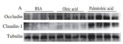
Figure 7 Effect of djulis hull crude extract on (A) LPS in serum and (B) the protein expression of tight junctions in the colon of high-fat diet-induced hyperglycaemia. ND: normal diet; HFD: high-fat diet; HCE: high dosage of crude extract; LPS: lipopolysaccharide. Values represent the mean ± SEM (n = 6). The statistical methods used one-way ANOVA, and the values with different letters are significantly different at p < 0.05.

10). Allyl isothiocyanate mitigates airway inflammation and constriction in a house dust mite-induced allergic asthma model via upregulation of tight junction proteins and the TRPA1 modulation. Biomedicine & Pharmacotherapy, 2023 (PubMed: 37634475) [IF=6.9]

Application: WB    Species: Mouse    Sample: lung
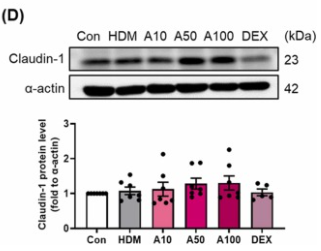
Fig. 4. Effects of AITC on tight junction-related protein in lung. AITC treatments on the modulation of tight junction-related protein expression. (A) E-cadherin, (B), ZO-1, (C) Occludin, (D) Claudin-1. Data are expressed in mean ± SEM (n = 5–7). Significant difference presented as different symbol. *p 

もっと読みます

Restrictive clause


Affinity Biosciences tests all products strictly. Citations are provided as a resource for additional applications that have not been validated by Affinity Biosciences. Please choose the appropriate format for each application and consult Materials and Methods sections for additional details about the use of any product in these publications.

For Research Use Only.
Not for use in diagnostic or therapeutic procedures. Not for resale. Not for distribution without written consent. Affinity Biosciences will not be held responsible for patent infringement or other violations that may occur with the use of our products. Affinity Biosciences, Affinity Biosciences Logo and all other trademarks are the property of Affinity Biosciences LTD.