CD4 Antibody - #DF6451
| 製品: | CD4 Antibody |
| カタログ: | DF6451 |
| タンパク質の説明: | Rabbit polyclonal antibody to CD4 |
| アプリケーション: | WB |
| Cited expt.: | |
| 反応性: | Human, Mouse |
| 予測: | Pig, Horse, Rabbit, Dog |
| 分子量: | 55kDa; 51kD(Calculated). |
| ユニプロット: | P01730 |
| RRID: | AB_2838414 |
製品説明
*The optimal dilutions should be determined by the end user. For optimal experimental results, antibody reuse is not recommended.
*Tips:
WB: For western blot detection of denatured protein samples. IHC: For immunohistochemical detection of paraffin sections (IHC-p) or frozen sections (IHC-f) of tissue samples. IF/ICC: For immunofluorescence detection of cell samples. ELISA(peptide): For ELISA detection of antigenic peptide.
引用形式: Affinity Biosciences Cat# DF6451, RRID:AB_2838414.
折りたたみ/展開
CD 4; CD4 (L3T4); CD4; CD4 antigen (p55); CD4 antigen; CD4 molecule; CD4 receptor; CD4+ Lymphocyte deficiency, included; CD4_HUMAN; CD4mut; L3T4; Leu3; Ly-4; Lymphocyte antigen CD4; MGC165891; OTTHUMP00000238897; p55; T cell antigen T4; T cell antigen T4/LEU3; T cell differentiation antigen L3T4; T cell OKT4 deficiency, included; T cell surface antigen T4/Leu 3; T cell surface antigen T4/Leu3; T cell surface glycoprotein CD4; T-cell surface antigen T4/Leu-3; T-cell surface glycoprotein CD4; W3/25; W3/25 antigen;
免疫原
A synthesized peptide derived from human CD4, corresponding to a region within C-terminal amino acids.
Highly expressed in T-helper cells. The presence of CD4 is a hallmark of T-helper cells which are specialized in the activation and growth of cytotoxic T-cells, regulation of B cells, or activation of phagocytes. CD4 is also present in other immune cells such as macrophages, dendritic cells or NK cells.
- P01730 CD4_HUMAN:
- Protein BLAST With
- NCBI/
- ExPASy/
- Uniprot
MNRGVPFRHLLLVLQLALLPAATQGKKVVLGKKGDTVELTCTASQKKSIQFHWKNSNQIKILGNQGSFLTKGPSKLNDRADSRRSLWDQGNFPLIIKNLKIEDSDTYICEVEDQKEEVQLLVFGLTANSDTHLLQGQSLTLTLESPPGSSPSVQCRSPRGKNIQGGKTLSVSQLELQDSGTWTCTVLQNQKKVEFKIDIVVLAFQKASSIVYKKEGEQVEFSFPLAFTVEKLTGSGELWWQAERASSSKSWITFDLKNKEVSVKRVTQDPKLQMGKKLPLHLTLPQALPQYAGSGNLTLALEAKTGKLHQEVNLVVMRATQLQKNLTCEVWGPTSPKLMLSLKLENKEAKVSKREKAVWVLNPEAGMWQCLLSDSGQVLLESNIKVLPTWSTPVQPMALIVLGGVAGLLLFIGLGIFFCVRCRHRRRQAERMSQIKRLLSEKKTCQCPHRFQKTCSPI
種類予測
Score>80(red) has high confidence and is suggested to be used for WB detection. *The prediction model is mainly based on the alignment of immunogen sequences, the results are for reference only, not as the basis of quality assurance.
High(score>80) Medium(80>score>50) Low(score<50) No confidence
研究背景
Integral membrane glycoprotein that plays an essential role in the immune response and serves multiple functions in responses against both external and internal offenses. In T-cells, functions primarily as a coreceptor for MHC class II molecule:peptide complex. The antigens presented by class II peptides are derived from extracellular proteins while class I peptides are derived from cytosolic proteins. Interacts simultaneously with the T-cell receptor (TCR) and the MHC class II presented by antigen presenting cells (APCs). In turn, recruits the Src kinase LCK to the vicinity of the TCR-CD3 complex. LCK then initiates different intracellular signaling pathways by phosphorylating various substrates ultimately leading to lymphokine production, motility, adhesion and activation of T-helper cells. In other cells such as macrophages or NK cells, plays a role in differentiation/activation, cytokine expression and cell migration in a TCR/LCK-independent pathway. Participates in the development of T-helper cells in the thymus and triggers the differentiation of monocytes into functional mature macrophages.
(Microbial infection) Primary receptor for human immunodeficiency virus-1 (HIV-1). Down-regulated by HIV-1 Vpu. Acts as a receptor for Human Herpes virus 7/HHV-7.
Palmitoylation and association with LCK contribute to the enrichment of CD4 in lipid rafts.
Phosphorylated by PKC; phosphorylation at Ser-433 plays an important role for CD4 internalization.
Cell membrane>Single-pass type I membrane protein.
Note: Localizes to lipid rafts (PubMed:12517957, PubMed:9168119). Removed from plasma membrane by HIV-1 Nef protein that increases clathrin-dependent endocytosis of this antigen to target it to lysosomal degradation. Cell surface expression is also down-modulated by HIV-1 Envelope polyprotein gp160 that interacts with, and sequesters CD4 in the endoplasmic reticulum.
Highly expressed in T-helper cells. The presence of CD4 is a hallmark of T-helper cells which are specialized in the activation and growth of cytotoxic T-cells, regulation of B cells, or activation of phagocytes. CD4 is also present in other immune cells such as macrophages, dendritic cells or NK cells.
研究領域
· Environmental Information Processing > Signaling molecules and interaction > Cell adhesion molecules (CAMs). (View pathway)
· Human Diseases > Immune diseases > Primary immunodeficiency.
· Organismal Systems > Immune system > Antigen processing and presentation. (View pathway)
· Organismal Systems > Immune system > Hematopoietic cell lineage. (View pathway)
· Organismal Systems > Immune system > Th1 and Th2 cell differentiation. (View pathway)
· Organismal Systems > Immune system > Th17 cell differentiation. (View pathway)
· Organismal Systems > Immune system > T cell receptor signaling pathway. (View pathway)
参考文献
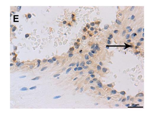
Application: IHC Species: Human Sample: tumor tissues
Restrictive clause
Affinity Biosciences tests all products strictly. Citations are provided as a resource for additional applications that have not been validated by Affinity Biosciences. Please choose the appropriate format for each application and consult Materials and Methods sections for additional details about the use of any product in these publications.
For Research Use Only.
Not for use in diagnostic or therapeutic procedures. Not for resale. Not for distribution without written consent. Affinity Biosciences will not be held responsible for patent infringement or other violations that may occur with the use of our products. Affinity Biosciences, Affinity Biosciences Logo and all other trademarks are the property of Affinity Biosciences LTD.

