製品: Claudin 2 Antibody
カタログ: AF0128
タンパク質の説明: Rabbit polyclonal antibody to Claudin 2
アプリケーション: WB IHC IF/ICC
Cited expt.: WB, IHC, IF/ICC
反応性: Human, Mouse, Rat
予測: Pig, Bovine, Horse, Sheep, Rabbit, Dog
分子量: 25kDa; 25kD(Calculated).
ユニプロット: P57739
RRID: AB_2833312

類似製品を見る>>

   サイズ 価格 在庫状況
 100ul $280 在庫あり
 200ul $350 在庫あり

リードタイム: 当日配達

For pricing and ordering contact:
お問い合わせ先

製品説明

ソース:
Rabbit
アプリケーション:
WB 1:500-1:3000, IHC 1:50-1:200, IF/ICC 1:200
*The optimal dilutions should be determined by the end user. For optimal experimental results, antibody reuse is not recommended.
*Tips:

WB: For western blot detection of denatured protein samples. IHC: For immunohistochemical detection of paraffin sections (IHC-p) or frozen sections (IHC-f) of tissue samples. IF/ICC: For immunofluorescence detection of cell samples. ELISA(peptide): For ELISA detection of antigenic peptide.

反応性:
Human,Mouse,Rat
予測:
Pig(100%), Bovine(100%), Horse(100%), Sheep(100%), Rabbit(100%), Dog(100%)
クローナリティ:
Polyclonal
特異性:
Claudin 2 Antibody detects endogenous levels of total Claudin 2.
RRID:
AB_2833312
引用形式: Affinity Biosciences Cat# AF0128, RRID:AB_2833312.
コンジュゲート:
Unconjugated.
精製:
The antiserum was purified by peptide affinity chromatography using SulfoLink™ Coupling Resin (Thermo Fisher Scientific).
保存:
Rabbit IgG in phosphate buffered saline , pH 7.4, 150mM NaCl, sodium azide and glycerol. Store at -20 °C. Stable for 12 months from date of receipt.
別名:

折りたたみ/展開

Claudin-2; Claudin2; CLD2_HUMAN; CLDN 2; CLDN2; OTTHUMP00000023793; SP 82; SP82;

免疫原

免疫原:

A synthesized peptide derived from human Claudin 2, corresponding to a region within C-terminal amino acids.

Uniprot:
遺伝子(ID):
タンパク質の説明:
claudin 2 Plays a major role in tight junction-specific obliteration of the intercellular space, through calcium- independent cell-adhesion activity. Belongs to the claudin family. Can form homo- and heteropolymers with other CLDN. Homopolymers interact with CLDN3, but not CLDN1, homopolymers. Directly interacts with TJP1/ZO-1, TJP2/ZO-2 and TJP3/ZO-3
タンパク質配列:
MASLGLQLVGYILGLLGLLGTLVAMLLPSWKTSSYVGASIVTAVGFSKGLWMECATHSTGITQCDIYSTLLGLPADIQAAQAMMVTSSAISSLACIISVVGMRCTVFCQESRAKDRVAVAGGVFFILGGLLGFIPVAWNLHGILRDFYSPLVPDSMKFEIGEALYLGIISSLFSLIAGIILCFSCSSQRNRSNYYDAYQAQPLATRSSPRPGQPPKVKSEFNSYSLTGYV

種類予測

種類予測:

Score>80(red) has high confidence and is suggested to be used for WB detection. *The prediction model is mainly based on the alignment of immunogen sequences, the results are for reference only, not as the basis of quality assurance.

Species
Results
Score
Pig
100
Horse
100
Bovine
100
Sheep
100
Dog
100
Rabbit
100
Chicken
78
Xenopus
0
Zebrafish
0
Model Confidence:
High(score>80) Medium(80>score>50) Low(score<50) No confidence

研究背景

機能:

Plays a major role in tight junction-specific obliteration of the intercellular space, through calcium-independent cell-adhesion activity.

PTMs:

The disulfide bond is necessary for pore formation, but is not required for correct protein trafficking.

細胞の位置付け:

Cell junction>Tight junction. Cell membrane>Multi-pass membrane protein.

Extracellular region or secreted Cytosol Plasma membrane Cytoskeleton Lysosome Endosome Peroxisome ER Golgi apparatus Nucleus Mitochondrion Manual annotation Automatic computational assertionSubcellular location
タンパク質ファミリー:

Belongs to the claudin family.

研究領域

· Cellular Processes > Cellular community - eukaryotes > Tight junction.   (View pathway)

· Environmental Information Processing > Signaling molecules and interaction > Cell adhesion molecules (CAMs).   (View pathway)

· Human Diseases > Infectious diseases: Viral > Hepatitis C.

· Organismal Systems > Immune system > Leukocyte transendothelial migration.   (View pathway)

参考文献

1). Beneficial Effects of Celastrol on Immune Balance by Modulating Gut Microbiota in Experimental Ulcerative Colitis Mice. Genomics, proteomics & bioinformatics, 2022 (PubMed: 35609771) [IF=11.5]

2). Metasilicate-based alkaline mineral water confers diarrhea resistance in maternally separated piglets via the microbiota-gut interaction. Pharmacological research, 2023 (PubMed: 36436708) [IF=9.1]

Application: IF/ICC    Species: piglets    Sample:
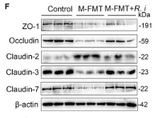
Fig. 7. Effect of SMP water on intestinal barrier integrity in MS piglets under weaning stress. (A) Western blotting measurements of the protein levels of MLCK/p-MLC signaling and tight/adherens junction proteins in small intestine from MS piglets (Occludin, 60 kDa; ZO-1, 195 kDa; Claudin 5, 23 kDa; α-Catenin, 100 kDa; E-Catenin, 130 kDa; MLC, 50 kDa; p-MLC, 50, β-actin, 42 kDa). (B) Occludin/β-actin. (C) ZO-1/β-actin. (D) Claudin5/β-actin. (E) α-Catenin/β-actin. (F) E-Cadherin/β-actin. (G) The mRNA levels of MLCK in duodenum tissue. (H) MLC/β-actin. (I) p-MLC/β-actin. (J) Immunofluorescence images of ZO-1, Claudin2 staining (green) and DAPI (blue) of duodenum in MS piglets. (K-L) Mean density of (K) ZO-1 and (L) Claudin2 in duodenum of MS piglets. (M) Correlation analysis between MLCK/p-MLC signaling and its-mediated tight/adherens junction proteins and NF-κB signaling pathway. (N) Factors interaction network analysis of NF-κB signaling pathway, AQPs, fluid absorption/secretion proteins, MLCK/p-MLC signaling and tight/adherens junction proteins in small intestine from MS piglets under weaning stress. Data are presented as the mean ± SD. ns P > 0.05, * P 

3). Poria cocos polysaccharides ameliorate AOM/DSS-induced colorectal cancer in mice by remodeling intestinal microbiota composition and enhancing intestinal barrier function. International journal of biological macromolecules, 2025 (PubMed: 40419041) [IF=7.7]

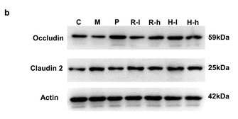
4). Commensal cow Roseburia reduces gut-dysbiosis-induced mastitis through inhibiting bacterial translocation by producing butyrate in mice. Cell Reports, 2022 (PubMed: 36417859) [IF=7.5]

Application: WB    Species: Mouse    Sample:

Figure 5Commensal Roseburia ameliorates gut-dysbiosis-induced mastitis in mice

5). Exposure to okadaic acid could disrupt the colonic microenvironment in rats. Ecotoxicology and environmental safety, 2023 (PubMed: 37597294) [IF=6.2]

Application: IF/ICC    Species: Rat    Sample:

Fig. 4. Impaired cell junction in colonic epithelium after exposure to OA. (A) Representative immunofluorescent images of claudin2 (CLDN2, Orange), occluding (OCLN, Yellow), tight junction protein 1 (TJP1, Magenta) and cadherin 1 (CDH1, Cyan) in distal colon sections under laser scanning confocal microscope. Nuclei is stained blue (DAPI). Scale bars, 100 µm. (B) Relative expression of Cldn2, Ocln, Tjp1 and Cdh1 revealed by qPCR at the mRNA level. All data represent the mean ± sem (n = 6). *p

6). Effects of Dietary Colostrum Basic Protein on Bone Growth and Calcium Absorption in Mice. Nutrients, 2024 (PubMed: 38474792) [IF=5.9]

Application: WB    Species: Mouse    Sample:

Figure 5 Effect of CBP on calcium absorption and related proteins in mice. (a) Apparent absorption rate of calcium. (b–f) TRPV6, PMCA1, CaBP-9k, CLD2, and CLD12 in jejunum, (g,h) CLD2 and CLD12 in ileum levels in the five groups. Data are presented as mean ± SEM (n = 3). Statistical significance between groups is represented by different lowercase letters (p < 0.05).

7). Knockout of IL-6 mitigates cold water-immersion restraint stress-induced intestinal epithelial injury and apoptosis. Frontiers in immunology, 2022 (PubMed: 36505466) [IF=5.7]

Application: WB    Species: Mouse    Sample:
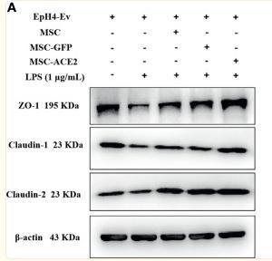
Figure 3 CWIR damaged intestinal epithelial tight junctions, which was improved by IL-6 KO. (A), Representative TEM images of jujunal tissues. Red arrow indicates the wider interval between epithelial cells. The scale bar means 10 μm. N=3. (B), Serum FITC-dextran concentration was evaluated in each group. N=6. (C), Relative protein expressions of occludin, claudin 1, and claudin 2 in the jejunum tissues among groups were checked and quantified. N=4-5, Data analysis was performed by two-way ANOVA with the Tukey post-hoc test.

8). Mesenchymal Stem Cells Overexpressing ACE2 Favorably Ameliorate LPS-Induced Inflammatory Injury in Mammary Epithelial Cells. Frontiers in Immunology, 2022 (PubMed: 35095873) [IF=5.7]

Application: WB    Species: Mice    Sample: EpH4-Ev cells

Figure 8 MSC-ACE2 upregulates blood-milk barrier-related protein expression to suppress LPS-induced inflammation in Eph4-EV cells. (A) Detection of relative protein expression levels of ZO-1, Claudin-1, Claudin-2 by Western blot; (B) Statistics of the ZO-1, Claudin-1, Claudin-2 Western blot results. Experiments were repeated three times and data were presented as the mean ± SEM (n = 4). * P < 0.05 vs. EpH4-Ev; # P < 0.05 vs. LPS; $ P < 0.05 vs. MSC; & P < 0.05 vs. MSC-GFP.

9). Long non-coding RNA ANRIL/p65 negative feedback loop protects intestinal barrier function in inflammatory bowel disease. Non-coding RNA research, 2025 (PubMed: 40242051) [IF=5.0]

Application: WB    Species: Mouse    Sample:

Fig. 2. ANRIL suppresses the expression of inflammatory factors and protects intestinal barrier function in DSS-induced mouse colitis. (A–D) RT-qPCR detection of expression levels of ANRIL, IL-6, TNF-α, and IL-1β in colon tissues of each mouse group. (E–G) ELISA detection of expression levels of IL-6, TNF-α, and IL-1β in colon tissues of each mouse group. (H) Assessment of intestinal permeability using the Fluorescein Isothiocyanate (FITC)-Dextran Permeability Assay. (I–L) Western blotting (WB) detection of expression levels of intestinal barrier-related proteins ZO-1, Claudin-2, and Occludin in colon tissues of each mouse group. (M–O) Immunofluorescence detection of expression of ZO-1, Claudin-2, and Occludin in colon tissues of each mouse group. ∗p < 0.05, ∗∗p < 0.01, ∗∗∗p < 0.001, ∗∗∗∗p < 0.0001. Data are expressed as mean ± standard deviation.

Application: IF/ICC    Species: Mouse    Sample:
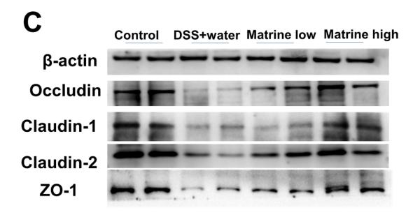
Fig. 2. ANRIL suppresses the expression of inflammatory factors and protects intestinal barrier function in DSS-induced mouse colitis. (A–D) RT-qPCR detection of expression levels of ANRIL, IL-6, TNF-α, and IL-1β in colon tissues of each mouse group. (E–G) ELISA detection of expression levels of IL-6, TNF-α, and IL-1β in colon tissues of each mouse group. (H) Assessment of intestinal permeability using the Fluorescein Isothiocyanate (FITC)-Dextran Permeability Assay. (I–L) Western blotting (WB) detection of expression levels of intestinal barrier-related proteins ZO-1, Claudin-2, and Occludin in colon tissues of each mouse group. (M–O) Immunofluorescence detection of expression of ZO-1, Claudin-2, and Occludin in colon tissues of each mouse group. ∗p < 0.05, ∗∗p < 0.01, ∗∗∗p < 0.001, ∗∗∗∗p < 0.0001. Data are expressed as mean ± standard deviation.

10). Matrine protects against DSS-induced murine colitis by improving gut barrier integrity, inhibiting the PPAR-α signaling pathway, and modulating gut microbiota. International Immunopharmacology, 2021 (PubMed: 34474274) [IF=4.8]

Application: WB    Species: Mice    Sample: colon tissues
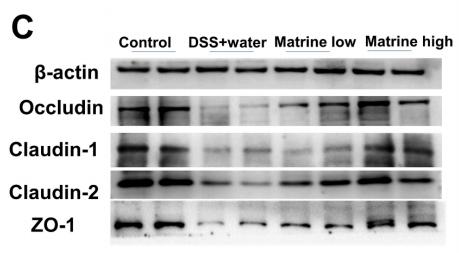
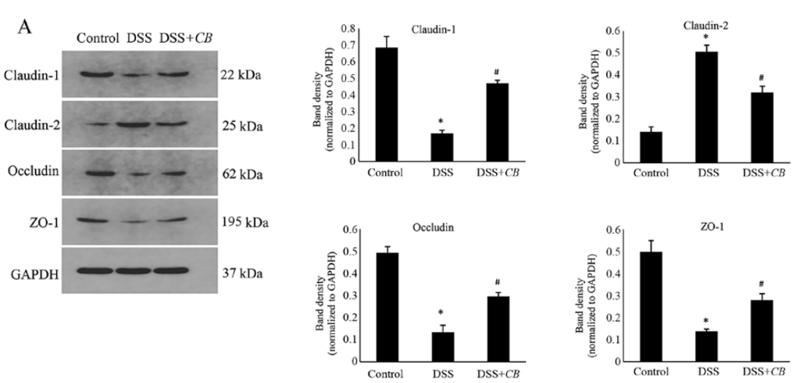
Fig. 4. Matrine improved gut barrier integrity in DSS-induced UC mice. (A) AB/ PAS and immunofluorescence imaging detected the mucin-producing cells and the expression of mucin-2, respectively. (B) Analysis of the expression levels of the mucin-producing cells and mucin-2. (C) The expressions of occludin, claudin-1,2, and ZO- 1 protein were verified by western blot. (D) Analysis of the expression levels of occludin, claudin-1,2, and ZO-1. Data are expressed as means ± SEM; n = 5 for each group. * P < 0.05, ** P < 0.01, *** P < 0.001, and ****P < 0.0001.

Application: WB    Species: mouse    Sample: colon

Fig. 4. |Matrine improved gut barrier integrity in DSS-induced UC mice.(C) The expressions of occludin, claudin-1,2, and ZO1 protein were verified by western blot.

もっと読みます

Restrictive clause


Affinity Biosciences tests all products strictly. Citations are provided as a resource for additional applications that have not been validated by Affinity Biosciences. Please choose the appropriate format for each application and consult Materials and Methods sections for additional details about the use of any product in these publications.

For Research Use Only.
Not for use in diagnostic or therapeutic procedures. Not for resale. Not for distribution without written consent. Affinity Biosciences will not be held responsible for patent infringement or other violations that may occur with the use of our products. Affinity Biosciences, Affinity Biosciences Logo and all other trademarks are the property of Affinity Biosciences LTD.