製品: Claudin 3 Antibody
カタログ: AF0129
タンパク質の説明: Rabbit polyclonal antibody to Claudin 3
アプリケーション: WB IHC IF/ICC
反応性: Human, Mouse, Rat
予測: Pig, Bovine, Sheep, Dog, Xenopus
分子量: 25kDa; 23kD(Calculated).
ユニプロット: O15551
RRID: AB_2833313

類似製品を見る>>

   サイズ 価格 在庫状況
 100ul $280 在庫あり
 200ul $350 在庫あり

リードタイム: 当日配達

For pricing and ordering contact:
お問い合わせ先

製品説明

ソース:
Rabbit
アプリケーション:
WB 1:500-1:3000, IHC 1:50-1:200, IF/ICC: 1:100-1:500
*The optimal dilutions should be determined by the end user.
*Tips:

WB: For western blot detection of denatured protein samples. IHC: For immunohistochemical detection of paraffin sections (IHC-p) or frozen sections (IHC-f) of tissue samples. IF/ICC: For immunofluorescence detection of cell samples. ELISA(peptide): For ELISA detection of antigenic peptide.

反応性:
Human,Mouse,Rat
予測:
Pig(88%), Bovine(88%), Sheep(88%), Dog(88%), Xenopus(88%)
クローナリティ:
Polyclonal
特異性:
Claudin 3 Antibody detects endogenous levels of total Claudin 3.
RRID:
AB_2833313
引用形式: Affinity Biosciences Cat# AF0129, RRID:AB_2833313.
コンジュゲート:
Unconjugated.
精製:
The antiserum was purified by peptide affinity chromatography using SulfoLink™ Coupling Resin (Thermo Fisher Scientific).
保存:
Rabbit IgG in phosphate buffered saline , pH 7.4, 150mM NaCl, 0.02% sodium azide and 50% glycerol. Store at -20 °C. Stable for 12 months from date of receipt.
別名:

折りたたみ/展開

C7orf1; Claudin-3; Claudin3; CLD3_HUMAN; CLDN 3; Cldn3; Clostridium perfringens enterotoxin receptor 2; CPE R2; CPE receptor 2; CPE-R 2; CPE-receptor 2; CPETR 2; CPETR2; HRVP 1; HRVP1; Rat ventral prostate 1 like protein; Rat ventral prostate.1 protein homolog; RVP1; Ventral prostate.1 like protein; Ventral prostate.1 protein homolog;

免疫原

免疫原:
Uniprot:
遺伝子(ID):
タンパク質の説明:
claudin 3 Plays a major role in tight junction-specific obliteration of the intercellular space, through calcium- independent cell-adhesion activity. Belongs to the claudin family. Can form homo- and heteropolymers with other CLDN. Homopolymers interact with CLDN1 and CLDN2 homopolymers. Directly interacts with TJP1/ZO-1, TJP2/ZO-2 and TJP3/ZO-3.
タンパク質配列:
MSMGLEITGTALAVLGWLGTIVCCALPMWRVSAFIGSNIITSQNIWEGLWMNCVVQSTGQMQCKVYDSLLALPQDLQAARALIVVAILLAAFGLLVALVGAQCTNCVQDDTAKAKITIVAGVLFLLAALLTLVPVSWSANTIIRDFYNPVVPEAQKREMGAGLYVGWAAAALQLLGGALLCCSCPPREKKYTATKVVYSAPRSTGPGASLGTGYDRKDYV

種類予測

種類予測:

Score>80(red) has high confidence and is suggested to be used for WB detection. *The prediction model is mainly based on the alignment of immunogen sequences, the results are for reference only, not as the basis of quality assurance.

Species
Results
Score
Pig
88
Bovine
88
Sheep
88
Dog
88
Xenopus
88
Chicken
57
Horse
0
Zebrafish
0
Rabbit
0
Model Confidence:
High(score>80) Medium(80>score>50) Low(score<50) No confidence

PTMs - O15551 基板として

Site PTM Type Enzyme
Y191 Phosphorylation
T192 Phosphorylation P17612 (PRKACA)
T194 Phosphorylation
K195 Ubiquitination
Y198 Phosphorylation
S199 Phosphorylation
S203 Phosphorylation
T204 Phosphorylation
S209 Phosphorylation
T212 Phosphorylation
Y214 Phosphorylation
Y219 Phosphorylation

研究背景

機能:

Plays a major role in tight junction-specific obliteration of the intercellular space, through calcium-independent cell-adhesion activity.

細胞の位置付け:

Cell junction>Tight junction. Cell membrane>Multi-pass membrane protein.

Extracellular region or secreted Cytosol Plasma membrane Cytoskeleton Lysosome Endosome Peroxisome ER Golgi apparatus Nucleus Mitochondrion Manual annotation Automatic computational assertionSubcellular location
サブユニット構造:

Can form homo- and heteropolymers with other CLDN. Homopolymers interact with CLDN1 and CLDN2 homopolymers. Directly interacts with TJP1/ZO-1, TJP2/ZO-2 and TJP3/ZO-3 (By similarity).

タンパク質ファミリー:

Belongs to the claudin family.

研究領域

· Cellular Processes > Cellular community - eukaryotes > Tight junction.   (View pathway)

· Environmental Information Processing > Signaling molecules and interaction > Cell adhesion molecules (CAMs).   (View pathway)

· Human Diseases > Infectious diseases: Viral > Hepatitis C.

· Organismal Systems > Immune system > Leukocyte transendothelial migration.   (View pathway)

参考文献

1). USP5 promotes epithelial-mesenchymal transition by stabilizing SLUG in hepatocellular carcinoma. Theranostics, 2019 (PubMed: 30809294) [IF=12.4]

Application: WB    Species: human    Sample: PLC-PRF-5 and Hep3B cells

Figure 3. |USP5 promotes EMT in hepatocellular carcinoma. (A) Motif analysis of SLUG ChIP-Seq cited from ChIPBASE. (B) PLC-PRF-5 and Hep3B cells were treated with different amounts of USP5 siRNA. Cellular extracts were prepared for ChIP assays with anti-SLUG. (C) PLC-PRF-5 and Hep3B cells were transfected with E-cadherin - dependent reporter gene plasmids. Luciferase activity was measured when cells overexpressed or knocked down USP5. (D) WB analysis of USP5, SLUG and EMT related markers in PLC-PRF-5 and Hep3B cells under USP5 knocked down or overexpressed treatment.

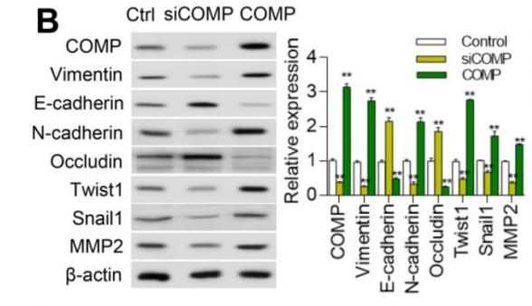
2). Cartilage Oligomeric Matrix Protein promotes epithelial-mesenchymal transition by interacting with Transgelin in Colorectal Cancer. Theranostics, 2020 (PubMed: 32754278) [IF=12.4]

Application: WB    Species: Human    Sample: HCT116 cells

Figure 4. COMP promotes EMT in colorectal cancer. (A) Cell phenotypic changes in cells treated with COMP siRNA and COMP overexpression. (B) WB analysis of COMP and EMT-related markers in HCT116 cells under COMP knockdown or overexpression. (C) Cell wound scratch assay of HCT116, HCT-8, and SW620 cells treated with COMP siRNA or COMP overexpression vectors. (D) Transwell assay of HCT116, HCT-8, and SW620 cells treated with COMP siRNA or COMP overexpression vectors. (E) Immunofluorescence assay of HCT116, HCT-8, and SW620 cells treated with COMP siRNA or COMP overexpression vectors. Image J software was used to analyze the relative intensity of E-cadherin and Vimentin. (F) HCT116 and HCT-8 cells with knocked down or overexpressed COMP were transplanted on nude mice. Tumor volumes were measured every 4 days. (G) Tumor weight in the control, COMP knockdown, and COMP overexpression groups. (H) The in situ spleen model of the colorectal cancer cell line SW620 showed that overexpression of COMP promoted liver metastasis of colorectal cancer, while downregulation of COMP inhibited liver metastasis of colorectal cancer. (I) IHC staining to identify EMT biomarkers and COMP-related proteins in the control, COMP knockdown, and COMP overexpression groups. COMP knockdown displayed strong E-cadherin staining and reduced Vimentin, Snail1, Twist1, and MMP-2 staining. The expression levels of other proteins were identical to those observed in WB experiments. (J) Staining indices of COMP, E-cadherin, Vimentin, Snail1, Twist1, and MMP2. The error bars in all graphs represented SD, and each experiment was repeated three times. * and ** stand for P<0.05 and P<0.01, respectively.

3). Gut microbiota-mediated secondary bile acid alleviates Staphylococcus aureus-induced mastitis through the TGR5-cAMP-PKA-NF-κB/NLRP3 pathways in mice. npj Biofilms and Microbiomes, 2023 (PubMed: 36755021) [IF=9.2]

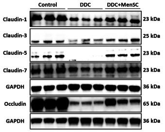
4). Human menstrual blood-derived stem cell transplantation suppresses liver injury in DDC-induced chronic cholestasis. Stem Cell Research & Therapy, 2022 (PubMed: 35123555) [IF=7.5]

Application: WB    Species: Mice    Sample: liver tissue

Fig. 5 MenSCs promoted TJ- and bile transport function-related protein expression and reduced fibrosis-related protein expression. A Western blot analysis protein levels of Claudin-1, Claudin-3, Claudin-5, Claudin-7 and Occludin in liver tissue of different groups (n = 3 for each group). B BSEP, OATP2 and NTCP1 expression in liver tissues of different groups (n = 3 for each group). C, D Liver COL1A1, α-SMA, TGF-β1 and β-catenin expression among different groups (n = 3 for each group). *p < 0.05, **p < 0.01, ***p < 0.001, ****p < 0.0001

5). Commensal cow Roseburia reduces gut-dysbiosis-induced mastitis through inhibiting bacterial translocation by producing butyrate in mice. Cell Reports, 2022 (PubMed: 36417859) [IF=7.5]

Application: WB    Species: Mouse    Sample:

Figure 5Commensal Roseburia ameliorates gut-dysbiosis-induced mastitis in mice

6). Disturbances of the gut microbiota-derived tryptophan metabolites as key actors in vagotomy-induced mastitis in mice. Cell reports, 2024 (PubMed: 39110590) [IF=7.5]

Application: WB    Species: Mouse    Sample:

Figure 1 Unilateral Vag induces mastitis and disrupts the BMB in mice (A) Experimental design. The mice were randomly divided into three groups: the control group, the Vag group, and the vagus nerve stimulation group (n = 7). After 3 days, the mice were sacrificed for the collection of mammary tissue. (B) Representative H&E-stained images of mammary tissues from three different groups (scale bar, 50 μm). (C) Histopathological score of the mammary gland (n = 7). ns, not significant. (D–F) Inflammatory cytokines, including TNF-α (D) and IL-1β (E) concentrations and MPO activity (F) in the three groups of mammary tissues, were detected by the kit (n = 7). (G–J) The expression levels of the TJ proteins ZO-1, Occludin, and Claudin-3 in representative mammary tissues as detected by western blotting (G) and the relative intensity of the three proteins using β-actin as an internal reference (H–J) (n = 3). (K) The localization and expression of TJ proteins in the mammary gland was determined by immunohistochemistry (scale bar, 20 μm). (L and M) Serum levels of TNF-α (L) and IL-1β (M) (n = 7). Data are presented as means ± SDs (C–F, H–J, L, and M), and one-way ANOVA was performed for statistical analysis (B–F and H–J). ∗p < 0.05; ∗∗p < 0.01; ∗∗∗p < 0.001; and ∗∗∗∗p < 0.0001 indicate significant differences. See also Figures S1 and S2.

Application: IHC    Species: Mouse    Sample:

Figure 1 Unilateral Vag induces mastitis and disrupts the BMB in mice (A) Experimental design. The mice were randomly divided into three groups: the control group, the Vag group, and the vagus nerve stimulation group (n = 7). After 3 days, the mice were sacrificed for the collection of mammary tissue. (B) Representative H&E-stained images of mammary tissues from three different groups (scale bar, 50 μm). (C) Histopathological score of the mammary gland (n = 7). ns, not significant. (D–F) Inflammatory cytokines, including TNF-α (D) and IL-1β (E) concentrations and MPO activity (F) in the three groups of mammary tissues, were detected by the kit (n = 7). (G–J) The expression levels of the TJ proteins ZO-1, Occludin, and Claudin-3 in representative mammary tissues as detected by western blotting (G) and the relative intensity of the three proteins using β-actin as an internal reference (H–J) (n = 3). (K) The localization and expression of TJ proteins in the mammary gland was determined by immunohistochemistry (scale bar, 20 μm). (L and M) Serum levels of TNF-α (L) and IL-1β (M) (n = 7). Data are presented as means ± SDs (C–F, H–J, L, and M), and one-way ANOVA was performed for statistical analysis (B–F and H–J). ∗p < 0.05; ∗∗p < 0.01; ∗∗∗p < 0.001; and ∗∗∗∗p < 0.0001 indicate significant differences. See also Figures S1 and S2.

7). Protective effect of synbiotic combination of Lactobacillus plantarum SC-5 and olive oil extract tyrosol in a murine model of ulcerative colitis. Journal of translational medicine, 2024 (PubMed: 38528541) [IF=7.4]

8). Enterogenic Stenotrophomonas maltophilia migrates to the mammary gland to induce mastitis by activating the calcium-ROS-AMPK-mTOR-autophagy pathway. Journal of animal science and biotechnology, 2023 (PubMed: 38124149) [IF=7.0]

Application: WB    Species: Mouse    Sample:

Fig. 2 Treatment with sf-GFP-S. maltophilia destroys the integrity of the blood-milk barrier. After the mice were treated with 1010 CFU S. maltophilia for a week, mammary gland tissues were collected and the integrity of blood-milk barrier was detected. A Expression of tight junction proteins ZO-1, Occludin, Claudin-3 in mouse mammary gland. B FITC-albumin fluorescence staining of mammary gland tissue to determine the degree of albumin infiltration. The data are presented as mean ± SD. One-way analysis of variance was performed for statistical analysis. *P < 0.05, **P < 0.01 and ***P < 0.001 indicate significance from each group. ns, no significance

9). Hexadecanamide alleviates Staphylococcus aureus-induced mastitis in mice by inhibiting inflammatory responses and restoring blood-milk barrier integrity. PLoS pathogens, 2023 (PubMed: 37948460) [IF=5.5]

Application: WB    Species: Mouse    Sample:

Fig 2 HEX improves S. aureus-caused disruption of the blood-milk barrier. A-D. Western blotting was performed to detect the protein levels of ZO-1, Occludin, and Claudin-3 in the mammary gland (n = 5). E. Representative immunohistochemical image of the mammary glands by ZO-1, Occludin, and Claudin-3 antibodies staining (scale bars, 20 μm). Data are expressed as mean ± SD (B-D). ns, no significance, *p < 0.05, **p < 0.01, and ***p < 0.001 indicate significant statistical difference by one-way ANOVA (B-D).

Application: IHC    Species: Mouse    Sample:

Fig 2 HEX improves S. aureus-caused disruption of the blood-milk barrier. A-D. Western blotting was performed to detect the protein levels of ZO-1, Occludin, and Claudin-3 in the mammary gland (n = 5). E. Representative immunohistochemical image of the mammary glands by ZO-1, Occludin, and Claudin-3 antibodies staining (scale bars, 20 μm). Data are expressed as mean ± SD (B-D). ns, no significance, *p < 0.05, **p < 0.01, and ***p < 0.001 indicate significant statistical difference by one-way ANOVA (B-D).

Application: IF/ICC    Species: Mouse    Sample: MMECs

Fig 3 HEX enhances barrier integrity and limits the NF-κB signaling pathway in MMECs. A-D. The protein levels of ZO-1, Occludin, and Claudin-3 were assessed after HEX pretreatment (0, 2.5, and 5 μM) for 2 h and S. aureus treatment (105 CFU) for 24 h (n = 5). E. Immunofluorescence staining was performed to detect TJs protein ZO-1, Occludin, and Claudin-3 expressions in MMECs (scale bars, 10 μm). F-H. Protein levels in the NF-κB signaling pathway, including IκB, p65, and phosphorylation levels of IκB and p65, were detected with HEX pretreatment (0, 2.5, and 5 μM) for 2 h and S. aureus treatment (105 CFU) for 24 h (n = 5). One-way ANOVA (B-D and G-H) was used, and values are presented as the mean ± SD (B-D and G-H). *p < 0.05, **p < 0.01, and ***p < 0.001 indicate significantly different from each group. ns, no significance.

10). A fiber-enriched diet alleviates Staphylococcus aureus-induced mastitis by activating the HDAC3-mediated antimicrobial program in macrophages via butyrate production in mice. PLoS Pathogens, 2023 (PubMed: 36656870) [IF=5.5]

もっと読みます

Restrictive clause


Affinity Biosciences tests all products strictly. Citations are provided as a resource for additional applications that have not been validated by Affinity Biosciences. Please choose the appropriate format for each application and consult Materials and Methods sections for additional details about the use of any product in these publications.

For Research Use Only.
Not for use in diagnostic or therapeutic procedures. Not for resale. Not for distribution without written consent. Affinity Biosciences will not be held responsible for patent infringement or other violations that may occur with the use of our products. Affinity Biosciences, Affinity Biosciences Logo and all other trademarks are the property of Affinity Biosciences LTD.