製品: Claudin 5 Antibody
カタログ: AF0130
タンパク質の説明: Rabbit polyclonal antibody to Claudin 5
アプリケーション: WB IHC IF/ICC
Cited expt.: WB, IF/ICC
反応性: Human, Mouse, Rat
予測: Pig, Bovine, Rabbit
分子量: 23kDa; 23kD(Calculated).
ユニプロット: O00501
RRID: AB_2833314

類似製品を見る>>

   サイズ 価格 在庫状況
 100ul $280 在庫あり
 200ul $350 在庫あり

リードタイム: 当日配達

For pricing and ordering contact:
お問い合わせ先

製品説明

ソース:
Rabbit
アプリケーション:
WB 1:500-1:3000, IHC 1:50-1:200, IF/ICC 1:200
*The optimal dilutions should be determined by the end user.
*Tips:

WB: For western blot detection of denatured protein samples. IHC: For immunohistochemical detection of paraffin sections (IHC-p) or frozen sections (IHC-f) of tissue samples. IF/ICC: For immunofluorescence detection of cell samples. ELISA(peptide): For ELISA detection of antigenic peptide.

反応性:
Human,Mouse,Rat
予測:
Pig(100%), Bovine(89%), Rabbit(89%)
クローナリティ:
Polyclonal
特異性:
Claudin 5 Antibody detects endogenous levels of total Claudin 5.
RRID:
AB_2833314
引用形式: Affinity Biosciences Cat# AF0130, RRID:AB_2833314.
コンジュゲート:
Unconjugated.
精製:
The antiserum was purified by peptide affinity chromatography using SulfoLink™ Coupling Resin (Thermo Fisher Scientific).
保存:
Rabbit IgG in phosphate buffered saline , pH 7.4, 150mM NaCl, 0.02% sodium azide and 50% glycerol. Store at -20 °C. Stable for 12 months from date of receipt.
別名:

折りたたみ/展開

Androgen withdrawal and apoptosis induced protein RVP1 like; AWAL; BEC 1; BEC1; Claudin 5 (transmembrane protein deleted in velocardiofacial syndrome); Claudin-5; Claudin5; CLD5_HUMAN; CLDN 5; Cldn5; CPETR L1; CPETRL 1; CPETRL1; TMDVCF; TMVCF; Transmembrane protein deleted in VCFS; Transmembrane protein deleted in velocardiofacial syndrome;

免疫原

免疫原:

A synthesized peptide derived from human Claudin 5, corresponding to a region within C-terminal amino acids.

Uniprot:
遺伝子(ID):
タンパク質の説明:
claudin 5 Plays a major role in tight junction-specific obliteration of the intercellular space. Belongs to the claudin family. Directly interacts with TJP1/ZO-1, TJP2/ZO-2 and TJP3/ZO- 3. Interacts with MPDZ.
タンパク質配列:
MGSAALEILGLVLCLVGWGGLILACGLPMWQVTAFLDHNIVTAQTTWKGLWMSCVVQSTGHMQCKVYDSVLALSTEVQAARALTVSAVLLAFVALFVTLAGAQCTTCVAPGPAKARVALTGGVLYLFCGLLALVPLCWFANIVVREFYDPSVPVSQKYELGAALYIGWAATALLMVGGCLLCCGAWVCTGRPDLSFPVKYSAPRRPTATGDYDKKNYV

種類予測

種類予測:

Score>80(red) has high confidence and is suggested to be used for WB detection. *The prediction model is mainly based on the alignment of immunogen sequences, the results are for reference only, not as the basis of quality assurance.

Species
Results
Score
Pig
100
Bovine
89
Rabbit
89
Chicken
78
Xenopus
75
Horse
0
Sheep
0
Dog
0
Zebrafish
0
Model Confidence:
High(score>80) Medium(80>score>50) Low(score<50) No confidence

研究背景

機能:

Plays a major role in tight junction-specific obliteration of the intercellular space.

細胞の位置付け:

Cell junction>Tight junction. Cell membrane>Multi-pass membrane protein.

Extracellular region or secreted Cytosol Plasma membrane Cytoskeleton Lysosome Endosome Peroxisome ER Golgi apparatus Nucleus Mitochondrion Manual annotation Automatic computational assertionSubcellular location
タンパク質ファミリー:

Belongs to the claudin family.

研究領域

· Cellular Processes > Cellular community - eukaryotes > Tight junction.   (View pathway)

· Environmental Information Processing > Signaling molecules and interaction > Cell adhesion molecules (CAMs).   (View pathway)

· Human Diseases > Infectious diseases: Viral > Hepatitis C.

· Organismal Systems > Immune system > Leukocyte transendothelial migration.   (View pathway)

参考文献

1). Engagement of circular RNA HECW2 in the nonautophagic role of ATG5 implicated in the endothelial-mesenchymal transition. Autophagy, 2018 (PubMed: 29260931) [IF=14.6]

Application: WB    Species: human    Sample: HBMECs

Figure 3|Role of ATG5 in LPS-induced EndoMT. Scale bar: 5 μm. Mice were administered LPS (0.83 mg/kg) once per day for 7 d. n=6 animals/group. *P<0.05,**P<0.01, ***P<0.001 versus saline group using the Mann-Whitney test. Transfection of HBMECs with siRNA ATG5 significantly inhibited the decreases in the TJP expression levels and the increases in the mesenchymal cell marker expression .

2). The endothelium permeability after bioresorbable scaffolds implantation caused by the heterogeneous expression of tight junction proteins. Materials today. Bio, 2022 (PubMed: 36090609) [IF=8.7]

Application: WB    Species: Rat    Sample:

Fig. 2. The differential expression of TJPs in the neointima of abdominal aorta after implantation of PLLA scaffold in rats. (A) Immunofluorescence staining of paraffin sections of abdominal aorta of rats implanted with PLLA scaffold (white arrows: ECs with higher expression level of TJPs, S: the scaffold strut, L: the lumen); (B-E) statistics of immunofluorescence staining intensity of ZO-1(B), Claudin-5(C), Occludin(D) and Tricellulin(E) in the neointima of sections. < 0.05, ∗∗p ​< ​0.01, ∗∗∗p ​< ​0.001.

3). A novel function of claudin-5 in maintaining the structural integrity of the heart and its implications in cardiac pathology. Biochimica et biophysica acta. Molecular basis of disease, 2024 (PubMed: 38838411) [IF=4.2]

Application: WB    Species: human    Sample:

Fig. 1. Myocardial dilation, myocyte atrophy and Cldn5 reduction in the left atrium of AF patients (A) Exemplar images of electrocardiogram (ECG) and apical four-chamber view of the heart in AF patient and non-AF control. Red box indicates left atrium (LA). (B) Representative western blot of Cldn5 in left appendages of AF patients compared to those of donors. Human lung tissue excised during lung cancer resection is used as positive control. (C) Quantitative analysis of the protein level of Cldn5 in AF and non-AF groups. * P < 0.05 vs. Non-AF; n = 4. (D)Exemplar left atrial appendage section stained with anti-Cldn5, CD31 and TOMM20 antibodies. Anti-Cldn5 primary antibody is Alexa Fluor 488 conjugated (Green) while CD31 and TOMM20 antibodies is visualized by Fluorescein donkey anti-rabbit IgG Alexa Fluor 594 (Red). DAPI is used to stain nucleus (Blue). Scale bar, 20 μm. (E) Quantification of immunofluorescent staining of Cldn5 in cardiomyocytes. * P < 0.05 vs. Non-AF; n = 4. (F) Exemplar left atrial appendage section stained with wheat germ agglutinin (WGA) for assessing cardiomyocyte's cross section. Scale bar, 20 μm. (G) Quantification of cross-section of cardiomyocytes. * P < 0.05 vs. Non-AF; n = 4. (For interpretation of the references to colour in this figure legend, the reader is referred to the web version of this article.)

4). Axl deficiency promotes the neuroinvasion of Japanese encephalitis virus by enhancing IL-1α production from pyroptotic macrophages. Journal of Virology, 2020 (PubMed: 32611752) [IF=4.0]

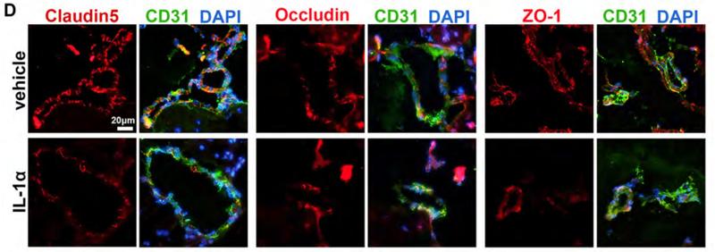
Application: IF/ICC    Species: mouse    Sample: brains

Fig.9. |Effects of IL-1α alone on BBB permeability and tight junction integrity.(D) IF staining of claudin-5, occludin, and ZO-1 (red), CD31 (microvascular endothelial cell marker, green), and DAPI (nucleus, blue), scale bar=20 μm.

5). Activation of Sigma-1 Receptor Enhanced Pericyte Survival via the Interplay Between Apoptosis and Autophagy: Implications for Blood-Brain Barrier Integrity in Stroke. Translational Stroke Research, 2020 (PubMed: 31290080) [IF=3.8]

Application: WB    Species: mouse    Sample: brain

Fig. 1| Knockdown of σ-1R aggravated cerebral ischemia-induced injury in pMCAO mice.h, i The decrease in TJP expression in the ipsilateral side of the brain observed at 24 h after pMCAO surgery was aggravated in σ-1R KO mice. TJP expression levels were determined via western blot analysis (h) and quantified through densitometric analysis.

6). The truncated AXIN1 isoform promotes hepatocellular carcinoma metastasis through SRSF9-mediated exon 9 skipping. Molecular and cellular biochemistry, 2025 (PubMed: 38748384) [IF=3.5]

Restrictive clause


Affinity Biosciences tests all products strictly. Citations are provided as a resource for additional applications that have not been validated by Affinity Biosciences. Please choose the appropriate format for each application and consult Materials and Methods sections for additional details about the use of any product in these publications.

For Research Use Only.
Not for use in diagnostic or therapeutic procedures. Not for resale. Not for distribution without written consent. Affinity Biosciences will not be held responsible for patent infringement or other violations that may occur with the use of our products. Affinity Biosciences, Affinity Biosciences Logo and all other trademarks are the property of Affinity Biosciences LTD.