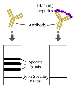
製品: THBS1 Antibody
カタログ: DF6848
タンパク質の説明: Rabbit polyclonal antibody to THBS1
アプリケーション: WB IHC IF/ICC
Cited expt.: WB, IHC, IF/ICC
反応性: Human, Mouse, Rat
予測: Horse, Rabbit, Dog
分子量: 150-180kD; 129kD(Calculated).
ユニプロット: P07996
RRID: AB_2838807

類似製品を見る>>

   サイズ 価格 在庫状況
 100ul $280 在庫あり
 200ul $350 在庫あり

リードタイム: 当日配達

For pricing and ordering contact:
お問い合わせ先

製品説明

ソース:
Rabbit
アプリケーション:
WB 1:500-1:2000, IHC 1:50-1:200, IF/ICC 1:100-1:500
*The optimal dilutions should be determined by the end user. For optimal experimental results, antibody reuse is not recommended.
*Tips:

WB: For western blot detection of denatured protein samples. IHC: For immunohistochemical detection of paraffin sections (IHC-p) or frozen sections (IHC-f) of tissue samples. IF/ICC: For immunofluorescence detection of cell samples. ELISA(peptide): For ELISA detection of antigenic peptide.

反応性:
Human,Mouse,Rat
予測:
Horse(89%), Rabbit(100%), Dog(100%)
クローナリティ:
Polyclonal
特異性:
THBS1 Antibody detects endogenous levels of total THBS1.
RRID:
AB_2838807
引用形式: Affinity Biosciences Cat# DF6848, RRID:AB_2838807.
コンジュゲート:
Unconjugated.
精製:
The antiserum was purified by peptide affinity chromatography using SulfoLink™ Coupling Resin (Thermo Fisher Scientific).
保存:
Rabbit IgG in phosphate buffered saline , pH 7.4, 150mM NaCl, 0.02% sodium azide and 50% glycerol. Store at -20 °C. Stable for 12 months from date of receipt.
別名:

折りたたみ/展開

THBS 1; THBS; Thbs1; Thrombospondin 1; Thrombospondin-1; TSP 1; TSP; TSP1; TSP1_HUMAN;

免疫原

免疫原:

A synthesized peptide derived from human THBS1, corresponding to a region within the internal amino acids.

Uniprot:
遺伝子(ID):
タンパク質の説明:
TSP1, also named as THBS1, THBS and TSP, is an adhesive glycoprotein that mediates cell-to-cell and cell-to-matrix interactions, and which can bind to fibrinogen, fibronectin, laminin, type V collagen and integrins alpha-V/beta-1, alpha-V/beta-3 and alpha-IIb/beta-3. TSP1 is a regulator of many biological processes including cell growth, adhesion, migration, platelet aggregation, and fibrin deposition and lysis. It interacts with a number of plasma proteins such as fibrinogen and plasminogen and co-polymerizes with fibrin in clot formation.
タンパク質配列:
MGLAWGLGVLFLMHVCGTNRIPESGGDNSVFDIFELTGAARKGSGRRLVKGPDPSSPAFRIEDANLIPPVPDDKFQDLVDAVRAEKGFLLLASLRQMKKTRGTLLALERKDHSGQVFSVVSNGKAGTLDLSLTVQGKQHVVSVEEALLATGQWKSITLFVQEDRAQLYIDCEKMENAELDVPIQSVFTRDLASIARLRIAKGGVNDNFQGVLQNVRFVFGTTPEDILRNKGCSSSTSVLLTLDNNVVNGSSPAIRTNYIGHKTKDLQAICGISCDELSSMVLELRGLRTIVTTLQDSIRKVTEENKELANELRRPPLCYHNGVQYRNNEEWTVDSCTECHCQNSVTICKKVSCPIMPCSNATVPDGECCPRCWPSDSADDGWSPWSEWTSCSTSCGNGIQQRGRSCDSLNNRCEGSSVQTRTCHIQECDKRFKQDGGWSHWSPWSSCSVTCGDGVITRIRLCNSPSPQMNGKPCEGEARETKACKKDACPINGGWGPWSPWDICSVTCGGGVQKRSRLCNNPTPQFGGKDCVGDVTENQICNKQDCPIDGCLSNPCFAGVKCTSYPDGSWKCGACPPGYSGNGIQCTDVDECKEVPDACFNHNGEHRCENTDPGYNCLPCPPRFTGSQPFGQGVEHATANKQVCKPRNPCTDGTHDCNKNAKCNYLGHYSDPMYRCECKPGYAGNGIICGEDTDLDGWPNENLVCVANATYHCKKDNCPNLPNSGQEDYDKDGIGDACDDDDDNDKIPDDRDNCPFHYNPAQYDYDRDDVGDRCDNCPYNHNPDQADTDNNGEGDACAADIDGDGILNERDNCQYVYNVDQRDTDMDGVGDQCDNCPLEHNPDQLDSDSDRIGDTCDNNQDIDEDGHQNNLDNCPYVPNANQADHDKDGKGDACDHDDDNDGIPDDKDNCRLVPNPDQKDSDGDGRGDACKDDFDHDSVPDIDDICPENVDISETDFRRFQMIPLDPKGTSQNDPNWVVRHQGKELVQTVNCDPGLAVGYDEFNAVDFSGTFFINTERDDDYAGFVFGYQSSSRFYVVMWKQVTQSYWDTNPTRAQGYSGLSVKVVNSTTGPGEHLRNALWHTGNTPGQVRTLWHDPRHIGWKDFTAYRWRLSHRPKTGFIRVVMYEGKKIMADSGPIYDKTYAGGRLGLFVFSQEMVFFSDLKYECRDP

種類予測

種類予測:

Score>80(red) has high confidence and is suggested to be used for WB detection. *The prediction model is mainly based on the alignment of immunogen sequences, the results are for reference only, not as the basis of quality assurance.

Species
Results
Score
Rabbit
100
Dog
100
Horse
89
Bovine
78
Sheep
78
Chicken
78
Pig
0
Xenopus
0
Zebrafish
0
Model Confidence:
High(score>80) Medium(80>score>50) Low(score<50) No confidence

研究背景

機能:

Adhesive glycoprotein that mediates cell-to-cell and cell-to-matrix interactions. Binds heparin. May play a role in dentinogenesis and/or maintenance of dentin and dental pulp (By similarity). Ligand for CD36 mediating antiangiogenic properties. Plays a role in ER stress response, via its interaction with the activating transcription factor 6 alpha (ATF6) which produces adaptive ER stress response factors (By similarity).

細胞の位置付け:

Secreted. Cell surface. Secreted>Extracellular space>Extracellular matrix. Endoplasmic reticulum. Sarcoplasmic reticulum.
Note: Secreted by thrombin-activated platelets and binds to the cell surface in the presence of extracellular Ca(2+) (PubMed:6777381). Incorporated into the extracellular matrix of fibroblasts (PubMed:6341993). Also detected in the endoplasmic reticulum and sarcoplasmic reticulum where it plays a role in the ER stress response (By similarity).

Extracellular region or secreted Cytosol Plasma membrane Cytoskeleton Lysosome Endosome Peroxisome ER Golgi apparatus Nucleus Mitochondrion Manual annotation Automatic computational assertionSubcellular location
タンパク質ファミリー:

Belongs to the thrombospondin family.

研究領域

· Cellular Processes > Cell growth and death > p53 signaling pathway.   (View pathway)

· Cellular Processes > Transport and catabolism > Phagosome.   (View pathway)

· Cellular Processes > Cellular community - eukaryotes > Focal adhesion.   (View pathway)

· Environmental Information Processing > Signal transduction > Rap1 signaling pathway.   (View pathway)

· Environmental Information Processing > Signal transduction > PI3K-Akt signaling pathway.   (View pathway)

· Environmental Information Processing > Signal transduction > TGF-beta signaling pathway.   (View pathway)

· Environmental Information Processing > Signaling molecules and interaction > ECM-receptor interaction.   (View pathway)

· Human Diseases > Infectious diseases: Parasitic > Malaria.

· Human Diseases > Infectious diseases: Viral > Human papillomavirus infection.

· Human Diseases > Cancers: Overview > Proteoglycans in cancer.

· Human Diseases > Cancers: Overview > MicroRNAs in cancer.

· Human Diseases > Cancers: Specific types > Bladder cancer.   (View pathway)

参考文献

1). Comprehensive single-cell RNA analysis reveals intertumoral microenvironment heterogeneity and hub niche of carcinogenesis in thyroid cancer. NPJ precision oncology, 2025 (PubMed: 41298873) [IF=7.9]

Application: IF/ICC    Species: human    Sample:

Fig. 5: Cell-to-cell communications among thyrocyte and immune cells predicted by the CellChat software in PTC and ATC. A Circle plot summarizing the maximum number of interactions among individual cell types in PTC. The thickness of the lines connecting cells indicates the interaction strength. B The same plot as in (A) but applied to ATC. C Differences of number and strength overall information flow within the inferred networks between PTC and ATC. D The potential outgoing and incoming interaction strength of each cellular cluster in PTC and ATC. Heatmap showing the relative signaling contribution of each cell group based on the number and strength compared PTC with ATC. E Heatmap showing the relative signaling contribution of each cell group based on the number and strength compared PTC with ATC. F Differential strength of network centrality based on APOE+ macrophages (PTC vs. ATC). G Bubble heatmap showing the cell–cell communication of selected ligand–receptor pairs between APOE+ macrophage and CD8+ PDCD1+ T cells. Dot size indicates P value, colored by communication probability. H Multiplex IHC shows the cross-talk between APOE+ macrophages and T cells via the ligand–receptor of THBS1-CD47.

2). Intravenous tenecteplase bridging reperfusion ameliorates cerebral ischemia/reperfusion injury by improving microvascular circulation in rats. Journal of thrombosis and haemostasis : JTH, 2025 (PubMed: 39826801) [IF=5.5]

3). Chemogenetic activation of astrocytic Gi signaling promotes spinogenesis and motor functional recovery after stroke. Glia, 2024 (PubMed: 38436489) [IF=5.4]

4). HIF-1α depletion regulates autophagy to improve arteriosclerosis obliterans of the lower extremities via the TSP-1/CD47 axis. Life sciences, 2025 (PubMed: 40543809) [IF=5.2]

5). Thrombospondin-1 promotes mechanical stress-mediated ligamentum flavum hypertrophy through the TGFβ1/Smad3 signaling pathway. Matrix biology : journal of the International Society for Matrix Biology, 2024 (PubMed: 38281553) [IF=4.5]

Application: WB    Species: human    Sample: LF cells

Fig. 3. Fibrosis phenotype and THBS1/TGFβ1 expression were significantly elevated in the LFH group. (A) The thickness of LF was measured by MRI before operation. The thickness of LF at the L4/5 facet joint level was indicated by red arrow. (B) Representative tissues of LF acquired from patients with spinal surgery. The top three were from non-LFH patients and the bottom three were from LFH patients. Scale bar = 5 mm. (C) Representative images of H&E, MASSON, and EVG staining of the non-LFH group (n = 6) and the LFH group (n = 6). The yellow dotted area represents collagen fibers. Scale bar = 20 μm. (D) Immunohistochemical staining of THBS1, TGFβ1, COL1A2, and α-SMA in the LF samples from LFH group (n = 6) and non-LFH group (n = 6), and the percentage of positive cells was quantified. Scale bar = 20 μm. (E) TEM of LF ultrastructure of non-LFH and LFH groups. The yellow area represents elastin fibers. Scale bar = 8 μm. (F) The qRT-PCR analysis results showing that LFH group (n = 6) had a higher mRNA level of THBS1, TGFβ1, ACTA2 (α-SMA), and COL1A2 compared with non-LFH group (n = 6). The 2−ΔΔCt method was used to normalized with GAPDH. (G) Western blotting assay of THBS1, TGFβ1, COL1A2, and α-SMA protein levels of LF samples of the two groups. GAPDH served as the loading control. EVG: verhoeff's van gieson, TEM: transmission electron microscope, EF: elastic fiber, CF: collagen fiber. **p < 0.01, ***p < 0.001.

Application: IHC    Species: Mouse    Sample:
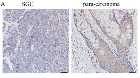
Fig. 7. The mechanical stress-THBS1 axis promotes hypertrophy and fibrosis of LF in vivo. (A) H&E staining of tissues from mice (n = 6 mice per group), and the quantitative analysis of LF area. Scale bar = 50 μm. (B) EVG staining of tissues from mice, and the quantitative analysis of volume fraction of elastin fibers in LF. Scale bar = 20 μm. (C, D) Immunohistochemical staining of α-SMA and THBS1 in mice tissues, and the percentage of positive cells was quantified. Scale bar = 20 μm. Error bars: mean ± S.D. ns, not significant. *p < 0.05, **p < 0.01, ***p < 0.001.

6). Allicin induces cell cycle arrest and apoptosis of breast cancer cells in vitro via modulating the p53 pathway. Molecular Biology Reports, 2021 (PubMed: 34626309) [IF=2.6]

7). MicroRNA‑3907 promotes the proliferation and migration of sebaceous gland carcinoma of the eyelid by targeting thrombospondin 1. Oncology Letters, 2021 (PubMed: 34691259) [IF=2.5]

Application: IHC    Species: Human    Sample: SGC tissues
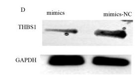
Figure 2. THBS1 expression levels in SGC tissues and cells. (A) Immunohistochemical staining of THBS1 in SGC and para-carcinoma tissues. Expression of THBS1 was negative in SGC tissues, but positive in para-carcinoma tissues. Scale bar, 50 µm. (B) Positive expression rate of THBS1 in eyelid SGC and para-carcinoma formalin-fixed paraffin-embedded samples. (C) Relative mRNA expression levels of THBS1 in SGC cells were lower than those in normal SG cells. **P<0.01 and ***P<0.001. SGC, sebaceous gland cancer; THBS1, thrombospondin 1.

Application: WB    Species: Human    Sample: SGC cells
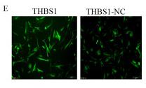
Figure 4. miR-3907 negatively regulates THBS1 expression. (A) Predicted binding sites for miR-3907 within the wild-type and mutant 3'UTR of THBS1. (B) Quantitative analysis of the relative luciferase activity of cells with reporter vectors containing the wild type or mutant THBS1 3'UTR, following miR-3907 co-transfection. (C) Reverse transcription-quantitative PCR and (D) western blotting with semi-quantification analysis revealed decreased THBS1 expression following miR-3907 mimics transfection. *P<0.05 and ***P<0.001. miR, microRNA; THBS1, thrombospondin 1; UTR, untranslated region; NC, negative control.

Application: IF/ICC    Species: Human    Sample: SGC cells

Figure 5. Inhibitory effects of THBS1 on SGC cell proliferation and migration. (A) THBS1-knockdown was induced by transfecting SGC cells with siRNA-THBS1. THBS1 expression level was determined by reverse transcription-quantitative PCR. Results of (B) CCK-8 and (C) wound-healing assays after transfection with siRNA-THBS1; siRNA-THBS1 increased the proliferation and migration of SGC cells. (D) THBS1 overexpression was induced by transfecting lentiviral vectors; THBS1 expression level was determined by reverse transcription-quantitative PCR. (E) Fluorescence assay results revealed that the THBS1 lentivirus vector was successfully transfected. (F) CCK-8 and (G) wound-healing assays results after transfection with THBS1 lentivirus revealed that THBS1 decreased the proliferation and migration of SGC cells. Scale bar, 500 µm. *P<0.05, **P<0.01 and ***P<0.001. SGC, sebaceous gland cancer; siRNA, small interfering RNA; CCK-8, Cell Counting Kit-8; THBS1, thrombospondin 1; NC, negative control.

8). Proteomics reveals the regulation of ALG1 expression by lentivirus-mediated THBS1 overexpression and its mechanism in meibomian gland carcinoma. Oncology letters, 2025 (PubMed: 40980150) [IF=2.5]

Application: WB    Species: human    Sample:

Figure 2. - Proteomic analysis following THBS1 overexpression. (A) Fluorescent imaging (×40; scale bar, 200 µm) of NCoe and THBS1oe groups after lentiviral-mediated THBS1 overexpression. (B) Evaluation of THBS1 overexpression using reverse transcription-quantitative PCR and western blot analysis. (C) Hierarchical clustering heatmap of differentially expressed proteins. (D) Volcano plot of DEPs: Green dots indicate downregulated proteins, and red dots indicate upregulated proteins. (E) GO and (F) KEGG pathway enrichment analysis of DEPs. **P

Restrictive clause


Affinity Biosciences tests all products strictly. Citations are provided as a resource for additional applications that have not been validated by Affinity Biosciences. Please choose the appropriate format for each application and consult Materials and Methods sections for additional details about the use of any product in these publications.

For Research Use Only.
Not for use in diagnostic or therapeutic procedures. Not for resale. Not for distribution without written consent. Affinity Biosciences will not be held responsible for patent infringement or other violations that may occur with the use of our products. Affinity Biosciences, Affinity Biosciences Logo and all other trademarks are the property of Affinity Biosciences LTD.