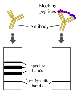
製品: IRF3 Antibody
カタログ: DF6895
タンパク質の説明: Rabbit polyclonal antibody to IRF3
アプリケーション: WB IHC IF/ICC
Cited expt.: WB
反応性: Human, Mouse, Rat
予測: Pig, Bovine, Horse, Sheep, Dog
分子量: 49kDa; 47kD(Calculated).
ユニプロット: Q14653
RRID: AB_2838854

類似製品を見る>>

   サイズ 価格 在庫状況
 100ul $280 在庫あり
 200ul $350 在庫あり

リードタイム: 当日配達

For pricing and ordering contact:
お問い合わせ先

製品説明

ソース:
Rabbit
アプリケーション:
WB 1:500-1:2000, IHC 1:50-1:200, IF/ICC 1:100-1:500
*The optimal dilutions should be determined by the end user. For optimal experimental results, antibody reuse is not recommended.
*Tips:

WB: For western blot detection of denatured protein samples. IHC: For immunohistochemical detection of paraffin sections (IHC-p) or frozen sections (IHC-f) of tissue samples. IF/ICC: For immunofluorescence detection of cell samples. ELISA(peptide): For ELISA detection of antigenic peptide.

反応性:
Human,Mouse,Rat
予測:
Pig(100%), Bovine(100%), Horse(88%), Sheep(100%), Dog(100%)
クローナリティ:
Polyclonal
特異性:
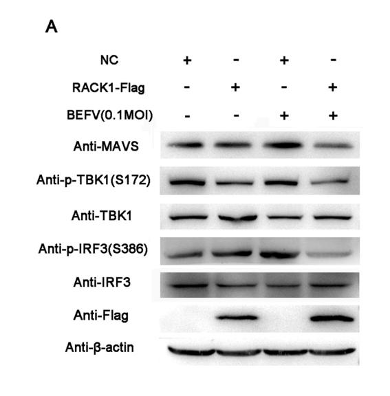
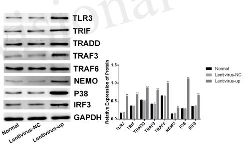
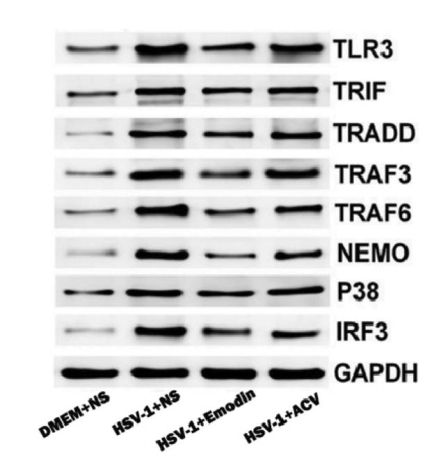
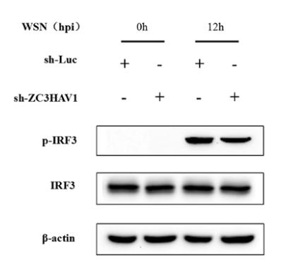
IRF3 Antibody detects endogenous levels of total IRF3.
RRID:
AB_2838854
引用形式: Affinity Biosciences Cat# DF6895, RRID:AB_2838854.
コンジュゲート:
Unconjugated.
精製:
The antiserum was purified by peptide affinity chromatography using SulfoLink™ Coupling Resin (Thermo Fisher Scientific).
保存:
Rabbit IgG in phosphate buffered saline , pH 7.4, 150mM NaCl, 0.02% sodium azide and 50% glycerol. Store at -20 °C. Stable for 12 months from date of receipt.
別名:

折りたたみ/展開

IIAE7; Interferon regulatory factor 3; IRF 3; IRF-3; IRF3; IRF3_HUMAN; MGC94729;

免疫原

免疫原:

A synthesized peptide derived from human IRF3, corresponding to a region within C-terminal amino acids.

Uniprot:
遺伝子(ID):
発現特異性:
Q14653 IRF3_HUMAN:

Expressed constitutively in a variety of tissues.

タンパク質の説明:
Interferon regulatory factors (IRFs) comprise a family of transcription factors that function within the Jak/Stat pathway to regulate interferon (IFN) and IFN-inducible gene expression in response to viral infection (1). IRFs play an important role in pathogen defense, autoimmunity, lymphocyte development, cell growth, and susceptibility to transformation. The IRF family includes nine members: IRF-1, IRF-2, ISGF3γ/p48, IRF-3, IRF-4 (Pip/LSIRF/ICSAT), IRF-5, IRF-6, IRF-7, and IRF-8/ICSBP. All IRF proteins share homology in their amino-terminal DNA-binding domains. IRF family members regulate transcription through interactions with proteins that share similar DNA-binding motifs, such as IFN-stimulated response elements (ISRE), IFN consensus sequences (ICS), and IFN regulatory elements (IRF-E) (2). IRF-3 can inhibit cell growth and plays a critical role in controlling the expression of genes in the innate immune response (1-4). In unstimulated cells, IRF-3 is present in the cytoplasm. Viral infection results in phosphorylation of IRF-3 and leads to its translocation to the nucleus where it activates promoters containing IRF-3-binding sites. Phosphorylation of IRF-3 occurs at a cluster of C-terminal Ser and Thr residues (between 385 and 405), leading to its association with the p300/CBP coactivator protein that promotes DNA binding and transcriptional activity (5). During infection, IRF-3 is likely activated through a pathway that includes activation of Toll-like receptors and a kinase complex that includes IKKε and TBK1 (6,7). IRF-3 is phosphorylated at Ser396 following viral infection, expression of viral nucleocapsid, and double-stranded RNA treatment. These events likely play a role in activation of IRF-3 (8).
タンパク質配列:
MGTPKPRILPWLVSQLDLGQLEGVAWVNKSRTRFRIPWKHGLRQDAQQEDFGIFQAWAEATGAYVPGRDKPDLPTWKRNFRSALNRKEGLRLAEDRSKDPHDPHKIYEFVNSGVGDFSQPDTSPDTNGGGSTSDTQEDILDELLGNMVLAPLPDPGPPSLAVAPEPCPQPLRSPSLDNPTPFPNLGPSENPLKRLLVPGEEWEFEVTAFYRGRQVFQQTISCPEGLRLVGSEVGDRTLPGWPVTLPDPGMSLTDRGVMSYVRHVLSCLGGGLALWRAGQWLWAQRLGHCHTYWAVSEELLPNSGHGPDGEVPKDKEGGVFDLGPFIVDLITFTEGSGRSPRYALWFCVGESWPQDQPWTKRLVMVKVVPTCLRALVEMARVGGASSLENTVDLHISNSHPLSLTSDQYKAYLQDLVEGMDFQGPGES

種類予測

種類予測:

Score>80(red) has high confidence and is suggested to be used for WB detection. *The prediction model is mainly based on the alignment of immunogen sequences, the results are for reference only, not as the basis of quality assurance.

Species
Results
Score
Pig
100
Bovine
100
Sheep
100
Dog
100
Horse
88
Xenopus
0
Zebrafish
0
Chicken
0
Rabbit
0
Model Confidence:
High(score>80) Medium(80>score>50) Low(score<50) No confidence

研究背景

機能:

Key transcriptional regulator of type I interferon (IFN)-dependent immune responses which plays a critical role in the innate immune response against DNA and RNA viruses. Regulates the transcription of type I IFN genes (IFN-alpha and IFN-beta) and IFN-stimulated genes (ISG) by binding to an interferon-stimulated response element (ISRE) in their promoters. Acts as a more potent activator of the IFN-beta (IFNB) gene than the IFN-alpha (IFNA) gene and plays a critical role in both the early and late phases of the IFNA/B gene induction. Found in an inactive form in the cytoplasm of uninfected cells and following viral infection, double-stranded RNA (dsRNA), or toll-like receptor (TLR) signaling, is phosphorylated by IKBKE and TBK1 kinases. This induces a conformational change, leading to its dimerization and nuclear localization and association with CREB binding protein (CREBBP) to form dsRNA-activated factor 1 (DRAF1), a complex which activates the transcription of the type I IFN and ISG genes. Can activate distinct gene expression programs in macrophages and can induce significant apoptosis in primary macrophages.

PTMs:

Constitutively phosphorylated on many Ser/Thr residues. Activated following phosphorylation by TBK1 and IKBKE. Innate adapter protein MAVS, STING1 or TICAM1 are first activated by viral RNA, cytosolic DNA, and bacterial lipopolysaccharide (LPS), respectively, leading to activation of the kinases TBK1 and IKBKE. These kinases then phosphorylate the adapter proteins on the pLxIS motif, leading to recruitment of IRF3, thereby licensing IRF3 for phosphorylation by TBK1. Phosphorylated IRF3 dissociates from the adapter proteins, dimerizes, and then enters the nucleus to induce IFNs.

(Microbial infection) Phosphorylation and subsequent activation of IRF3 is inhibited by vaccinia virus protein E3.

Ubiquitinated; ubiquitination involves RBCK1 leading to proteasomal degradation. Polyubiquitinated; ubiquitination involves TRIM21 leading to proteasomal degradation.

ISGylated by HERC5 resulting in sustained IRF3 activation and in the inhibition of IRF3 ubiquitination by disrupting PIN1 binding. The phosphorylation state of IRF3 does not alter ISGylation.

細胞の位置付け:

Cytoplasm. Nucleus.
Note: Shuttles between cytoplasmic and nuclear compartments, with export being the prevailing effect (PubMed:10805757). When activated, IRF3 interaction with CREBBP prevents its export to the cytoplasm (PubMed:10805757).

Extracellular region or secreted Cytosol Plasma membrane Cytoskeleton Lysosome Endosome Peroxisome ER Golgi apparatus Nucleus Mitochondrion Manual annotation Automatic computational assertionSubcellular location
組織特異性:

Expressed constitutively in a variety of tissues.

タンパク質ファミリー:

Belongs to the IRF family.

研究領域

· Human Diseases > Infectious diseases: Bacterial > Pertussis.

· Human Diseases > Infectious diseases: Viral > Hepatitis C.

· Human Diseases > Infectious diseases: Viral > Hepatitis B.

· Human Diseases > Infectious diseases: Viral > Measles.

· Human Diseases > Infectious diseases: Viral > Influenza A.

· Human Diseases > Infectious diseases: Viral > Human papillomavirus infection.

· Human Diseases > Infectious diseases: Viral > Herpes simplex infection.

· Human Diseases > Infectious diseases: Viral > Epstein-Barr virus infection.

· Human Diseases > Cancers: Overview > Viral carcinogenesis.

· Organismal Systems > Immune system > Toll-like receptor signaling pathway.   (View pathway)

· Organismal Systems > Immune system > NOD-like receptor signaling pathway.   (View pathway)

· Organismal Systems > Immune system > RIG-I-like receptor signaling pathway.   (View pathway)

· Organismal Systems > Immune system > Cytosolic DNA-sensing pathway.   (View pathway)

参考文献

1). Fully Biocompatible Tantalum-Based Antioxidant Nanoshields for Proximal Tubule Epithelial Cells-Targeted Mitochondrial Holistic Protection in Acute Kidney Injury. Advanced materials (Deerfield Beach, Fla.), 2025 (PubMed: 41122030) [IF=27.4]

2). Coupling Mitochondrial Homeostasis to Oxi-Inflamm-Aging Network Disruption via Peptide-Functionalized Nanocomposite Hydrogel for Osteoarthritis Intervention. ADVANCED MATERIALS, 2025 [IF=26.8]

3). Supramolecular lipid nanoparticle reprograms tumor microenvironment by cucurbit[7]uril-based host–guest recognition for STING-activating cancer immunotherapy. Materials Today, 2025 [IF=22.0]

4). Molybdenum-bridged endo-exogenous antioxidant synergy reverses acute kidney injury via mitochondrial homeostasis reconstruction. Bioactive Materials, 2025 [IF=20.3]

Application: WB    Species: human    Sample: HK-2 cells

Fig. 6. NMDs inhibited mtDNA leakage-induced cGAS-STING pathway activation. (A) Fluorescence staining results of dsDNA (green), TOM20 (red), and DAPI (blue) in HK-2 cells from different treatment groups. (B) Levels of 2′-3′cGAMP in kidneys from different treatment groups. (C) WB analysis of cGAS-STING related protein expression in HK-2 cells. (D) Schematic illustration of NMDs inhibiting mtDNA leakage-induced cGAS-STING pathway activation. (E–G) Levels of inflammatory cytokines TNF-α (E), IL-6 (F) and chemokines CXCL1 (G) in HK-2 cells from different treatment groups. (H) Fluorescence staining results of dsDNA (green), TOM20 (red), and DAPI (blue) in kidneys from different treatment groups. (I) IHC results of cGAS and STING in kidneys from different treatment groups. (J–L) Levels of inflammatory cytokines TNF-α (J), IL-6 (K) and chemokines CXCL1 (L) in kidneys from different treatment groups. (M) IHC results of F4/80 and Ly-6G in kidneys from different treatment groups. Data are presented as mean ± SD. One-way ANOVA followed by SNK test was used for analysis. n = 3, ∗P < 0.05, ∗∗P < 0.01, ∗∗∗P < 0.001, ∗∗∗∗P < 0.0001; ns, not significant (P > 0.05).

5). Hierarchical Targeting Nanodrug with Holistic DNA Protection for Effective Treatment of Acute Kidney Injury. Advanced science (Weinheim, Baden-Wurttemberg, Germany), 2025 (PubMed: 39703158) [IF=15.1]

6). Nanosensitizer for enhanced radiotherapy via tumor microenvironment reshaping and ROS amplification. Theranostics, 2025 [IF=13.3]

7). Novel intravenous formulation for radiosensitization in osteosarcoma treatment. Materials today. Bio, 2025 (PubMed: 40206141) [IF=8.7]

Application: WB    Species: Mouse    Sample: K7M2 cells

Fig. 5. HPTM with NIR and RT induced accumulation of cytosolic DNA, activation of cGAS-STING and ICD. (A) Representative fluorescence images and (B) quantitative analysis of Picogreen(green) staining of K7M2 cells treated by different treatments, including HPTM, HP + NIR + RT, RT, HPTM + NIR, HPTM + RT, HPTM + NIR + RT. (C) Activation of cGAS-STING related protein was measured by Western Blot. (D) Cytokine levels of IFN-γ in K7M2 cells by ELISA on day 3 post various treatments. (E) Released ATP and (F) HMGB1 were quantified through ATP detection kits and ELISA kits, respectively. (G) Calreticulin immunofluorescence on the outer surface of K7M2RR cells was imaged by LSCM. All experiments were carried out with 3 biological replicates. # indicates a group which was compared with other groups. All values are the mean ± SD, n = 3. P values, ∗p < 0.05, ∗∗p < 0.01, ∗∗∗p < 0.001,∗∗∗∗p < 0.0001. ns, not significant.

8). Up-regulated lncRNA CYLD as a ceRNA of miR-2383 facilitates bovine viral diarrhea virus replication by promoting CYLD expression to counteract RIG-I-mediated type-I IFN production. International journal of biological macromolecules, 2023 (PubMed: 37839600) [IF=7.7]

9). Commensal cow Roseburia reduces gut-dysbiosis-induced mastitis through inhibiting bacterial translocation by producing butyrate in mice. Cell Reports, 2022 (PubMed: 36417859) [IF=7.5]

10). TRIM25 triggers pyroptosis through mitochondrial DNA release in intestinal ischemia-reperfusion injury. Free radical biology & medicine, 2026 (PubMed: 41679564) [IF=7.1]

もっと読みます

Restrictive clause


Affinity Biosciences tests all products strictly. Citations are provided as a resource for additional applications that have not been validated by Affinity Biosciences. Please choose the appropriate format for each application and consult Materials and Methods sections for additional details about the use of any product in these publications.

For Research Use Only.
Not for use in diagnostic or therapeutic procedures. Not for resale. Not for distribution without written consent. Affinity Biosciences will not be held responsible for patent infringement or other violations that may occur with the use of our products. Affinity Biosciences, Affinity Biosciences Logo and all other trademarks are the property of Affinity Biosciences LTD.