製品: Claudin 1 Antibody
カタログ: DF6919
タンパク質の説明: Rabbit polyclonal antibody to Claudin 1
アプリケーション: WB IHC IF/ICC
反応性: Human, Mouse, Rat
予測: Pig, Bovine, Horse, Sheep, Rabbit, Chicken
分子量: 23kDa; 23kD(Calculated).
ユニプロット: O95832
RRID: AB_2838878

類似製品を見る>>

   サイズ 価格 在庫状況
 100ul $280 在庫あり
 200ul $350 在庫あり

リードタイム: 当日配達

For pricing and ordering contact:
お問い合わせ先

製品説明

ソース:
Rabbit
アプリケーション:
WB 1:500-1:2000, IHC 1:50-1:200, IF/ICC 1:100-500
*The optimal dilutions should be determined by the end user.
*Tips:

WB: For western blot detection of denatured protein samples. IHC: For immunohistochemical detection of paraffin sections (IHC-p) or frozen sections (IHC-f) of tissue samples. IF/ICC: For immunofluorescence detection of cell samples. ELISA(peptide): For ELISA detection of antigenic peptide.

反応性:
Human,Mouse,Rat
予測:
Pig(100%), Bovine(100%), Horse(100%), Sheep(100%), Rabbit(100%), Chicken(83%)
クローナリティ:
Polyclonal
特異性:
Claudin 1 Antibody detects endogenous levels of total Claudin 1.
RRID:
AB_2838878
引用形式: Affinity Biosciences Cat# DF6919, RRID:AB_2838878.
コンジュゲート:
Unconjugated.
精製:
The antiserum was purified by peptide affinity chromatography using SulfoLink™ Coupling Resin (Thermo Fisher Scientific).
保存:
Rabbit IgG in phosphate buffered saline , pH 7.4, 150mM NaCl, 0.02% sodium azide and 50% glycerol. Store at -20 °C. Stable for 12 months from date of receipt.
別名:

折りたたみ/展開

Claudin-1; Claudin1; CLD 1; CLD1; CLD1_HUMAN; CLDN 1; Cldn1; ILVASC; SEMP 1; SEMP1; Senescence associated epithelial membrane protein 1; Senescence associated epithelial membrane protein; Senescence-associated epithelial membrane protein;

免疫原

免疫原:
Uniprot:
遺伝子(ID):
発現特異性:
O95832 CLD1_HUMAN:

Strongly expressed in liver and kidney. Expressed in heart, brain, spleen, lung and testis.

タンパク質の説明:
Tight junctions, or zonula occludens, form a continuous barrier to fluids across the epithelium and endothelium. They function in regulation of paracellular permeability and in the maintenance of cell polarity, blocking the movement of transmembrane proteins between the apical and the basolateral cell surfaces. Tight junctions are composed of claudin and occludin proteins, which join the junctions to the cytoskeleton (1,2). The claudin family is composed of 23 integral membrane proteins, and their expression, which varies among tissue types, may determine both the strength and properties of the epithelial barrier. Alteration in claudin protein expression pattern is associated with several types of cancer (2,3). Claudin-1 is expressed primarily in keratinocytes (4) and normal mammary epithelial cells, but is absent or reduced in breast carcinomas and breast cancer cell lines (5,6).
タンパク質配列:
MANAGLQLLGFILAFLGWIGAIVSTALPQWRIYSYAGDNIVTAQAMYEGLWMSCVSQSTGQIQCKVFDSLLNLSSTLQATRALMVVGILLGVIAIFVATVGMKCMKCLEDDEVQKMRMAVIGGAIFLLAGLAILVATAWYGNRIVQEFYDPMTPVNARYEFGQALFTGWAAASLCLLGGALLCCSCPRKTTSYPTPRPYPKPAPSSGKDYV

種類予測

種類予測:

Score>80(red) has high confidence and is suggested to be used for WB detection. *The prediction model is mainly based on the alignment of immunogen sequences, the results are for reference only, not as the basis of quality assurance.

Species
Results
Score
Pig
100
Horse
100
Bovine
100
Sheep
100
Rabbit
100
Chicken
83
Dog
0
Xenopus
0
Zebrafish
0
Model Confidence:
High(score>80) Medium(80>score>50) Low(score<50) No confidence

PTMs - O95832 基板として

Site PTM Type Enzyme
K115 Ubiquitination
T153 Phosphorylation
K189 Ubiquitination
T191 O-Glycosylation
T191 Phosphorylation
S192 O-Glycosylation
S192 Phosphorylation
T195 Phosphorylation
K201 Ubiquitination
S205 O-Glycosylation
S205 Phosphorylation
S206 O-Glycosylation
S206 Phosphorylation
K208 Ubiquitination
Y210 Phosphorylation

研究背景

機能:

Claudins function as major constituents of the tight junction complexes that regulate the permeability of epithelia. While some claudin family members play essential roles in the formation of impermeable barriers, others mediate the permeability to ions and small molecules. Often, several claudin family members are coexpressed and interact with each other, and this determines the overall permeability. CLDN1 is required to prevent the paracellular diffusion of small molecules through tight junctions in the epidermis and is required for the normal barrier function of the skin. Required for normal water homeostasis and to prevent excessive water loss through the skin, probably via an indirect effect on the expression levels of other proteins, since CLDN1 itself seems to be dispensable for water barrier formation in keratinocyte tight junctions.

(Microbial infection) Acts as a receptor for hepatitis C virus (HCV) in hepatocytes. Associates with CD81 and the CLDN1-CD81 receptor complex is essential for HCV entry into host cell. Acts as a receptor for dengue virus.

細胞の位置付け:

Cell junction>Tight junction. Cell membrane>Multi-pass membrane protein. Basolateral cell membrane.
Note: Associates with CD81 and the CLDN1-CD81 complex localizes to the basolateral cell membrane.

Extracellular region or secreted Cytosol Plasma membrane Cytoskeleton Lysosome Endosome Peroxisome ER Golgi apparatus Nucleus Mitochondrion Manual annotation Automatic computational assertionSubcellular location
組織特異性:

Strongly expressed in liver and kidney. Expressed in heart, brain, spleen, lung and testis.

サブユニット構造:

Homopolymers interact with CLDN3, but not CLDN2, homopolymers. Can form homo- and heteropolymers with other claudin family members. Directly interacts with TJP1/ZO-1, TJP2/ZO-2 and TJP3/ZO-3. Interacts with MPDZ and PATJ (By similarity). Interacts with OCLN, CLDN4, CLDN6 and CLDN9. Interacts with CD81.

(Microbial infection) Interacts with hepatitis c virus E1 and E2 proteins.

(Microbial infection) Interacts with dengue virus small envelope protein M.

タンパク質ファミリー:

Belongs to the claudin family.

研究領域

· Cellular Processes > Cellular community - eukaryotes > Tight junction.   (View pathway)

· Environmental Information Processing > Signaling molecules and interaction > Cell adhesion molecules (CAMs).   (View pathway)

· Human Diseases > Infectious diseases: Bacterial > Pathogenic Escherichia coli infection.

· Human Diseases > Infectious diseases: Viral > Hepatitis C.

· Organismal Systems > Immune system > Leukocyte transendothelial migration.   (View pathway)

参考文献

1). Capsaicin shapes gut microbiota and pre-metastatic niche to facilitate cancer metastasis to liver. Pharmacological Research, 2023 (PubMed: 36608780) [IF=9.3]

2). Polysaccharide from Strongylocentrotus nudus eggs regulates intestinal epithelial autophagy through CD36/PI3K-Akt pathway to ameliorate inflammatory bowel disease. International Journal of Biological Macromolecules, 2023 (PubMed: 37327932) [IF=8.2]

3). Hypoxia activates SUMO-1-HIF-1α signaling pathway to upregulate pro-inflammatory cytokines and permeability in human tonsil epithelial cells. LIFE SCIENCES, 2021 (PubMed: 33794253) [IF=6.1]

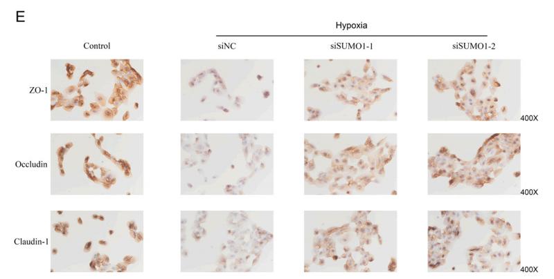
Application: IHC    Species: Human    Sample: HTEC cells

Fig. 3. SUMO1 silencing contributes to reduction in permeability of HTEC cells under hypoxic condition. (A). Knockdown of SUMO1 inhibited the secretion of IL-6, IL-8 and TNF-α in HTEC cells treated under hypoxic condition for 24 h. ***p < 0.001 vs control, !!p < 0.01 vs siNC. (B). SUMO1 silencing promoted the TEER value in HTEC cells treated under hypoxic condition for 24 h. ***p < 0.001 vs control, !!!p < 0.001 vs siNC. (C). SUMO-1 silencing reduced the permeability of HTEC cells treated under hypoxic condition for 24 h. *p < 0.05 vs control, ***p < 0.001 vs siNC; !!!p < 0.001 vs siNC. (D). Western blotting was used to examine the protein levels of SUMO1, HIF-1α, VEGF, Occluding, Claudin-1 and ZO-1 in cells as indicated. (E). Immunohistochemistry staining assay was performed to determine the levels of ZO-1, Occluding and Claudin-1 in cells as indicated above.

Application: WB    Species: Human    Sample: HTEC cells

Fig. 3. SUMO1 silencing contributes to reduction in permeability of HTEC cells under hypoxic condition. (A). Knockdown of SUMO1 inhibited the secretion of IL-6, IL-8 and TNF-α in HTEC cells treated under hypoxic condition for 24 h. ***p < 0.001 vs control, !!p < 0.01 vs siNC. (B). SUMO1 silencing promoted the TEER value in HTEC cells treated under hypoxic condition for 24 h. ***p < 0.001 vs control, !!!p < 0.001 vs siNC. (C). SUMO-1 silencing reduced the permeability of HTEC cells treated under hypoxic condition for 24 h. *p < 0.05 vs control, ***p < 0.001 vs siNC; !!!p < 0.001 vs siNC. (D). Western blotting was used to examine the protein levels of SUMO1, HIF-1α, VEGF, Occluding, Claudin-1 and ZO-1 in cells as indicated. (E). Immunohistochemistry staining assay was performed to determine the levels of ZO-1, Occluding and Claudin-1 in cells as indicated above.

4). Longan Polysaccharides with Covalent Selenylation Combat the Fumonisin B1-Induced Cell Toxicity and Barrier Disruption in Intestinal Epithelial (IEC-6) Cells. Nutrients, 2023 (PubMed: 37960333) [IF=5.9]

Application: WB    Species: Rat    Sample: IEC-6 cells

Figure 7 Expression of ZO-1, occludin, and claudin-1 in IEC-6 cells treated by the medium or polysaccharide products (LP, SeLP1, and SeLP2, 10 μg/mL) for 24 h, followed by 40 μmol/L FB1 exposure for 48 h.

5). "Yajieshaba" prevents acute alcoholic liver injury and repairs the intestinal mucosal barrier. Journal of ethnopharmacology, 2024 (PubMed: 37490990) [IF=5.4]

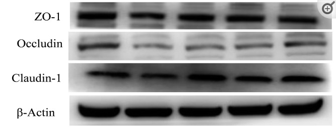
Application: WB    Species: Mouse    Sample:

Fig. 9. YJSB promoted the expression of alcohol-stimulated ileal mucosal proteins (n = 3). Panel G represents Western blot analysis shown in Fig. 8 , panel H–J represents data shown in Fig. 8 . A indicates the normal group, B indicates the model group, C indicates the Bifendate group, D indicates the YJSB 0.39 g/kg dose group, E indicates the YJSB 1.17 g/kg dose group, F indicates the YJSB 3.51 g/kg dose group, H indicates the expression of claudin1 in the ileum, I indicates the expression of occludin in the ileum, J indicates the expression of ZO-1 in the ileum. YJSB pretreatment upregulated the expression of claudin1, occludin, and ZO-1 in the ileal mucosa; ##p < 0.01 compared to the normal group; **p < 0.01 and *p < 0.05 compared to the model group.

Restrictive clause


Affinity Biosciences tests all products strictly. Citations are provided as a resource for additional applications that have not been validated by Affinity Biosciences. Please choose the appropriate format for each application and consult Materials and Methods sections for additional details about the use of any product in these publications.

For Research Use Only.
Not for use in diagnostic or therapeutic procedures. Not for resale. Not for distribution without written consent. Affinity Biosciences will not be held responsible for patent infringement or other violations that may occur with the use of our products. Affinity Biosciences, Affinity Biosciences Logo and all other trademarks are the property of Affinity Biosciences LTD.