製品: Tri-Methyl-Histone H3 (Lys27)/H3K27me3 Antibody
カタログ: DF6941
タンパク質の説明: Rabbit polyclonal antibody to Tri-Methyl-Histone H3 (Lys27)/H3K27me3
アプリケーション: WB IHC IF/ICC IP CHIP
反応性: Human, Mouse, Rat
分子量: 15kDa; 16kD(Calculated).
ユニプロット: Q16695
RRID: AB_2838900

類似製品を見る>>

   サイズ 価格 在庫状況
 100ul $280 在庫あり
 200ul $350 在庫あり

リードタイム: 当日配達

For pricing and ordering contact:
お問い合わせ先

製品説明

ソース:
Rabbit
アプリケーション:
WB 1:500-1:2000, IHC 1:50-1:200, IF/ICC 1:50-1:200, IP 1:50-1:200, CHIP 1:50-1:200
*The optimal dilutions should be determined by the end user.
*Tips:

WB: For western blot detection of denatured protein samples. IHC: For immunohistochemical detection of paraffin sections (IHC-p) or frozen sections (IHC-f) of tissue samples. IF/ICC: For immunofluorescence detection of cell samples. ELISA(peptide): For ELISA detection of antigenic peptide.

反応性:
Human,Mouse,Rat
クローナリティ:
Polyclonal
特異性:
Tri-Methyl-Histone H3 (Lys27)/H3K27me3 Antibody detects endogenous levels of Tri-Methyl-Histone H3 only when methylated at Lys27.
RRID:
AB_2838900
引用形式: Affinity Biosciences Cat# DF6941, RRID:AB_2838900.
コンジュゲート:
Unconjugated.
精製:
The antiserum was purified by peptide affinity chromatography using SulfoLink™ Coupling Resin (Thermo Fisher Scientific).
保存:
Rabbit IgG in phosphate buffered saline , pH 7.4, 150mM NaCl, 0.02% sodium azide and 50% glycerol. Store at -20 °C. Stable for 12 months from date of receipt.
別名:

折りたたみ/展開

;H3.3A; HIST1 cluster, H3E; H3 histone family, member A; H3.1; H3/l; H3F3; H3FF; H3FJ; H3FL; Histone gene cluster 1, H3 histone family, member E; histone H3.1t; Histone H3/o; FLJ92264; H 3; H3; H3 histone family, member B; H3 histone family, member C; H3 histone family, member D; H3 histone family, member F; H3 histone family, member H; H3 histone family, member I; H3 histone family, member J; H3 histone family, member K; H3 histone family, member L; H3 histone family, member T; H3 histone, family 3A; H3/A; H3/b; H3/c; H3/d; h3/f; H3/h; H3/i; H3/j; H3/k; H3/t; H31_HUMAN; H3F1K; H3F3A; H3FA; H3FB; H3FC; H3FD; H3FH; H3FI; H3FK; HIST1 cluster, H3A; HIST1 cluster, H3B; HIST1 cluster, H3C; HIST1 cluster, H3D; HIST1 cluster, H3F; HIST1 cluster, H3G; HIST1 cluster, H3H; HIST1 cluster, H3I; HIST1 cluster, H3J; HIST1H3A; HIST1H3B; HIST1H3C; HIST1H3D; HIST1H3E; HIST1H3F; HIST1H3G; HIST1H3H; HIST1H3I; HIST1H3J; HIST3H3; Histone 1, H3a; Histone 1, H3b; Histone 1, H3c; Histone 1, H3d; Histone 1, H3e; Histone 1, H3f; Histone 1, H3g; Histone 1, H3h; Histone 1, H3i; Histone 3, H3; histone cluster 1 H3 family member a; histone cluster 1 H3 family member b; histone cluster 1 H3 family member c; histone cluster 1 H3 family member d; histone cluster 1 H3 family member e; histone cluster 1 H3 family member f; histone cluster 1 H3 family member g; histone cluster 1 H3 family member h; histone cluster 1 H3 family member i; histone cluster 1 H3 family member j; Histone cluster 1, H3a; Histone cluster 1, H3b; Histone cluster 1, H3c; Histone cluster 1, H3d; Histone cluster 1, H3e; Histone cluster 1, H3f; Histone cluster 1, H3g; Histone cluster 1, H3i; Histone cluster 1, H3j; Histone gene cluster 1, H3 histone family, member A; Histone gene cluster 1, H3 histone family, member B; Histone gene cluster 1, H3 histone family, member C; Histone gene cluster 1, H3 histone family, member D; Histone gene cluster 1, H3 histone family, member F; Histone gene cluster 1, H3 histone family, member G; Histone gene cluster 1, H3 histone family, member H; Histone gene cluster 1, H3 histone family, member I; Histone gene cluster 1, H3 histone family, member J; Histone gene cluster 1, H3A; Histone gene cluster 1, H3B; Histone gene cluster 1, H3C; Histone gene cluster 1, H3D; Histone gene cluster 1, H3E; Histone gene cluster 1, H3F; Histone gene cluster 1, H3G; Histone gene cluster 1, H3H; Histone gene cluster 1, H3I; Histone gene cluster 1, H3J; Histone H 3; Histone H3.1; Histone H3.2; Histone H3.3; Histone H3/a; Histone H3/b; Histone H3/c; Histone H3/d; Histone H3/f; Histone H3/h; Histone H3/i; Histone H3/j; Histone H3/k; Histone H3/l; Histone H3/m;

免疫原

免疫原:
Uniprot:
遺伝子(ID):
発現特異性:
Q16695 H31T_HUMAN:

Expressed in testicular cells.

タンパク質の説明:
Modulation of chromatin structure plays an important role in the regulation of transcription in eukaryotes. The nucleosome, made up of DNA wound around eight core histone proteins (two each of H2A, H2B, H3, and H4), is the primary building block of chromatin (1). The amino-terminal tails of core histones undergo various post-translational modifications, including acetylation, phosphorylation, methylation, and ubiquitination (2-5). These modifications occur in response to various stimuli and have a direct effect on the accessibility of chromatin to transcription factors and, therefore, gene expression (6). In most species, histone H2B is primarily acetylated at Lys5, 12, 15, and 20 (4,7). Histone H3 is primarily acetylated at Lys9, 14, 18, 23, 27, and 56. Acetylation of H3 at Lys9 appears to have a dominant role in histone deposition and chromatin assembly in some organisms (2,3). Phosphorylation at Ser10, Ser28, and Thr11 of histone H3 is tightly correlated with chromosome condensation during both mitosis and meiosis (8-10). Phosphorylation at Thr3 of histone H3 is highly conserved among many species and is catalyzed by the kinase haspin. Immunostaining with phospho-specific antibodies in mammalian cells reveals mitotic phosphorylation at Thr3 of H3 in prophase and its dephosphorylation during anaphase (11).
タンパク質配列:
MARTKQTARKSTGGKAPRKQLATKVARKSAPATGGVKKPHRYRPGTVALREIRRYQKSTELLIRKLPFQRLMREIAQDFKTDLRFQSSAVMALQEACESYLVGLFEDTNLCVIHAKRVTIMPKDIQLARRIRGERA

PTMs - Q16695 基板として

Site PTM Type Enzyme
K5 Methylation
K10 Acetylation
K10 Methylation
S11 Phosphorylation Q13557 (CAMK2D) , O75582 (RPS6KA5) , P51812 (RPS6KA3)
T12 Phosphorylation
K15 Acetylation
K15 Methylation
K15 Sumoylation
K15 Ubiquitination
K19 Acetylation
K24 Acetylation
K24 Methylation
K28 Acetylation
K28 Methylation
K28 Ubiquitination
S29 Phosphorylation O75582 (RPS6KA5)
T33 Phosphorylation
K37 Acetylation
K37 Methylation
K37 Ubiquitination
K38 Methylation
K38 Ubiquitination
R41 Methylation
Y42 Phosphorylation
R43 Methylation
T46 Phosphorylation
R50 Methylation
K57 Acetylation
K57 Sumoylation
K57 Ubiquitination
S58 Phosphorylation
T59 Phosphorylation
R64 Methylation
K65 Methylation
K80 Acetylation
K80 Methylation
K80 Sumoylation
K80 Ubiquitination
T81 Phosphorylation
R84 Methylation
S87 Phosphorylation
S88 Phosphorylation
S99 Phosphorylation
T108 Phosphorylation
T119 Phosphorylation
K123 Acetylation
K123 Sumoylation
K123 Ubiquitination
R129 Methylation

研究背景

機能:

Core component of nucleosome. Nucleosomes wrap and compact DNA into chromatin, limiting DNA accessibility to the cellular machineries which require DNA as a template. Histones thereby play a central role in transcription regulation, DNA repair, DNA replication and chromosomal stability. DNA accessibility is regulated via a complex set of post-translational modifications of histones, also called histone code, and nucleosome remodeling.

PTMs:

Acetylation is generally linked to gene activation. Acetylation on Lys-10 (H3K9ac) impairs methylation at Arg-9 (H3R8me2s). Acetylation on Lys-19 (H3K18ac) and Lys-24 (H3K24ac) favors methylation at Arg-18 (H3R17me). Acetylation at Lys-123 (H3K122ac) by EP300/p300 plays a central role in chromatin structure: localizes at the surface of the histone octamer and stimulates transcription, possibly by promoting nucleosome instability (By similarity).

Citrullination at Arg-9 (H3R8ci) and/or Arg-18 (H3R17ci) by PADI4 impairs methylation and represses transcription.

Asymmetric dimethylation at Arg-18 (H3R17me2a) by CARM1 is linked to gene activation. Symmetric dimethylation at Arg-9 (H3R8me2s) by PRMT5 is linked to gene repression. Asymmetric dimethylation at Arg-3 (H3R2me2a) by PRMT6 is linked to gene repression and is mutually exclusive with H3 Lys-5 methylation (H3K4me2 and H3K4me3). H3R2me2a is present at the 3' of genes regardless of their transcription state and is enriched on inactive promoters, while it is absent on active promoters (By similarity).

Methylation at Lys-5 (H3K4me), Lys-37 (H3K36me) and Lys-80 (H3K79me) are linked to gene activation. Methylation at Lys-5 (H3K4me) facilitates subsequent acetylation of H3 and H4. Methylation at Lys-80 (H3K79me) is associated with DNA double-strand break (DSB) responses and is a specific target for TP53BP1. Methylation at Lys-10 (H3K9me) and Lys-28 (H3K27me) are linked to gene repression. Methylation at Lys-10 (H3K9me) is a specific target for HP1 proteins (CBX1, CBX3 and CBX5) and prevents subsequent phosphorylation at Ser-11 (H3S10ph) and acetylation of H3 and H4. Methylation at Lys-5 (H3K4me) and Lys-80 (H3K79me) require preliminary monoubiquitination of H2B at 'Lys-120'. Methylation at Lys-10 (H3K9me) and Lys-28 (H3K27me) are enriched in inactive X chromosome chromatin. Monomethylation at Lys-57 (H3K56me1) by EHMT2/G9A in G1 phase promotes interaction with PCNA and is required for DNA replication (By similarity).

Phosphorylated at Thr-4 (H3T3ph) by HASPIN during prophase and dephosphorylated during anaphase. Phosphorylation at Ser-11 (H3S10ph) by AURKB is crucial for chromosome condensation and cell-cycle progression during mitosis and meiosis. In addition phosphorylation at Ser-11 (H3S10ph) by RPS6KA4 and RPS6KA5 is important during interphase because it enables the transcription of genes following external stimulation, like mitogens, stress, growth factors or UV irradiation and result in the activation of genes, such as c-fos and c-jun. Phosphorylation at Ser-11 (H3S10ph), which is linked to gene activation, prevents methylation at Lys-10 (H3K9me) but facilitates acetylation of H3 and H4. Phosphorylation at Ser-11 (H3S10ph) by AURKB mediates the dissociation of HP1 proteins (CBX1, CBX3 and CBX5) from heterochromatin. Phosphorylation at Ser-11 (H3S10ph) is also an essential regulatory mechanism for neoplastic cell transformation. Phosphorylated at Ser-29 (H3S28ph) by MAP3K20 isoform 1, RPS6KA5 or AURKB during mitosis or upon ultraviolet B irradiation. Phosphorylation at Thr-7 (H3T6ph) by PRKCB is a specific tag for epigenetic transcriptional activation that prevents demethylation of Lys-5 (H3K4me) by LSD1/KDM1A. At centromeres, specifically phosphorylated at Thr-12 (H3T11ph) from prophase to early anaphase, by DAPK3 and PKN1. Phosphorylation at Thr-12 (H3T11ph) by PKN1 is a specific tag for epigenetic transcriptional activation that promotes demethylation of Lys-10 (H3K9me) by KDM4C/JMJD2C. Phosphorylation at Tyr-42 (H3Y41ph) by JAK2 promotes exclusion of CBX5 (HP1 alpha) from chromatin (By similarity).

Ubiquitinated.

Lysine deamination at Lys-5 (H3K4all) to form allysine is mediated by LOXL2. Allysine formation by LOXL2 only takes place on H3K4me3 and results in gene repression (By similarity).

Butyrylation of histones marks active promoters and competes with histone acetylation. It is present during late spermatogenesis.

Succinylation at Lys-80 (H3K79succ) by KAT2A takes place with a maximum frequency around the transcription start sites of genes. It gives a specific tag for epigenetic transcription activation. Desuccinylation at Lys-123 (H3K122succ) by SIRT7 in response to DNA damage promotes chromatin condensation and double-strand breaks (DSBs) repair.

Serine ADP-ribosylation constitutes the primary form of ADP-ribosylation of proteins in response to DNA damage. Serine ADP-ribosylation at Ser-11 (H3S10ADPr) is mutually exclusive with phosphorylation at Ser-11 (H3S10ph) and impairs acetylation at Lys-10 (H3K9ac).

細胞の位置付け:

Nucleus. Chromosome.

Extracellular region or secreted Cytosol Plasma membrane Cytoskeleton Lysosome Endosome Peroxisome ER Golgi apparatus Nucleus Mitochondrion Manual annotation Automatic computational assertionSubcellular location
組織特異性:

Expressed in testicular cells.

サブユニット構造:

The nucleosome is a histone octamer containing two molecules each of H2A, H2B, H3 and H4 assembled in one H3-H4 heterotetramer and two H2A-H2B heterodimers. The octamer wraps approximately 147 bp of DNA.

タンパク質ファミリー:

Belongs to the histone H3 family.

研究領域

· Human Diseases > Substance dependence > Alcoholism.

· Human Diseases > Cancers: Overview > Transcriptional misregulation in cancer.

· Human Diseases > Immune diseases > Systemic lupus erythematosus.

参考文献

1). QDPR deficiency drives immune suppression in pancreatic cancer. Cell metabolism, 2024 (PubMed: 38642552) [IF=27.7]

2). P21 and P27 promote tumorigenesis and progression via cell cycle acceleration in seminal vesicles of TRAMP mice. International Journal of Biological Sciences, 2019 (PubMed: 31592235) [IF=8.2]

3). Triclocarban exposure affects mouse oocyte in vitro maturation through inducing mitochondrial dysfunction and oxidative stress. Environmental Pollution, 2020 (PubMed: 32135433) [IF=7.6]

Application: IF/ICC    Species: mouse    Sample: oocytes

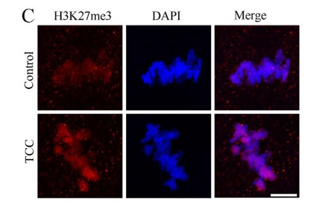
Fig. 8. TCC exposure altered epigenetic modification in mouse oocytes. (A) Images depicting H3K27me2 in control and TCC-treated oocytes. H3K27me2, red. Bar, 10 mm. (B) The fluorescence intensity of H3K27me2 in control and TCC- treated oocytes. Control, n ¼ 20; 12 mM, n ¼ 19. ***Significantly different (P < 0.0001). (C) Images depicting H3K27me3 in control and TCC-treated oocytes. H3K27me2, red. Bar, 10 mm. (D) The fluorescence intensity of H3K27me3 in control and TCC- treated oocytes. Control, n ¼ 30; 12 mM, n ¼ 24. ***Significantly different (P < 0.0001). (For interpretation of the references to colour in this figure legend, the reader is referred to the Web version of this article.)

4). Chlorothalonil exposure compromised mouse oocyte in vitro maturation through inducing oxidative stress and activating MAPK pathway. Ecotoxicology and environmental safety, 2024 (PubMed: 38367607) [IF=6.2]

Application: IF/ICC    Species: Mouse    Sample:

Fig. 6. CTL exposure perturbed the levels of H3K4me3 and H3K27me3 in mouse oocytes. The control and CTL-treated oocytes were cultured for 2 h and discarded the ones could not accomplish GVBD, while the remainders were cultured for 8 h for immunostaining. (A and C) Representative images of H3K4me3 and H3K27me3 in control and CTL-treated oocytes, respectively. DNA, blue; H3K4me3 and H3K27me3, red. Scale bar, 10 µm. (B) The fluorescence intensity of H3K4me3 in control (n = 26) and CTL-treated (n = 25) oocytes. (D) The fluorescence intensity of H3K27me3 in control (n = 30) and CTL-treated (n = 26) oocytes.

5). TNIK drives castration-resistant prostate cancer via phosphorylating EGFR. iScience, 2024 (PubMed: 38226156) [IF=5.8]

Application: WB    Species: Mouse    Sample:

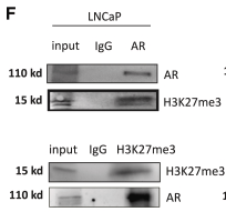
Figure 3 The essential role of the AR and H3K27me3 complex in the regulation of TNIK expression (A) LNCaP cells were cultured in steroid-depleted medium for 24 h and subsequently treated with DHT (10 nM) for the indicated time. Protein levels were analyzed by western blot. (B) The levels of TNIK protein were measured by western blot in LNCaP and C4-2 cells upon AR knockdown. (C) qPCR analysis mRNA expression of TNIK after LNCaP cells were treated with MDV3100 for 24 h. Data are shown as mean ± SD. ∗∗∗p < 0.005. (D) The levels of TNIK protein were measured in LNCaP cells treated for 24 h with the AR antagonist MDV3100 (enzalutamide, 100 nM). (E) Coimmunofluorescence (co-IF) analysis of TNIK and AR proteins in LNCaP treated with MDV3100. Scale bars: 50 μm. (F) Immunoprecipitation of AR or H3K27me3 in LNCaP and C4-2 cells followed by immunoblot analysis of H3K27me3 or AR. IgG represents a control antibody used for IPs. (G) ChIP–PCR analysis of H3K27me3 and AR binding to the TNIK gene promoter in LNCaP cells treated with MDV3100. ChIP–PCR primer in Table S3. Data are shown as mean ± SD. ∗∗∗p < 0.005. (H and I) LNCaP and C4-2 cells were treated with DZNep (0, 5, 10 μm) and GSKJ1 (0, 5, 10 μm) for 8 h, WB analysis of TNIK protein expression. (J) Western blot showed that treatment of the indicated LNCaP or C4-2 cells with GSK-J1 could reverse the effects of AR knockdown on TNIK.

6). EPC1/2 regulate hematopoietic stem and progenitor cell proliferation by modulating H3 acetylation and DLST. iScience, 2024 (PubMed: 38439957) [IF=5.8]

Application: WB    Species: Human    Sample:

Fig S7. Effects of epc1a and epc2 deficiency on H3 or H4 acetylated protein, related to Figures 2 and 3. (A) Enriched GO terms for acetylation-related DEGs in epc1a-/- mutants. (B) The quantification of H3K9Ac (B1), H3K27Ac (B2) and H3K56Ac (B3) proteins in control, epc1a-/- and epc2-/- embryos. (C) H4K5/K8/K12/K16Ac protein level in the control, epc1a-/- and epc2-/- embryos at 33 hpf (C1), and quantification analysis (C2). (D, E) Western blotting analysis of H3K4me1 (D1) and H3K27me3 (E1) in the control, epc1a-/- and epc2-/- embryos, with Actin and PCNA as internal controls, and quantification of H3K4me1 (D2) and H3K27me3 (E2). (F, G) Double staining of runx1-GFP with anti-H3K9Ac (F1-F12) and H3K56Ac (G1-G12) in the CHT in the control, epc1a-/- and epc2-/- embryos at 72 hpf, and quantification of runx1+ cells (F13, G13), runx1+H3K9Ac+ (F14) and runx1+H3K56Ac+ cells (G14), with white arrowheads indicating double-positive cells. F4, F8, F12, G4, G8 and G12 show the magnified images of F3, F7, F11, G3, G7 and G11, respectively. (H) Protein level of H3K27Ac in the control, epc1a-/- and epc2-/- embryos and the corresponding groups treated with VPA (H1), and quantification analysis (H2). Each experiment was repeated three times, and a representative result is shown. All embryos are shown in lateral view, anterior to the left, and dorsal to the up. Scale bars, 100 μm (F1-F12, G1-G12). Data are presented as mean ± SD (n ≥ 3). t-test, *P < 0.05, **P < 0.01, ***P < 0.001, NS, not significant.

7). HBx-mediated decrease of AIM2 contributes to hepatocellular carcinoma metastasis. Molecular Oncology, 2017 (PubMed: 28580773) [IF=5.0]

Application: WB    Species:    Sample:

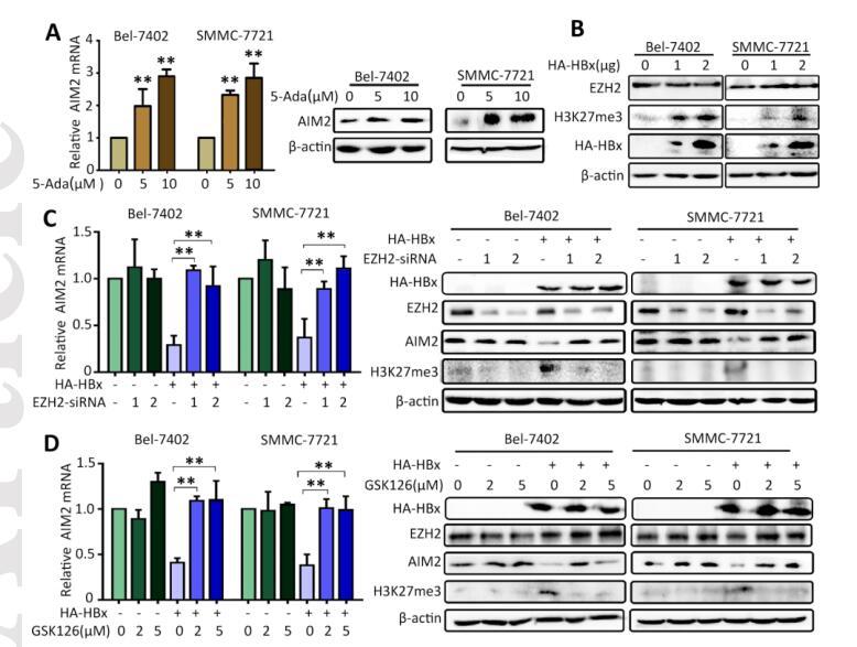
Figure 3. EZH2 is required for HBx-induced AIM2 decrease. A. AIM2 mRNA and protein expression levels were tested in Bel-7402 and SMMC-7721 cells treated with 5-Aza for 24 h. B. EZH2 and H3K27me3 level was detected in Bel-7402 and SMMC-7721 cells transfected with HBx. C. EZH2 was transiently knocked down by siRNAs in HBx-expressing Bel-7402 and SMMC-7721 cells. AIM2 expression was determined. D. EZH2 activity was blocked by its inhibitor GSK-126 in cells with or without the presence of HBx. The impact of EZH2 on AIM2 expression was checked.

8). Bisphenol B exposure disrupts mouse oocyte meiotic maturation in vitro through affecting spindle assembly and chromosome alignment. Frontiers in Cell and Developmental Biology, 2020 (PubMed: 33392205) [IF=4.6]

Application: IF/ICC    Species: mouse    Sample: oocytes

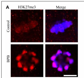
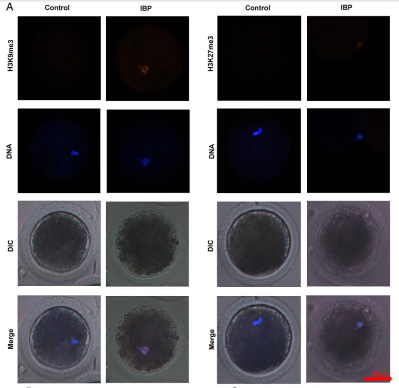
FIGURE 7 | BPB exposure altered epigenetic modification in mouse oocytes.(A) Images depicting H3K27me3 in control and BPB-treated oocytes.H3K27me3, red. Bar, 10 µm.

9). Bisphenol F exposure affects mouse oocyte in vitro maturation through inducing oxidative stress and DNA damage. ENVIRONMENTAL TOXICOLOGY, 2022 (PubMed: 35218298) [IF=4.4]

10). High-dose synthetic phenolic antioxidant propyl gallate impairs mouse oocyte meiotic maturation through inducing mitochondrial dysfunction and DNA damage. Environmental Toxicology, 2023 (PubMed: 37052413) [IF=4.4]

もっと読みます

Restrictive clause


Affinity Biosciences tests all products strictly. Citations are provided as a resource for additional applications that have not been validated by Affinity Biosciences. Please choose the appropriate format for each application and consult Materials and Methods sections for additional details about the use of any product in these publications.

For Research Use Only.
Not for use in diagnostic or therapeutic procedures. Not for resale. Not for distribution without written consent. Affinity Biosciences will not be held responsible for patent infringement or other violations that may occur with the use of our products. Affinity Biosciences, Affinity Biosciences Logo and all other trademarks are the property of Affinity Biosciences LTD.