製品: FOXM1 Antibody
カタログ: DF6962
タンパク質の説明: Rabbit polyclonal antibody to FOXM1
アプリケーション: WB IHC
反応性: Human, Mouse, Rat
予測: Pig, Zebrafish, Bovine, Horse, Sheep, Rabbit, Dog, Xenopus
分子量: 84kD, 100-130kD; 84kD(Calculated).
ユニプロット: Q08050
RRID: AB_2838918

類似製品を見る>>

   サイズ 価格 在庫状況
 100ul $280 在庫あり
 200ul $350 在庫あり

リードタイム: 当日配達

For pricing and ordering contact:
お問い合わせ先

製品説明

ソース:
Rabbit
アプリケーション:
WB 1:500-1:2000, IHC 1:50-1:200
*The optimal dilutions should be determined by the end user.
*Tips:

WB: For western blot detection of denatured protein samples. IHC: For immunohistochemical detection of paraffin sections (IHC-p) or frozen sections (IHC-f) of tissue samples. IF/ICC: For immunofluorescence detection of cell samples. ELISA(peptide): For ELISA detection of antigenic peptide.

反応性:
Human,Mouse,Rat
予測:
Pig(100%), Zebrafish(83%), Bovine(100%), Horse(100%), Sheep(100%), Rabbit(92%), Dog(100%), Xenopus(92%)
クローナリティ:
Polyclonal
特異性:
FOXM1 Antibody detects endogenous levels of total FOXM1.
RRID:
AB_2838918
引用形式: Affinity Biosciences Cat# DF6962, RRID:AB_2838918.
コンジュゲート:
Unconjugated.
精製:
The antiserum was purified by peptide affinity chromatography using SulfoLink™ Coupling Resin (Thermo Fisher Scientific).
保存:
Rabbit IgG in phosphate buffered saline , pH 7.4, 150mM NaCl, 0.02% sodium azide and 50% glycerol. Store at -20 °C. Stable for 12 months from date of receipt.
別名:

折りたたみ/展開

FKHL16; Forkhead box M1; Forkhead box protein M1; forkhead like 16; Forkhead-related protein FKHL16; FOX M1; Foxm1; FOXM1_HUMAN; FOXM1B; Hepatocyte nuclear factor 3 forkhead homolog 11; HFH-11; HFH11; HNF-3/fork-head homolog 11; HNF3; INS1; M phase phosphoprotein 2; M-phase phosphoprotein 2; MPHOSPH2; MPM-2 reactive phosphoprotein 2; MPP2; PIG29; TGT3; Transcription factor Trident; Trident; WIN; Winged-helix factor from INS-1 cells; Winged-helix factor from INS1 cells;

免疫原

免疫原:
Uniprot:
遺伝子(ID):
発現特異性:
Q08050 FOXM1_HUMAN:

Expressed in thymus, testis, small intestine, colon followed by ovary. Appears to be expressed only in adult organs containing proliferating/cycling cells or in response to growth factors. Also expressed in epithelial cell lines derived from tumors. Not expressed in resting cells. Isoform 2 is highly expressed in testis.

タンパク質の説明:
Forkhead box M1 (FoxM1) is a forkhead box family transcription factor that regulates a number of genes throughout the cell cycle to help control DNA replication, mitosis and cell proliferation. FoxM1 expression increases during G1 and S and reaches maximum levels in G2/M (1-3). Nuclear translocation occurs just before entry into G2/M and is associated with FoxM1 phosphorylation (4). Phosphorylation of FoxM1 by MAPK (Ser331 Ser704), Cyclin/Cdk (Thr600, Thr611, Thr672), Plk1 (Ser715, Ser724), and Chk2 (Ser376) stabilizes and activates FoxM1 (4-7). Forkhead box M1 is expressed in all embryonic tissues but is restricted to proliferating tissues in adults (8). Recent studies have shown that FoxM1 expression is negatively regulated by p53 (9,10). Upregulation of FoxM1 is associated with many human cancers, including prostate, breast, lung, ovary, colon, pancreas, stomach, bladder, liver, and kidney, and may be associated with p53 mutations in some tumors (10,11). As a result, FoxM1 inhibitors have become a topic of interest for potential cancer therapy (12).
タンパク質配列:
MKTSPRRPLILKRRRLPLPVQNAPSETSEEEPKRSPAQQESNQAEASKEVAESNSCKFPAGIKIINHPTMPNTQVVAIPNNANIHSIITALTAKGKESGSSGPNKFILISCGGAPTQPPGLRPQTQTSYDAKRTEVTLETLGPKPAARDVNLPRPPGALCEQKRETCADGEAAGCTINNSLSNIQWLRKMSSDGLGSRSIKQEMEEKENCHLEQRQVKVEEPSRPSASWQNSVSERPPYSYMAMIQFAINSTERKRMTLKDIYTWIEDHFPYFKHIAKPGWKNSIRHNLSLHDMFVRETSANGKVSFWTIHPSANRYLTLDQVFKPLDPGSPQLPEHLESQQKRPNPELRRNMTIKTELPLGARRKMKPLLPRVSSYLVPIQFPVNQSLVLQPSVKVPLPLAASLMSSELARHSKRVRIAPKVLLAEEGIAPLSSAGPGKEEKLLFGEGFSPLLPVQTIKEEEIQPGEEMPHLARPIKVESPPLEEWPSPAPSFKEESSHSWEDSSQSPTPRPKKSYSGLRSPTRCVSEMLVIQHRERRERSRSRRKQHLLPPCVDEPELLFSEGPSTSRWAAELPFPADSSDPASQLSYSQEVGGPFKTPIKETLPISSTPSKSVLPRTPESWRLTPPAKVGGLDFSPVQTSQGASDPLPDPLGLMDLSTTPLQSAPPLESPQRLLSSEPLDLISVPFGNSSPSDIDVPKPGSPEPQVSGLAANRSLTEGLVLDTMNDSLSKILLDISFPGLDEDPLGPDNINWSQFIPELQ

種類予測

種類予測:

Score>80(red) has high confidence and is suggested to be used for WB detection. *The prediction model is mainly based on the alignment of immunogen sequences, the results are for reference only, not as the basis of quality assurance.

Species
Results
Score
Pig
100
Horse
100
Bovine
100
Sheep
100
Dog
100
Xenopus
92
Rabbit
92
Zebrafish
83
Chicken
0
Model Confidence:
High(score>80) Medium(80>score>50) Low(score<50) No confidence

PTMs - Q08050 基板として

Site PTM Type Enzyme
S4 Phosphorylation Q00534 (CDK6) , P11802 (CDK4)
S35 Phosphorylation P11802 (CDK4) , Q00534 (CDK6)
S41 Phosphorylation
K57 Ubiquitination
K63 Acetylation
K189 Ubiquitination
S191 Phosphorylation
K201 Sumoylation
K218 Sumoylation
S251 Phosphorylation P06493 (CDK1)
K278 Methylation
K278 Ubiquitination
K282 Methylation
S331 Phosphorylation
T354 Phosphorylation
K356 Sumoylation
T357 Phosphorylation
S376 Phosphorylation O96017 (CHEK2)
K422 Acetylation
K422 Sumoylation
K422 Ubiquitination
K440 Acetylation
K440 Sumoylation
K440 Ubiquitination
S451 Phosphorylation Q00534 (CDK6) , P11802 (CDK4)
K460 Sumoylation
K478 Sumoylation
S481 Phosphorylation
S489 Phosphorylation Q00534 (CDK6) , P11802 (CDK4)
K495 Sumoylation
S501 Phosphorylation
S508 Phosphorylation Q00534 (CDK6) , P11802 (CDK4)
T510 Phosphorylation P11802 (CDK4) , Q00534 (CDK6)
Y517 Phosphorylation
S522 Phosphorylation Q00534 (CDK6) , P11802 (CDK4)
T524 Phosphorylation
T600 Phosphorylation P11802 (CDK4) , P06493 (CDK1) , Q00534 (CDK6)
K603 Acetylation
K603 Sumoylation
T611 Phosphorylation P11802 (CDK4) , P06493 (CDK1) , Q00534 (CDK6)
K614 Acetylation
T620 Phosphorylation Q00534 (CDK6) , P11802 (CDK4) , P06493 (CDK1)
S623 Phosphorylation
T627 Phosphorylation P11802 (CDK4) , Q00534 (CDK6)
S638 Phosphorylation
S672 Phosphorylation
S692 Phosphorylation
S693 Phosphorylation
S704 Phosphorylation Q00534 (CDK6) , P11802 (CDK4)
S717 Phosphorylation
T719 Phosphorylation
S730 Phosphorylation P53350 (PLK1)
S739 Phosphorylation P53350 (PLK1)

研究背景

機能:

Transcriptional factor regulating the expression of cell cycle genes essential for DNA replication and mitosis. Plays a role in the control of cell proliferation. Plays also a role in DNA breaks repair participating in the DNA damage checkpoint response.

PTMs:

Phosphorylated in M (mitotic) phase. Phosphorylation by the checkpoint kinase CHEK2 in response to DNA damage increases the FOXM1 protein stability probably stimulating the transcription of genes involved in DNA repair. Phosphorylated by CDK1 in late S and G2 phases, creating docking sites for the POLO box domains of PLK1. Subsequently, PLK1 binds and phosphorylates FOXM1, leading to activation of transcriptional activity and subsequent enhanced expression of key mitotic regulators.

細胞の位置付け:

Nucleus.

Extracellular region or secreted Cytosol Plasma membrane Cytoskeleton Lysosome Endosome Peroxisome ER Golgi apparatus Nucleus Mitochondrion Manual annotation Automatic computational assertionSubcellular location
組織特異性:

Expressed in thymus, testis, small intestine, colon followed by ovary. Appears to be expressed only in adult organs containing proliferating/cycling cells or in response to growth factors. Also expressed in epithelial cell lines derived from tumors. Not expressed in resting cells. Isoform 2 is highly expressed in testis.

タンパク質ファミリー:

Within the protein there is a domain which acts as a transcriptional activator. Insertion of a splicing sequence within it inactivates this transcriptional activity, as it is the case for isoform 4.

研究領域

· Cellular Processes > Cell growth and death > Cellular senescence.   (View pathway)

参考文献

1). Auxiliary antitumor effects of fungal proteins from Hericium erinaceus by target on the gut microbiota. Journal of Food Science, 2020 (PubMed: 32460371) [IF=3.9]

Application: WB    Species: mouse    Sample: tumor

Figure 12|–Effects of fungal proteins extracted from Hericium erinaceus on tumor tissues in xenografted cancer mice. (A-H) is the QT-PCR analysis results of some key genes in the tumor tissues; (I) is the western bolting analysis results of some key proteins in the tumor tissues; (J) is the level of LPS in serum;(K)is the plasma concentration of 5-Fu.

2). MFAP2 aggravates tumor progression through activating FOXM1/β‐catenin‐mediated glycolysis in ovarian cancer. KAOHSIUNG JOURNAL OF MEDICAL SCIENCES, 2022 (PubMed: 35546486) [IF=3.3]

Application: WB    Species: Human    Sample: A2780 and SKOV3 cells

FIGURE 4MFAP2 promotes FOXM1/β-catenin-mediated glycolysis signaling in ovarian cancer cells. (A) The levels of FOXM1 and glycolysis-related protein expression in A2780 and SKOV3 cells were determined by western blot assay; (B) the levels of Wnt/β-catenin signaling pathway and its downstream target genes in A2780 and SKOV3 cells were determined by western blot assay; (C) the levels of β-catenin in the nucleus and cytoplasm of A2780 and SKOV3 cells were analyzed by western blot assay. Data are shown as mean ± SD; *p < 0.05, **p < 0.01, and ***p < 0.001. MFAP2, Microfibril-associated protein 2

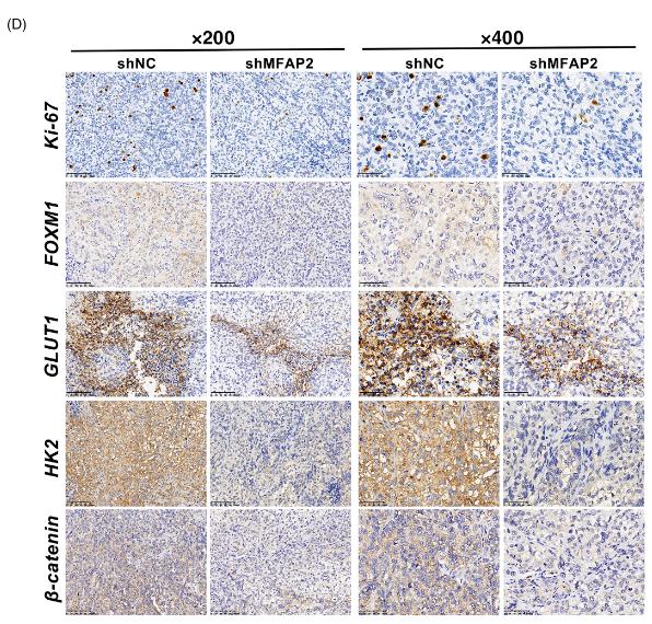
Application: IHC    Species: Mice    Sample: SKOV3 cells

FIGURE 5Knockdown of MFAP2 inhibits xenograft tumor growth of ovarian cancer cells in vivo (A) photographs of tumors excised from the mice injected with SKOV3 cells transfected with shNC or shMFAP2. (B,C) Tumor growth curves (B) and tumor weight (C) of mice injected with A2780 cells. (D) The levels of MFAP2, Ki-67, FOXM1, GLUT1, HK2, and β-catenin in tumors from mice injected with SKOV3 cells were evaluated by immunohistochemical (IHC) staining (scale bar = 100 μm, magnification, ×200; scale bar = 50 μm, magnification, ×400). Data are shown as mean ± SD; *p < 0.05, **p < 0.01, and ***p < 0.001. IHC, xxxxxx; MFAP2, Microfibril-associated protein 2

3). The Interaction of GLUT1 and FOXM1 Leads to a Poor Prognosis in Colorectal Cancer. Anti-Cancer Agents in Medicinal Chemistry, 2020 (PubMed: 32188390) [IF=2.8]

4). Fungal Proteins from Hericium Erinaceus Show Auxiliary Antitumor Effects with 5-Fluoro-2,4(1H,3H)-Pyrimidinedione by Improving the Gut Microbiota in Mice. , 2018

Restrictive clause


Affinity Biosciences tests all products strictly. Citations are provided as a resource for additional applications that have not been validated by Affinity Biosciences. Please choose the appropriate format for each application and consult Materials and Methods sections for additional details about the use of any product in these publications.

For Research Use Only.
Not for use in diagnostic or therapeutic procedures. Not for resale. Not for distribution without written consent. Affinity Biosciences will not be held responsible for patent infringement or other violations that may occur with the use of our products. Affinity Biosciences, Affinity Biosciences Logo and all other trademarks are the property of Affinity Biosciences LTD.